
Abstract
BACKGROUND AND PURPOSE: The levator claviculae muscle is an infrequently recognized variant in humans, occurring in 2% to 3% of the population, and has rarely been reported in the radiologic or anatomic literature. The importance of this muscle to radiologists is in distinguishing it from an abnormality; most commonly, cervical adenopathy. After discovering this muscle on the CT scans of two patients during routine clinical examinations, we conducted a study to determine the prevalence and appearance of the muscle on CT studies.
METHODS: We evaluated 300 CT scans that adequately depicted the expected location of the muscle. The most superior level in which the muscle could be identified and the apparent location of insertion on the clavicle were recorded for all subjects in whom the muscle was detected.
RESULTS: Seven levator claviculae muscles were identified in six subjects (2%). It was bilateral in one, on the left in four, and on the right in one. It was identified up to the level of the transverse process of C3 in all cases. The insertion was the middle third of the clavicle for two muscles and the lateral third of the clavicle for the remaining five muscles.
CONCLUSION: Because the levator claviculae muscle will most likely be encountered during a radiologist's career, it is important to recognize this muscle as a variant and not as an abnormality.
The levator claviculae muscle is a normal muscle in lower mammals but is an infrequently recognized variant in humans. It arises from the transverse processes of the upper cervical vertebrae and inserts in the lateral half of the clavicle. Although this anomaly was first described over 125 years ago, and has been reported to occur in 2% to 3% of the population (1), it has only rarely been described in the literature (2–4). The importance of this muscle to radiologists is in distinguishing it from an abnormality; most commonly, cervical adenopathy (2, 3). After discovering this muscle on the CT scans of two patients during routine clinical examinations, we conducted a study to determine the prevalence and appearance of the muscle on CT scans.
Methods
Consecutive CT studies of the neck were reviewed retrospectively to obtain 300 scans that clearly depicted the anatomy of the region of the levator claviculae muscle. To be included in the study, the scans had to have a section thickness of 5 mm or less and had to include, as a minimum, the neck from the level of C4 to the level of T1, the region covering the characteristic course of the muscle. Scans were obtained without angulation of the gantry. Fifty-nine scans were excluded because the region at which the muscle can be identified was obscured by neoplasm, prior surgery, trauma, or body habitus.
The scans were reviewed independently by two radiologists. The most superior extent of the muscle and its apparent insertion site on the clavicle was recorded for all subjects in whom the muscle was identified. In the two cases in which the radiologists disagreed, a consensus was reached when they viewed the scans together.
The short-axis dimension of each levator claviculae muscle was measured.
Results
Seven levator claviculae muscles were identified in six subjects. In four subjects, the muscle was on the left, in one it was on the right, and in one the muscle was found bilaterally (Figs 1 and 2). The muscle could be identified up to the level of the transverse process of C3 in all subjects. The muscle then coursed inferiorly and laterally to pass lateral to the scalene muscles and levator scapulae muscle while remaining medial to the sternocleidomastoid muscle. The levator claviculae muscle then passed posterior to the sternocleidomastoid muscle before inserting on the clavicle. Two of the muscles, including one of the bilateral muscles, appeared to insert near the middle of the clavicle. The remaining five muscles inserted on the lateral third of the clavicle. Three of these appeared to blend with the trapezius before inserting on the clavicle.

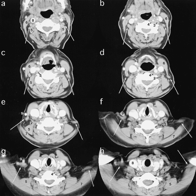
A–H, CT scans obtained from superior (a) to inferior (h) in a subject with a left levator claviculae muscle (arrows). At the most superior level at which it is seen, the muscle is located lateral to the transverse process of C3 and immediately anterior and lateral to the levator scapulae (5). Its medial margin is posterolateral to the longus capitis muscle (3). As the muscle descends inferiorly, it moves laterally, lying anterior and lateral to the levator scapulae (5). The muscle moves from medial to the sternocleidomastoid muscle (6) to posterior to it. Just above its insertion, the levator claviculae muscle lies close to the trapezius (7).
Key to Abbreviations
1 Anterior scalene muscle
2 Middle and/or posterior scalene muscle
3 Longus capitis muscle
4 Longus colli muscle
5 Levator scapulae muscle
6 Sternocleidomastoid muscle
7 Trapezius muscle
8 Internal jugular vein
9 External jugular vein
n Lymph node
→ Levator claviculae muscle

A–H, CT scans obtained from superior (a) to inferior (h) in the subject with bilateral levator claviculae muscles (arrows). The muscles have a similar course to the levator claviculae muscles in fig 1. On the right, the muscle lies anterior to the levator scapulae (5) and anteromedial to the trapezius (7), and appears to insert near the middle of the clavicle. On the left, the muscle has a more lateral course and insertion
The largest short-axis dimension of the levator claviculae muscle varied from 5 to 11 mm, with three muscles measuring 10 or 11 mm.
Discussion
We found the frequency of the levator claviculae muscle to be 2%, a figure similar to the prevalence reported more than a century ago (1). The inclusion of our initial two cases in the retrospective study may have biased our results toward a higher rate of occurrence, even though these cases were in the middle of the time period during which scans for this study were obtained. If these cases were excluded, the frequency would drop to 1.3%.
The levator claviculae muscle has a characteristic course through the neck. It has been reported to arise from the anterior portion of the transverse processes of the upper cervical vertebrae (1–4). We could not clearly see the origin of the muscle, as it is obscured by the belly of the longus capitis muscle and may be in a similar location as the upper origins of the longus capitis and longus coli muscles (5). The levator claviculae muscle could be identified most superiorly at the level of C3, where the muscle body could be seen; therefore, the origin is most likely above this level, although slips may arise at the C3 level as well. The levator claviculae muscle then headed inferiorly and laterally, coursing lateral to the scalene muscles, anterior or anterolateral to the levator scapulae muscle, and medial to the sternocleidomastoid muscle. It then passed posterior and lateral to the sternocleidomastoid muscle toward the lateral half of the clavicle. The insertion on the clavicle was not clearly seen, most likely because it inserts as a smaller muscle or tendon. The apparent insertion point on the clavicle was slightly variable and ranged from the middle of the clavicle to the most lateral portion, where it appeared to blend with the trapezius muscle. Recognition of the typical course makes for relatively easy identification of the muscle, which may vary in size. The course allows the differentiation of the muscle from lymphadenopathy or an unenhanced vessel.
Our experience is similar to previous descriptions, with the majority describing the origin of the levator claviculae muscle from the upper cervical vertebrae transverse processes and the insertion in the middle to lateral thirds of the clavicle (1–4). In our series, they all arose from the C3 level or more cranially, and the majority inserted in the lateral third of the clavicle. Our experience is also similar to that of others with regard to laterality. When the muscle occurs unilaterally, it is more common on the left. The series by Wood (1) had more bilateral occurrences (50% to our 17%) but the numbers in both series were small. Additional variants have been described, but are sufficiently different to possibly represent a different muscle (6).
Conclusion
Because the levator claviculae muscle occurs in about 2% of the population, it is a variant that the average radiologist will encounter during his or her career. Recognition of the levator claviculae muscle as a normal variant will prevent misinterpretation of the muscle as adenopathy or a thrombosed vein.
Footnotes
- Received June 25, 1998.
- Copyright © American Society of Neuroradiology


