
Article Figures & Data
Figures
- fig 1.
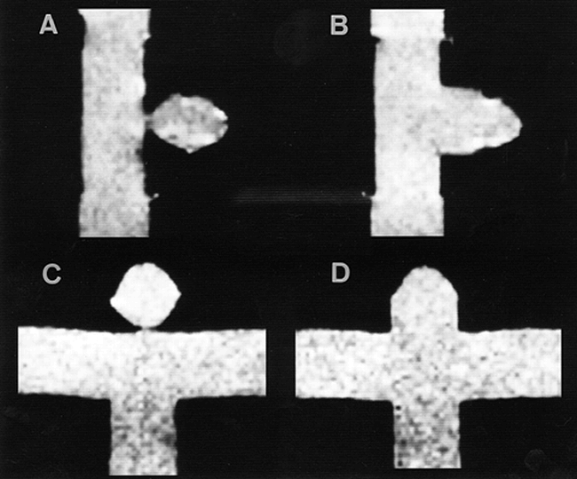
A–D, Spin-echo T1-weighted images (300/15/1) of lateral and terminal saccular aneurysm models with fluid but without active flow. The height and fundus diameter of the models were 10 mm; the neck diameters were 2.5 mm (A, C) and 10.0 mm (B, D).
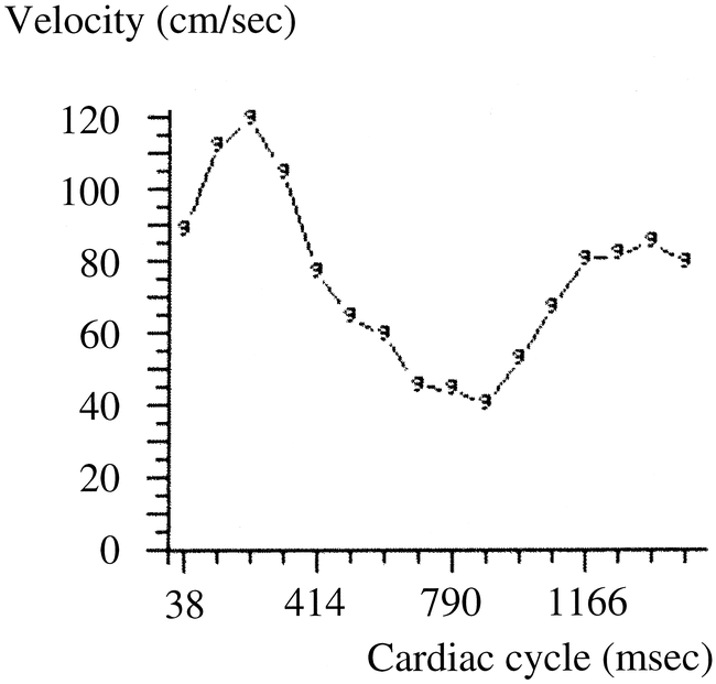
- fig 2.
Flow velocity during cardiac cycle.
- fig 3.
Tagged MR images of the lateral saccular aneurysm model with a neck size of 2.5 mm.
A–F, The time difference between images is 25 milliseconds (large arrows indicate the flow direction of the parent vessel; small arrows, the tag stripe).
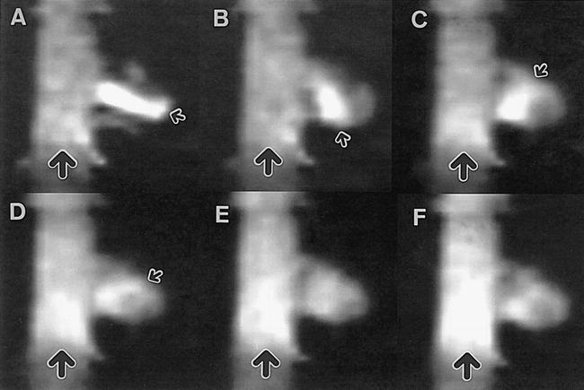
- fig 4.
Tagged MR images of the lateral saccular aneurysm model with a neck size of 10 mm.
A–F, The time difference between images is 25 milliseconds (large arrows indicate the flow direction of the parent vessel; small arrows, the high signal band between tag stripes).
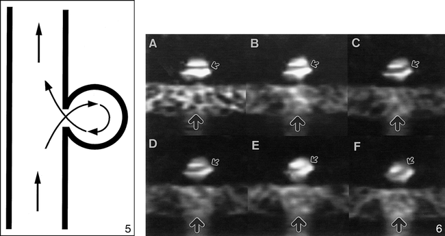
- fig 5.
Diagram of circulation in lateral saccular aneurysm models.fig 6. Tagged MR images of the terminal saccular aneurysm model with a neck size of 2.5 mm.
A–F, The time difference between images is 25 milliseconds (large arrows indicate the flow direction of the parent vessel; small arrows, the tag stripe).
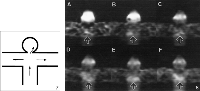
- fig 7.
Diagram of circulation in a terminal saccular aneurysm model with a neck size of 2.5 mm.fig 8. Tagged MR images of the terminal saccular aneurysm model with a neck size of 10 mm.
A–F, The time difference between images is 25 milliseconds (arrows indicate the flow direction of the parent vessel model).


