
Article Figures & Data
Figures
- fig 1.
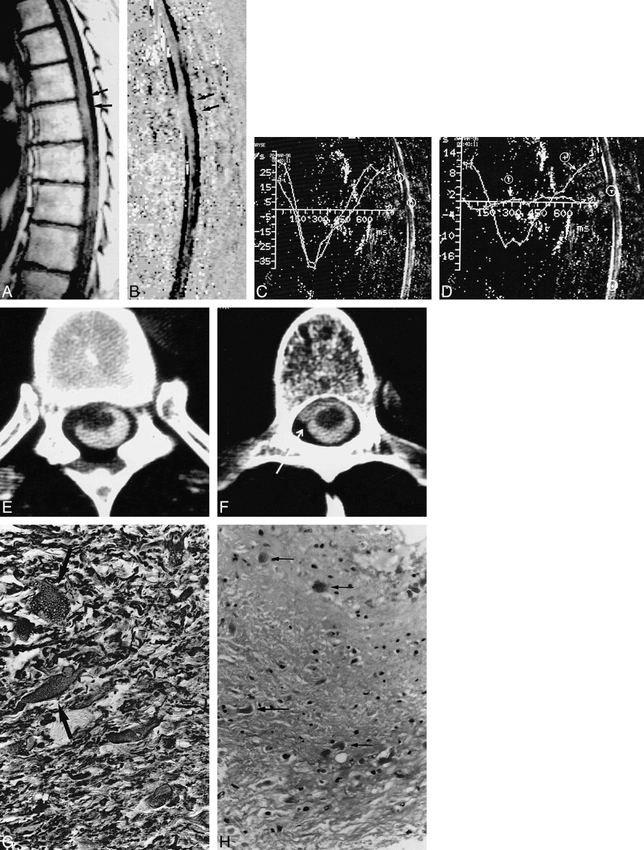
Case 1: spontaneous spinal cord herniation.
A, Sagittal T1-weighted MR image shows ventral displacement of the spinal cord at the T6 level (arrows).
B, Sagittal velocity image during systolic phase, 300 milliseconds after the R wave of the EKG. Dark areas correspond to craniocaudal flow. No flow is visible on the ventral side of the spinal cord at the T6 level. Normal flow is seen in the posterior subarachnoid spaces. A posterior arachnoid cyst is unlikely.
C and D, Evolution of CSF flow during the RR cycle of the ECG. By convention, caudal flow is represented as negative. CSF velocity curves measured in the anterior subarachnoid spaces superior to the cord herniation and in the posterior subarachnoid spaces at the level of the herniation show similar normal patterns (C). There is no evidence of an associated posterior arachnoid cyst. Cursors have been placed at the level of the herniation (T) and below it (+). There is limited cord motion at the level of the herniation, as manifested by a minimal deflection of the pulse wave, T, from zero, whereas normal cord motion is identified below the herniation (D).
E and F, CT myelography at the level of the cord herniation (E): the cord is ventrally displaced and protrudes into the left anterolateral epidural space. Just below the cord herniation, an anterior pseudomeningocele is filled with contrast medium (arrow, F).
G, On histopathologic section, hypertrophic astrocytes are visible in some areas (arrows) (alkaline phosphatase-antialkaline phosphatase method for GFAP, original magnification ×400).
H, Histologic section shows chronic reactive changes with Rosenthal fibers (arrows) and mild pilocytic gliosis (H and E, original magnification ×250).
- fig 2.
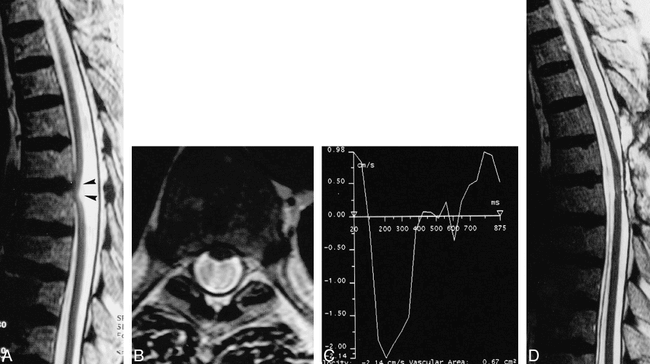
Case 2: ISCH.
A, Sagittal T2-weighted MR image shows ventral displacement of the spinal cord at the T5–T6 level (arrowheads).
B, Axial T2-weighted MR image at the level of the cord herniation shows anterior cord displacement and loss of the anterior subarachnoid spaces. Loss of phase coherence is responsible for the dark signal in the dorsal subarachnoid space.
C, CSF velocity curve measured in the posterior subarachnoid spaces at the level of the herniation shows a normal CSF velocity pattern. A posterior arachnoid cyst was considered unlikely.
D, Postoperative sagittal T2-weighted MR image shows normal location of the spinal cord.
- fig 3.
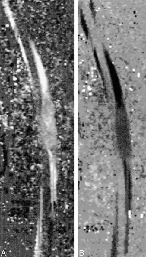
Arachnoid cyst.
A and B, Sagittal velocity images during diastolic (A) and systolic (B) phases show low CSF flow (gray areas). The contents of the distended arachnoid cyst are clearly visible along the dorsal side of the spinal cord.
In this issue


Jump to section
Related Articles
Cited By...
- Thoracic idiopathic spinal cord herniation in a young patient: a diagnostic and therapeutic challenge
- Clinical Reasoning: A case of slowly progressive painful paraparesis
- The thoracic anterior spinal cord adhesion syndrome
- Idiopathic Thoracic Spinal Cord Herniation: Retrospective Analysis Supporting a Mechanism of Diskogenic Dural Injury and Subsequent Tamponade
- The American Journal of Neuroradiology 1980-1999 Where We Have Been: Where We Are Going