Advertisement

Main menu

  • Home
  • Content
    • Current Issue
    • Accepted Manuscripts
    • Article Preview
    • Past Issue Archive
    • Video Articles
    • AJNR Case Collection
    • Case of the Week Archive
    • Case of the Month Archive
    • Classic Case Archive
  • Special Collections
    • AJNR Awards
    • ASNR Foundation Special Collection
    • Most Impactful AJNR Articles
    • Photon-Counting CT
    • Spinal CSF Leak Articles (Jan 2020-June 2024)
  • Multimedia
    • AJNR Podcasts
    • AJNR SCANtastic
    • Trainee Corner
    • MRI Safety Corner
    • Imaging Protocols
  • For Authors
    • Submit a Manuscript
    • Submit a Video Article
    • Submit an eLetter to the Editor/Response
    • Manuscript Submission Guidelines
    • Statistical Tips
    • Fast Publishing of Accepted Manuscripts
    • Graphical Abstract Preparation
    • Imaging Protocol Submission
    • Author Policies
  • About Us
    • About AJNR
    • Editorial Board
    • Editorial Board Alumni
  • More
    • Become a Reviewer/Academy of Reviewers
    • Subscribers
    • Permissions
    • Alerts
    • Feedback
    • Advertisers
    • ASNR Home

User menu

  • Alerts
  • Log in

Search

  • Advanced search
American Journal of Neuroradiology

American Journal of Neuroradiology

ASHNR American Society of Functional Neuroradiology ASHNR American Society of Pediatric Neuroradiology ASSR
  • Alerts
  • Log in

Advanced Search

  • Home
  • Content
    • Current Issue
    • Accepted Manuscripts
    • Article Preview
    • Past Issue Archive
    • Video Articles
    • AJNR Case Collection
    • Case of the Week Archive
    • Case of the Month Archive
    • Classic Case Archive
  • Special Collections
    • AJNR Awards
    • ASNR Foundation Special Collection
    • Most Impactful AJNR Articles
    • Photon-Counting CT
    • Spinal CSF Leak Articles (Jan 2020-June 2024)
  • Multimedia
    • AJNR Podcasts
    • AJNR SCANtastic
    • Trainee Corner
    • MRI Safety Corner
    • Imaging Protocols
  • For Authors
    • Submit a Manuscript
    • Submit a Video Article
    • Submit an eLetter to the Editor/Response
    • Manuscript Submission Guidelines
    • Statistical Tips
    • Fast Publishing of Accepted Manuscripts
    • Graphical Abstract Preparation
    • Imaging Protocol Submission
    • Author Policies
  • About Us
    • About AJNR
    • Editorial Board
    • Editorial Board Alumni
  • More
    • Become a Reviewer/Academy of Reviewers
    • Subscribers
    • Permissions
    • Alerts
    • Feedback
    • Advertisers
    • ASNR Home
  • Follow AJNR on Twitter
  • Visit AJNR on Facebook
  • Follow AJNR on Instagram
  • Join AJNR on LinkedIn
  • RSS Feeds

AJNR is seeking candidates for the AJNR Podcast Editor. Read the position description.

Research ArticleBRAIN

Hyperintensities of the Optic Radiation on T2-Weighted MR Images of Elderly Subjects

Mika Kitajima, Yukunori Korogi, Tomoko Okuda, Shin-ya Shiraishi, Osamu Ikeda, Shoji Morishita, Mutsumasa Takahashi and Komyo Eto
American Journal of Neuroradiology June 1999, 20 (6) 1009-1014;
Mika Kitajima
  • Find this author on Google Scholar
  • Find this author on PubMed
  • Search for this author on this site
Yukunori Korogi
  • Find this author on Google Scholar
  • Find this author on PubMed
  • Search for this author on this site
Tomoko Okuda
  • Find this author on Google Scholar
  • Find this author on PubMed
  • Search for this author on this site
Shin-ya Shiraishi
  • Find this author on Google Scholar
  • Find this author on PubMed
  • Search for this author on this site
Osamu Ikeda
  • Find this author on Google Scholar
  • Find this author on PubMed
  • Search for this author on this site
Shoji Morishita
  • Find this author on Google Scholar
  • Find this author on PubMed
  • Search for this author on this site
Mutsumasa Takahashi
  • Find this author on Google Scholar
  • Find this author on PubMed
  • Search for this author on this site
Komyo Eto
  • Find this author on Google Scholar
  • Find this author on PubMed
  • Search for this author on this site
  • Article
  • Figures & Data
  • Info & Metrics
  • Responses
  • References
  • PDF
Loading

Article Figures & Data

Figures

  • fig 1.
    • Download figure
    • Open in new tab
    • Download powerpoint
    fig 1.

    Age-specific prevalence (%) of hyperintensities of the optic radiation and/or its surrounding structures increases with age. HI-OR indicates hyperintensity at or around the optic radiation

  • fig 2.
    • Download figure
    • Open in new tab
    • Download powerpoint
    fig 2.

    A, Coronal high-resolution T2-weighted image (4000/128/2) of a 65-year-old woman shows linear hyperintensities of the tapetum on both sides (arrows).

    B, Coronal high-resolution T2-weighted image (4000/128/2) of a 78-year-old woman shows thick laminar hyperintensities of the optic radiation (short arrows) and other linear hyperintensities of the tapetum (arrowheads) on both sides. The hypointense structure between the optic radiation and the tapetum is the internal sagittal stratum (long arrows).

  • fig 3.
    • Download figure
    • Open in new tab
    • Download powerpoint
    fig 3.

    Relationship between the grade of white matter hyperintensities and the number of patients in each group. The frequency (%) of hyperintensity of the optic radiation and/or its surrounding structures correlated well with severity of other white matter hyperintensities. HI-OR indicates hyperintensity at or around the optic radiation
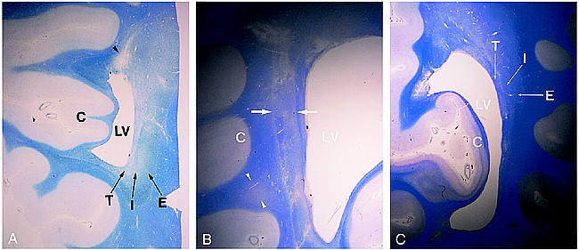
  • fig 4.
    • Download figure
    • Open in new tab
    • Download powerpoint
    fig 4.

    A, Coronal specimen from an 88-year-old woman shows blurring of the three layers: external sagittal stratum or the optic radiation (E), internal sagittal stratum (I), and tapetum (T). All three layers are pale relative to adjacent white matter. Infarcted area is also observed at the superior periventricular white matter (arrowhead). LV indicates lateral ventricle; C, medial occipital cortex. Klüver-Barrera method, original magnification × 4.

    B, Coronal specimen from a 73-year-old man shows loss of distinction between the external sagittal stratum or the optic radiation and the internal sagittal stratum (arrows). This area is pale relative to adjacent white matter. Patchy pallor lesions are also observed in the white matter adjacent to the external sagittal stratum (arrowheads). LV indicates lateral ventricle; C, lateral occipital cortex. Klüver-Barrera method, original magnification × 4.

    C, Coronal specimen from a 79-year-old man shows thinning of the three layers: external sagittal stratum or the optic radiation (E), internal sagittal stratum (I), and tapetum (T). LV indicates lateral ventricle; C, medial occipital cortex. Klüver-Barrera method, original magnification × 4.

PreviousNext
Back to top

In this issue

American Journal of Neuroradiology
Vol. 20, Issue 6
1 Jun 1999
  • Table of Contents
  • Index by author
Advertisement
Print
Download PDF
Email Article

Thank you for your interest in spreading the word on American Journal of Neuroradiology.

NOTE: We only request your email address so that the person you are recommending the page to knows that you wanted them to see it, and that it is not junk mail. We do not capture any email address.

Enter multiple addresses on separate lines or separate them with commas.
Hyperintensities of the Optic Radiation on T2-Weighted MR Images of Elderly Subjects
(Your Name) has sent you a message from American Journal of Neuroradiology
(Your Name) thought you would like to see the American Journal of Neuroradiology web site.
CAPTCHA
This question is for testing whether or not you are a human visitor and to prevent automated spam submissions.
Cite this article
Mika Kitajima, Yukunori Korogi, Tomoko Okuda, Shin-ya Shiraishi, Osamu Ikeda, Shoji Morishita, Mutsumasa Takahashi, Komyo Eto
Hyperintensities of the Optic Radiation on T2-Weighted MR Images of Elderly Subjects
American Journal of Neuroradiology Jun 1999, 20 (6) 1009-1014;

Citation Manager Formats

  • BibTeX
  • Bookends
  • EasyBib
  • EndNote (tagged)
  • EndNote 8 (xml)
  • Medlars
  • Mendeley
  • Papers
  • RefWorks Tagged
  • Ref Manager
  • RIS
  • Zotero
0 Responses
Respond to this article
Share
Bookmark this article
Hyperintensities of the Optic Radiation on T2-Weighted MR Images of Elderly Subjects
Mika Kitajima, Yukunori Korogi, Tomoko Okuda, Shin-ya Shiraishi, Osamu Ikeda, Shoji Morishita, Mutsumasa Takahashi, Komyo Eto
American Journal of Neuroradiology Jun 1999, 20 (6) 1009-1014;
del.icio.us logo Twitter logo Facebook logo Mendeley logo
  • Tweet Widget
  • Facebook Like
  • Google Plus One

Jump to section

  • Article
    • Abstract
    • Methods
    • Results
    • Discussion
    • Conclusion
    • Footnotes
    • References
  • Figures & Data
  • Info & Metrics
  • Responses
  • References
  • PDF

Related Articles

  • No related articles found.
  • PubMed
  • Google Scholar

Cited By...

  • Quantifying Tissue Properties of the Optic Radiations Using Strategically Acquired Gradient Echo Imaging and Enhancing the Contrast Using Diamagnetic Susceptibility Weighted Imaging
  • Disconnectome Associated with Progressive Ischemic Periventricular White Matter Lesions
  • Crossref
  • Google Scholar

This article has not yet been cited by articles in journals that are participating in Crossref Cited-by Linking.

More in this TOC Section

  • White Matter Alterations in the Brains of Patients with Active, Remitted, and Cured Cushing Syndrome: A DTI Study
  • Qualitative and Quantitative Analysis of MR Imaging Findings in Patients with Middle Cerebral Artery Stroke Implanted with Mesenchymal Stem Cells
  • Fast Contrast-Enhanced 4D MRA and 4D Flow MRI Using Constrained Reconstruction (HYPRFlow): Potential Applications for Brain Arteriovenous Malformations
Show more BRAIN

Similar Articles

Advertisement

Indexed Content

  • Current Issue
  • Accepted Manuscripts
  • Article Preview
  • Past Issues
  • Editorials
  • Editor's Choice
  • Fellows' Journal Club
  • Letters to the Editor
  • Video Articles

Cases

  • Case Collection
  • Archive - Case of the Week
  • Archive - Case of the Month
  • Archive - Classic Case

Special Collections

  • AJNR Awards
  • ASNR Foundation Special Collection
  • Most Impactful AJNR Articles
  • Photon-Counting CT
  • Spinal CSF Leak Articles (Jan 2020-June 2024)

More from AJNR

  • Trainee Corner
  • Imaging Protocols
  • MRI Safety Corner

Multimedia

  • AJNR Podcasts
  • AJNR Scantastics

Resources

  • Turnaround Time
  • Submit a Manuscript
  • Submit a Video Article
  • Submit an eLetter to the Editor/Response
  • Manuscript Submission Guidelines
  • Statistical Tips
  • Fast Publishing of Accepted Manuscripts
  • Graphical Abstract Preparation
  • Imaging Protocol Submission
  • Evidence-Based Medicine Level Guide
  • Publishing Checklists
  • Author Policies
  • Become a Reviewer/Academy of Reviewers
  • News and Updates

About Us

  • About AJNR
  • Editorial Board
  • Editorial Board Alumni
  • Alerts
  • Permissions
  • Not an AJNR Subscriber? Join Now
  • Advertise with Us
  • Librarian Resources
  • Feedback
  • Terms and Conditions
  • AJNR Editorial Board Alumni

American Society of Neuroradiology

  • Not an ASNR Member? Join Now

© 2025 by the American Society of Neuroradiology All rights, including for text and data mining, AI training, and similar technologies, are reserved.
Print ISSN: 0195-6108 Online ISSN: 1936-959X

Powered by HighWire