Advertisement

Main menu

  • Home
  • Content
    • Current Issue
    • Accepted Manuscripts
    • Article Preview
    • Past Issue Archive
    • Video Articles
    • AJNR Case Collection
    • Case of the Week Archive
    • Case of the Month Archive
    • Classic Case Archive
  • Special Collections
    • AJNR Awards
    • ASNR Foundation Special Collection
    • Most Impactful AJNR Articles
    • Photon-Counting CT
    • Spinal CSF Leak Articles (Jan 2020-June 2024)
  • Multimedia
    • AJNR Podcasts
    • AJNR SCANtastic
    • Trainee Corner
    • MRI Safety Corner
    • Imaging Protocols
  • For Authors
    • Submit a Manuscript
    • Submit a Video Article
    • Submit an eLetter to the Editor/Response
    • Manuscript Submission Guidelines
    • Statistical Tips
    • Fast Publishing of Accepted Manuscripts
    • Graphical Abstract Preparation
    • Imaging Protocol Submission
    • Author Policies
  • About Us
    • About AJNR
    • Editorial Board
    • Editorial Board Alumni
  • More
    • Become a Reviewer/Academy of Reviewers
    • Subscribers
    • Permissions
    • Alerts
    • Feedback
    • Advertisers
    • ASNR Home

User menu

  • Alerts
  • Log in

Search

  • Advanced search
American Journal of Neuroradiology

American Journal of Neuroradiology

ASHNR American Society of Functional Neuroradiology ASHNR American Society of Pediatric Neuroradiology ASSR
  • Alerts
  • Log in

Advanced Search

  • Home
  • Content
    • Current Issue
    • Accepted Manuscripts
    • Article Preview
    • Past Issue Archive
    • Video Articles
    • AJNR Case Collection
    • Case of the Week Archive
    • Case of the Month Archive
    • Classic Case Archive
  • Special Collections
    • AJNR Awards
    • ASNR Foundation Special Collection
    • Most Impactful AJNR Articles
    • Photon-Counting CT
    • Spinal CSF Leak Articles (Jan 2020-June 2024)
  • Multimedia
    • AJNR Podcasts
    • AJNR SCANtastic
    • Trainee Corner
    • MRI Safety Corner
    • Imaging Protocols
  • For Authors
    • Submit a Manuscript
    • Submit a Video Article
    • Submit an eLetter to the Editor/Response
    • Manuscript Submission Guidelines
    • Statistical Tips
    • Fast Publishing of Accepted Manuscripts
    • Graphical Abstract Preparation
    • Imaging Protocol Submission
    • Author Policies
  • About Us
    • About AJNR
    • Editorial Board
    • Editorial Board Alumni
  • More
    • Become a Reviewer/Academy of Reviewers
    • Subscribers
    • Permissions
    • Alerts
    • Feedback
    • Advertisers
    • ASNR Home
  • Follow AJNR on Twitter
  • Visit AJNR on Facebook
  • Follow AJNR on Instagram
  • Join AJNR on LinkedIn
  • RSS Feeds

AJNR is seeking candidates for the AJNR Podcast Editor. Read the position description.

Research ArticleBRAIN

Curved Planar Reformatted CT Angiography: Usefulness for the Evaluation of Aneurysms at the Carotid Siphon

Takashi Ochi, Kenji Shimizu, Yoshifumi Yasuhara, Toshirou Shigesawa, Teruhito Mochizuki and Junpei Ikezoe
American Journal of Neuroradiology June 1999, 20 (6) 1025-1030;
Takashi Ochi
  • Find this author on Google Scholar
  • Find this author on PubMed
  • Search for this author on this site
Kenji Shimizu
  • Find this author on Google Scholar
  • Find this author on PubMed
  • Search for this author on this site
Yoshifumi Yasuhara
  • Find this author on Google Scholar
  • Find this author on PubMed
  • Search for this author on this site
Toshirou Shigesawa
  • Find this author on Google Scholar
  • Find this author on PubMed
  • Search for this author on this site
Teruhito Mochizuki
  • Find this author on Google Scholar
  • Find this author on PubMed
  • Search for this author on this site
Junpei Ikezoe
  • Find this author on Google Scholar
  • Find this author on PubMed
  • Search for this author on this site
  • Article
  • Figures & Data
  • Info & Metrics
  • Responses
  • References
  • PDF
Loading

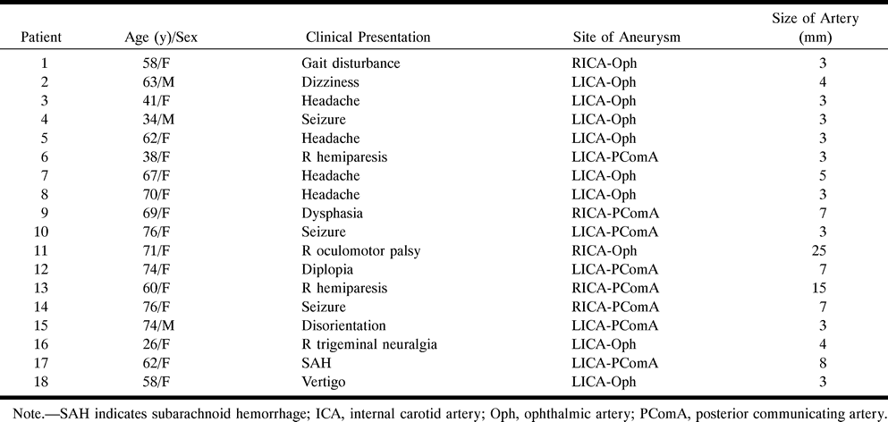
Article Figures & Data

Figures

  • Tables
  • fig 1.
    • Download figure
    • Open in new tab
    • Download powerpoint
    fig 1.

    Patient with an infraclinoid, small aneurysm of the left internal carotid artery–ophthalmic artery.

    A, 3D CT angiogram with SSD (superoposterior view) shows a slight laterally bulging contour (arrowhead); however, a significant saccular component cannot be identified.

    B, Composite sagittal CPR image clearly shows a small aneurysm (arrow) adherent to the anterior clinoid process.
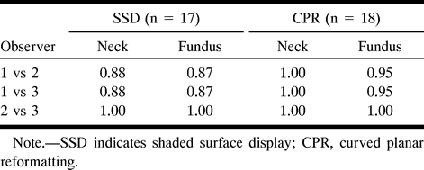
  • fig 2.
    • Download figure
    • Open in new tab
    • Download powerpoint
    fig 2.

    ;t1Case 1: Patient with an aneurysm of the right internal carotid artery–ophthalmic artery.

    A, Right carotid angiogram (left anterior oblique view) shows an aneurysm of the internal carotid artery–ophthalmic artery (arrow).

    B, 3D CT angiogram with SSD (superoposterior view) shows an aneurysm (arrow). The fundus cannot be correctly rendered with the SSD technique owing to partial volume effects of the skull base bone.

    C, Composite sagittal CPR image clearly shows the fundus and neck of the aneurysm (arrow).
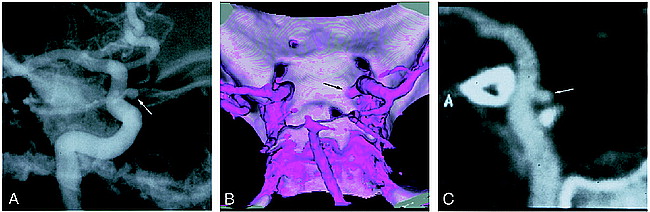
  • fig 3.
    • Download figure
    • Open in new tab
    • Download powerpoint
    fig 3.

    ;t1Case 11: Patient with an aneurysm of the right internal carotid artery–ophthalmic artery.

    A, Right carotid angiogram (left lateral view) shows a giant aneurysm.

    B, 3D CT angiogram with SSD (superoposterior view) shows an aneurysm. The neck cannot be identified with the SSD technique owing to the giant overlapping fundus.

    C, Composite sagittal CPR image clearly shows the neck of the aneurysm with the associated vessel of origin.
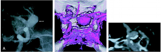
  • fig 4.
    • Download figure
    • Open in new tab
    • Download powerpoint
    fig 4.

    ;t1Case 13: Patient with an aneurysm of the internal carotid artery–posterior communicating artery.

    A, Right carotid angiogram (left anterior oblique view) shows an aneurysm (arrow).

    B, 3D CT angiogram with SSD (superior view) shows an aneurysm (arrow). Mural calcium cannot be well appreciated.

    C, Composite sagittal CPR image clearly shows calcification in the wall of the aneurysm (arrow) and parent artery.

Tables

  • Figures
  • Table 1:
    • View popup
    • Download powerpoint
    Table 1:

    Summary of 18 patients with an aneurysm at the carotid siphon

  • Table 2:
    • View popup
    • Download powerpoint
    Table 2:

    Number of visible aneurysmal necks and fundi

  • Table 3:
    • View popup
    • Download powerpoint
    Table 3:

    ;gk values for interobserver variability

PreviousNext
Back to top

In this issue

American Journal of Neuroradiology
Vol. 20, Issue 6
1 Jun 1999
  • Table of Contents
  • Index by author
Advertisement
Print
Download PDF
Email Article

Thank you for your interest in spreading the word on American Journal of Neuroradiology.

NOTE: We only request your email address so that the person you are recommending the page to knows that you wanted them to see it, and that it is not junk mail. We do not capture any email address.

Enter multiple addresses on separate lines or separate them with commas.
Curved Planar Reformatted CT Angiography: Usefulness for the Evaluation of Aneurysms at the Carotid Siphon
(Your Name) has sent you a message from American Journal of Neuroradiology
(Your Name) thought you would like to see the American Journal of Neuroradiology web site.
CAPTCHA
This question is for testing whether or not you are a human visitor and to prevent automated spam submissions.
Cite this article
Takashi Ochi, Kenji Shimizu, Yoshifumi Yasuhara, Toshirou Shigesawa, Teruhito Mochizuki, Junpei Ikezoe
Curved Planar Reformatted CT Angiography: Usefulness for the Evaluation of Aneurysms at the Carotid Siphon
American Journal of Neuroradiology Jun 1999, 20 (6) 1025-1030;

Citation Manager Formats

  • BibTeX
  • Bookends
  • EasyBib
  • EndNote (tagged)
  • EndNote 8 (xml)
  • Medlars
  • Mendeley
  • Papers
  • RefWorks Tagged
  • Ref Manager
  • RIS
  • Zotero
0 Responses
Respond to this article
Share
Bookmark this article
Curved Planar Reformatted CT Angiography: Usefulness for the Evaluation of Aneurysms at the Carotid Siphon
Takashi Ochi, Kenji Shimizu, Yoshifumi Yasuhara, Toshirou Shigesawa, Teruhito Mochizuki, Junpei Ikezoe
American Journal of Neuroradiology Jun 1999, 20 (6) 1025-1030;
del.icio.us logo Twitter logo Facebook logo Mendeley logo
  • Tweet Widget
  • Facebook Like
  • Google Plus One

Jump to section

  • Article
    • Abstract
    • Methods
    • Results
    • Discussion
    • Conclusion
    • Footnotes
    • References
  • Figures & Data
  • Info & Metrics
  • Responses
  • References
  • PDF

Related Articles

  • No related articles found.
  • PubMed
  • Google Scholar

Cited By...

  • Assessment of Intracranial Arterial Stenosis with Multidetector Row CT Angiography: A Postprocessing Techniques Comparison
  • Curved Planar Reformatted CT Angiography: Utility for the Evaluation of Aneurysms at the Carotid Siphon
  • Crossref
  • Google Scholar

This article has not yet been cited by articles in journals that are participating in Crossref Cited-by Linking.

More in this TOC Section

  • Usefulness of Quantitative Susceptibility Mapping for the Diagnosis of Parkinson Disease
  • White Matter Alterations in the Brains of Patients with Active, Remitted, and Cured Cushing Syndrome: A DTI Study
  • Qualitative and Quantitative Analysis of MR Imaging Findings in Patients with Middle Cerebral Artery Stroke Implanted with Mesenchymal Stem Cells
Show more BRAIN

Similar Articles

Advertisement

Indexed Content

  • Current Issue
  • Accepted Manuscripts
  • Article Preview
  • Past Issues
  • Editorials
  • Editor's Choice
  • Fellows' Journal Club
  • Letters to the Editor
  • Video Articles

Cases

  • Case Collection
  • Archive - Case of the Week
  • Archive - Case of the Month
  • Archive - Classic Case

Special Collections

  • AJNR Awards
  • ASNR Foundation Special Collection
  • Most Impactful AJNR Articles
  • Photon-Counting CT
  • Spinal CSF Leak Articles (Jan 2020-June 2024)

More from AJNR

  • Trainee Corner
  • Imaging Protocols
  • MRI Safety Corner

Multimedia

  • AJNR Podcasts
  • AJNR Scantastics

Resources

  • Turnaround Time
  • Submit a Manuscript
  • Submit a Video Article
  • Submit an eLetter to the Editor/Response
  • Manuscript Submission Guidelines
  • Statistical Tips
  • Fast Publishing of Accepted Manuscripts
  • Graphical Abstract Preparation
  • Imaging Protocol Submission
  • Evidence-Based Medicine Level Guide
  • Publishing Checklists
  • Author Policies
  • Become a Reviewer/Academy of Reviewers
  • News and Updates

About Us

  • About AJNR
  • Editorial Board
  • Editorial Board Alumni
  • Alerts
  • Permissions
  • Not an AJNR Subscriber? Join Now
  • Advertise with Us
  • Librarian Resources
  • Feedback
  • Terms and Conditions
  • AJNR Editorial Board Alumni

American Society of Neuroradiology

  • Not an ASNR Member? Join Now

© 2025 by the American Society of Neuroradiology All rights, including for text and data mining, AI training, and similar technologies, are reserved.
Print ISSN: 0195-6108 Online ISSN: 1936-959X

Powered by HighWire