
Article Figures & Data
Figures
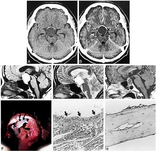
- fig 1.
Calcification in Rathke's cleft cyst in a 23-year-old woman.
A, Presurgical axial CT scan of head shows crescent-like calcification in wall of suprasellar cyst.
B, Postcontrast CT scan reveals thin-ring enhancement of wall of cyst.
C, Presurgical sagittal T1-weighted MR image (400/15/2 [TR/TE/excitations]) shows hyperintense cyst with small nodule in inferior wall of cavity. Character of cyst wall is not apparent on this image.
D, Sagittal T2-weighted MR image (5000/90/1) depicts a small, nodular lesion in the anterior ventral portion of cyst. Note that cyst wall is not well defined on MR images.
E, T1-weighted MR image (400/14/2), after contrast administration prior to second surgery, shows an intra- and suprasellar cyst with enhancing thin wall and septum.
F, lntraoperative photograph shows a bony plate (curved arrows) in cystic cavity underneath incised dura of sellar floor (transsphenoidal approach).
G, Photomicrographs of biopsy specimen. Cyst is lined by a layer of columnar cells with cilia (arrows) (hematoxylin-eosin–stained section, × 200).
H, Photomicrographs of a plate of mature bone removed from cyst cavity in hematoxylin-eosin–stained section (× 200).
In this issue


Jump to section
Related Articles
Cited By...
- No citing articles found.