
Article Figures & Data
Figures
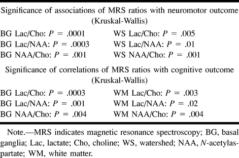
- fig 1.
Location of voxels and normal neonatal proton spectra.
A, The deep gray nuclei voxel includes most of the lentiform nucleus, the ventrolateral thalamus, and the posterior limb of the internal capsule. The spectrum reveals: 1) a small myoinositol peak, 2) a large choline peak, 3) two small creatine/phosphocreatine peaks, and 4) a medium-sized NAA peak.
B, The watershed voxel includes primarily white matter from the intravascular boundary zone. Note that the NAA and choline peaks are relatively smaller in the less mature watershed zone than in the more mature deep gray nuclei. Minimal or no lactate was seen in most patients who were developmentally normal at 12 months.
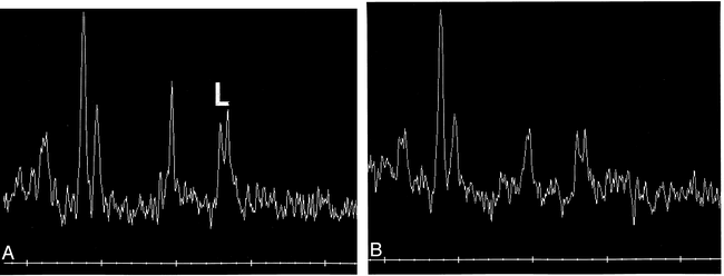
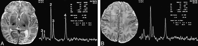
- fig 2.
Neonate with basal nuclei pattern of injury.
A and B, The basal nuclei voxel (A) shows marked elevation of the lactate peak (L) centered at 1.31 ppm. In this acute phase, the relative sizes of the choline, creatine, and NAA peaks are normal. The watershed voxel (B) shows less elevated lactate (compare with A).
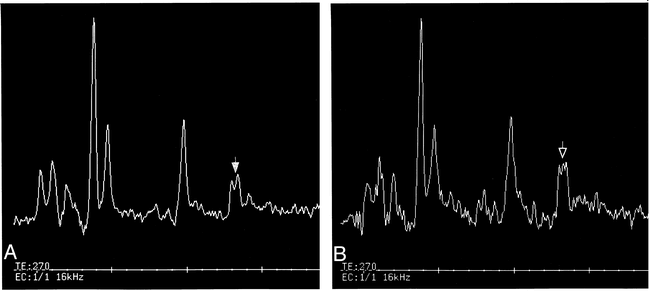
- fig 3.
Neonate with watershed pattern of injury.
A and B, Both spectra show some lactate elevation at 1.31 ppm. The spectrum from the basal nuclei voxel (A), however, shows a relatively smaller elevation of lactate (filled arrow) than the spectrum (open arrow) from the watershed voxel (B).
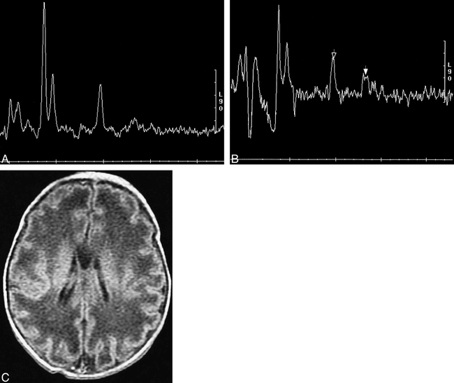
- fig 4.
False-positive spectra in an immature (36-week gestation) neonate at 2-day postnatal age.
A, Spectrum from the voxel in the basal nuclei voxel shows a relatively low NAA level, suggesting immaturity of the brain. Minimal or no lactate peak is seen in this voxel.
B, Spectrum from the less mature watershed voxel shows a very low NAA peak (open arrow) and a small- to moderate-sized lactate peak (solid arrow). This patient shows appropriate development at age 12 months, suggesting that the low NAA and high lactate are the result of immaturity, not injury.
C, Axial SE (500/12) image shows an immature gyral pattern, confirming the immaturity of the infant's brain.
- fig 5.
Normal spectra in a 2-day-old infant with neonatal encephalopathy and abnormal neurologic development at age 12 months.
A, Spectrum from voxel in the basal nuclei voxel shows normal NAA, choline, and creatine/phosphocreatine peaks (compare with fig 1A). Minimal lactate (arrow) is seen at 1.31 ppm; this level of lactate was seen in many infants who developed normally and was interpreted as normal.
B, Spectrum from the watershed zone voxel also shows normal relative peak areas for choline, creatine/phosphocreatine, and NAA (compare with fig 1B). No lactate is appreciated in this voxel.
C, Axial SE (500/12) image at the level of the basal nuclei shows globular hyperintensity (arrows) in the lentiform nuclei, indicative of an injury more than 1 week old.
D, Axial SE (500/12) image in a child with normal neonatal course and normal postnatal development shows the normal appearance of the neonatal basal nuclei. The hyperintensity in the vein of Galen and superior sagittal sinus is the result of inflow of unsaturated protons.


Tables
In this issue


Jump to section
Related Articles
Cited By...
- Use of magnetic resonance imaging in neuroprognostication after pediatric cardiac arrest: Survey of current practices
- Integrating neuroimaging biomarkers into the multicentre, high-dose erythropoietin for asphyxia and encephalopathy (HEAL) trial: rationale, protocol and harmonisation
- Magnetic Resonance Biomarkers in Neonatal Encephalopathy (MARBLE): a prospective multicountry study
- Brain Volume and Metabolism in Fetuses With Congenital Heart Disease: Evaluation With Quantitative Magnetic Resonance Imaging and Spectroscopy
- An MRI Study of Neurological Injury Before and After Congenital Heart Surgery
- Seizure-associated brain injury in term newborns with perinatal asphyxia
- Proton Spectroscopy and Diffusion Imaging on the First Day of Life after Perinatal Asphyxia: Preliminary Report
- 1H-MR spectroscopy is sensitive to subtle effects of perinatal asphyxia
- Three-dimensional Proton MR Spectroscopic Imaging of Premature and Term Neonates
- Line-Scan Diffusion Imaging of Term Neonates with Perinatal Brain Ischemia