
Abstract
Summary: We describe serial MR imaging findings in a patient with HTLV-I-associated myelopathy. The patient had acute progression of neurologic symptoms and exhibited swelling of the entire length of the spinal cord with increased T2 signal and contrast enhancement on MR imaging. The spinal cord became atrophic a few years later.
Human T lymphotropic virus type I (HTLV-I)-associated myelopathy (HAM) is a slowly progressive disease. MR findings reported for this disease are cerebral white matter lesions and spinal cord atrophy (1–3). We present a patient with HAM who exhibited swelling of the entire length of the spinal cord with increased T2 signal and contrast enhancement on MR images.
Case Report
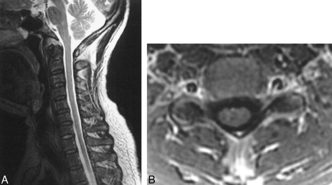
A 45-year-old man was admitted to our institution on November 18, 1994 with a 1-month history of progressive motor weakness of the lower extremities, difficulty in urination and defecation, and numbness below the lower thoracic level. He had had a low-grade fever and headache for 1 month in September 1994. He had no history of blood transfusion. Neurologic examination revealed spastic paraparesis and decreased vibration and temperature sensation below the second thoracic vertebral level. Laboratory examination revealed elevated anti-HTLV-I antibody titers in serum (> 4096×) and normal titers in CSF (< 16×). Other antiviral antibody titers in CSF were also normal. Antinuclear antibody was negative in his serum, and no oligoclonal IgG bands were observed in CSF. MR imaging revealed swelling of the entire length of the spinal cord with high signal on T2-weighted images and peripheral cord enhancement after intravenous contrast administration (Fig 1A–D). No brain abnormalities were visible.

MR images (Nov 21, 1994). Sagittal T2-weighted images reveal marked cord swelling and high intensity in the central portion over the entire length of the spinal cord (A and B). Peripheral enhancement is shown by a sagittal T1-weighted image (C) and an axial T1-weighted image at the C7/T1 level (D) with contrast material
The patient was treated with 1000 mg methylpredonisolone intravenously for 3 days, followed by 16 mg oral betamethasone therapy for 1 month. His signs and symptoms improved gradually and disappeared 1 month later. MR imaging revealed mild cord swelling with mild high signal on T2-weighted images and mild contrast enhancement 2 weeks after the initial MR imaging (Fig 2A–B).
MR images (Dec 3, 1994). Sagittal T2-weighted image reveals mild cord swelling with mild high signal in the central portion (A). Contrast-enhanced axial T1-weighted image at the C7/T1 level reveals mild cord swelling and mild contrast enhancement (B)
His neurologic symptoms relapsed 2 months after admission, after completion of corticosteroid therapy. Laboratory examination revealed elevated anti-HTLV-I antibody titers both in serum (> 4096×) and CSF (> 4096×) and elevated IgG in CSF (42.2mg/dL: normal < 5 mg/dL). Eight weeks after the second MR study, swelling of the entire length of the spinal cord, high signal on T2-weighted images, and peripheral cord enhancement again were observed on MR images (Fig 3A–B).

MR images (Jan 31, 1995). Sagittal T2-weighted image reveals a cord swelling with central high intensity (A). Contrast-enhanced axial T1-weighted image at T3 level reveals a swollen cord with peripheral enhancement (B)
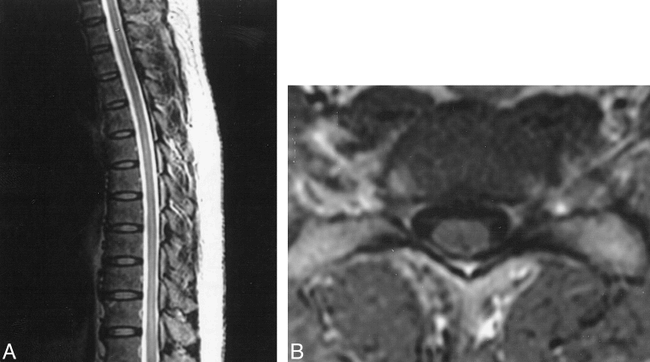
Subsequently, his signs and symptoms slowly progressed. He was diagnosed with HAM based on the diagnostic guidelines for HAM/tropical spastic paraparesis (TSP) submitted by the World Health Organization (4). MR examination 1 year later showed the spinal cord was normal in size and no abnormal signal or enhancement was visible (Fig 4A–B). His neurologic manifestations slowly progressed. Three years after the first admission, he required two-hand support in walking and had mild cord atrophy that exhibited neither signal abnormality nor enhancement on MR images (Fig 5A–B).

MR images (Dec 14, 1995). The spinal cord is normal in size and shows no abnormal signal on the T2-weighted image (A). Enhanced axial T1-weighted image at the C5/6 level shows no abnormal enhancement (B)

MR images (Mar 31, 1998). The spinal cord has no signal abnormality on the T2-weighted image (A). Mild cord atrophy is visible on the contrast-enhanced, axial T1-weighted image at the C5/6 level (B)
Discussion
Infection with HTLV-I is associated with a wide variety of human diseases including adult T-cell leukemia, neurologic disorders, uveitis and arthropathy. Although the global distribution of HTLV-I has not been fully determined, this virus is highly endemic in Japan and is also prevalent in Melanesia, the Caribbean, and some areas of Africa. It is transmitted through breast feeding, blood transfusion, sexual intercourse, and intravenous drug use (4). The main neurologic manifestation of HTLV-I infection is a myelopathy. HTLV-I-associated myelopathy (HAM) in Japan and HTLV-I-positive tropical spastic paraparesis (TSP) in the Caribbean are clinically and pathologically identical, and it is recommended that this disease entity be identified as HAM/TSP for the present (4). The incidence of HAM/TSP is estimated to be 0.25% among HTLV-I infected persons (5). The manifestations of HAM/TSP are believed to result from HTLV-I infection associating with an immunopathogenic or autoimmune mechanisms and host factors, including HLA, are considered important for disease manifestation (6). These might be responsible for the low reported incidence of HAM/TSP.
Cerebral white matter lesions and cord atrophy have been reported as MR findings of HAM/TSP (1–3). Fererraz et al reported that MR imaging revealed cerebral white matter lesions in 52% and spinal cord atrophy in 74% of patients with HAM (3).
The main neurologic manifestation of HAM/TSP is chronic spastic paraparesis, which is usually slowly progressive (4). Nakagawa et al found that 14 of 213 patients with HAM exhibited acute progression of symptoms 2 years before their first examination (7). They speculated that the acute progression of symptoms reflected an inflammatory process related to a cell-mediated immune process occurring in the spinal cord, because their 14 patients had high anti-HTLV-I titers and neopterin levels in CSF. Neopterin is a useful parameter of cell-mediated immune reaction in the CSF of HAM/TSP, for it is derived from macrophages stimulated by activated T cells (7). Neopterin levels in CSF of the patients in the chronic phase of HAM/TSP were elevated mildly, suggesting the presence of slight but persistent inflammation (7). Our patient exhibited acute progression of neurologic symptoms, and MR images revealed swelling of the entire length of the spinal cord with peripheral contrast enhancement and increased signal on T2-weighted images. The patient responded well to corticosteroid therapy. Our MR findings support Nakagawa's speculation that acute progression of symptoms reflects the inflammatory process in the spinal cord. MR study of our patient subsequently revealed cord atrophy. Although cord atrophy has been considered a typical MR finding of HAM (1), we believe that cord atrophy is a manifestation of the chronic phase of HAM/TSP.
Akizuki et al (8) reported histopathologic findings of the spinal cord in HAM/TSP; loss of myelin and axon was observed bilaterally mainly in the lateral and anterior columns, and occurred mainly along the tract. In addition, perivascular and parenchymal infiltration with lymphocytes and macrophages, as well as astrocytosis, were observed in the white and gray matter of the entire spinal cord (8). These findings are quite different from those of vacuolar myelopathy, which is the most common pathologic finding at postmortem examination of HIV-positive patients, although both HIV and HTLV-I are human retroviruses associated with myelopathies. Vacuolar myelopathy is characterized by extensive vacuolation of the white matter, especially of the posterior and lateral columns, which is visible as a region of high intensity on T2-weighted images (9). In vacuolar myelopathy, severe inflammatory reactive changes and axonal destruction are absent histopathologically, and no pathologically related contrast enhancement is observed on MR images (9). In HAM/TSP, inflammatory reactive changes have been reported in both the white and gray matter. Our patient, however, exhibited only peripheral enhancement. The reason for this is unclear. Gero et al (10) reported a case of inflammatory myelitis in which immediate postcontrast scans revealed peripheral enhancement but a delayed scan showed progressive enhancement of the spinal cord toward the center. Our patient might have exhibited central enhancement if a delayed enhancement study had been performed.
Relapsing-remitting myelopathy is also observed in patients with elevated anticardiolipin antibody (ACA). The MR findings for myelopathy with elevated ACA are reported to be widening of the cord with signal abnormalities, elongated lesions extending to multiple segments and frequently to a thoracic location, enhancement during relapses, and development of atrophy over time (11). Unfortunately, our patient did not undergo testing for ACA. Nevertheless, our case revealed diffuse extension of the spinal cord lesion, unlike reported cases of myelopathy with elevated ACA in which segmental extension was observed.
To our knowledge, spinal cord swelling associated with HAM/TSP has been described in only two reports. In one of the two, a 51-year-old woman with HAM presented with abrupt onset of neurologic symptoms and spinal cord swelling in the acute stage of CT myelography; disappearance of cord swelling was documented 4 months after corticosteroid therapy (12). In the other report, a 34-year-old man with TSP presented with progressive quadriparesis and cervical cord swelling, high signal on T2-weighted images, and peripheral cord enhancement (10). He was lost to follow-up. Isoda et al reported a case of transverse myelitis with HTLV-I infection with cord swelling and high intensity on T2-weighted images without cord enhancement (13). Their patient was not diagnosed with HAM/TSP, and the authors did not describe his clinical course or results of follow-up.
In conclusion, HAM/TSP should be included in the differential diagnosis in cases of swelling of the entire length of the spinal cord, high signal on T2-weighted images, and contrast enhancement on MR images.
Acknowledgments
We thank Yutaka Nemoto, M.D., Takahiko Tashiro, M.D., Hajime Ikeda, M.D. and Hiroyuki Shimada, M.D. for their helpful advice.
Footnotes
↵1 Address reprint requests to Miyuki Shakudo, MD, Department of Radiology, Osaka City University Medical School,1-4-3 Asahimachi, Abeno-ku, Osaka 545-8585, Japan.
References
- Received February 8, 1999.
- Accepted after revision March 31, 1999.
- Copyright © American Society of Neuroradiology


