
Abstract
Summary: We compared two MR imaging sequences, fast inversion recovery for myelin suppression (FIRMS) and echo-planar FIRMS (EP-FIRMS), for depicting gray/white matter contrast. In 18 patients, the frequency bandwidth (BW) was optimized for each sequence; in nine patients, the BW was held constant. In the BW-optimized group, the mean contrast-to-noise ratio (C/N) was three times higher with the FIRMS sequence. In the BW-constant group, the mean C/N was 27% higher with the EP-FIRMS sequence; however, geometric distortion degraded the EP-FIRMS images excessively. For optimal gray/white contrast, FIRMS appears to be the superior pulse sequence.
In an earlier study (1), a fast inversion recovery for myelin suppression (FIRMS) sequence with a TI of 250 was found to generate gray/white matter contrast superior to that seen with standard sequences, and therefore led to improved diagnosis of cortical dysplasias. The development of echo-planar (EP) imaging raises the question as to whether myelin suppression can be optimized further. The purpose of this study, therefore, was to compare the efficacy of FIRMS with that of a previously undescribed EP sequence, EP-FIRMS, for maximizing gray/white matter contrast.
Methods
Pulse Sequences
Twenty-seven patients underwent MR imaging of the brain with FIRMS and EP-FIRMS, using a 1.5-T Horizon unit (GE Medical Systems, Milwaukee, WI). Patients with white matter disease or seizures were excluded from the study. For all subjects, imaging was done with the following constant pulse sequence parameters: TR = 4500, TI = 250, matrix size = 256 × 256 pixels, number of signal averages = 1, section thickness = 5 mm with no intersection gap, and FOV = 28 × 21 cm. In a preliminary validation study, TIs of 250 and 300 were compared in four subjects by using contrast-to-noise ratios (C/N), an objective measure of gray/white contrast. No significant difference was noted between the two (15.2 for TI = 250 versus 15.3 for TI = 300), a finding also supported by a previous study (2). On the basis of these results, a TI of 250 was used throughout the study.
FIRMS is based on a fast spin-echo (FSE) sequence, and, as such, multiple lines of data acquisition are referred to in terms of echo train length (ETL). On the other hand, EP-FIRMS is based on a spin-echo/echo-planar sequence, and for this reason the data are acquired in terms of shots. In each case, the number of shots and the ETL were controlled by means of the following conversion: ETL = 256/shots. Subjects were divided into two groups: one in which the bandwidth (BW) was kept constant for both FIRMS and EP-FIRMS (BW-constant) sequences and one in which the BW was optimized for each sequence (BW-optimized).
For all subjects in the BW-constant group (n = 9), the receive BW was 31.3 kHz for both sequences (obtaining 28 sections), with an ETL of 32 for FIRMS and eight shots for EP-FIRMS. The TE for FIRMS was 40, with a scan time of 1:50. For EP-FIRMS, the TE was 45, and the scan time was 2:02. For the BW-optimized FIRMS sequences (n = 18), the receive BW was 16 kHz (to maximize C/N), with a TE of 28. A total of 26 sections were obtained, requiring a time of 2:43. For the BW-optimized EP-FIRMS sequences, the BW was 111 kHz to minimize geometric distortion from magnetic susceptibility, with a TE of 19.9. A total of 26 sections were obtained, requiring a scan time of 2:33. Patients in the BW-optimized group were further divided into two subgroups, one for whom an ETL of 16 was applied (n = 13) and one for whom the ETL was 32 (n = 5). Standard deviations were calculated for all groups.
Image Analysis (Objective)
Conspicuity was determined objectively by using region-of-interest (ROI) measurements of hippocampal gray matter, adjacent white matter, and noise. In all cases, precisely the same ROI was taken for both sequences without track-ball adjustments. In no case did the subject's position change between sequences. The area of the ROI was at least 50 mm2. The choice of the hippocampal region for measurement was based on the ease of defining a sufficiently large area of gray matter. Either the left or right hippocampus was selected without preference, depending on which had a larger homogeneous area of pixels that could be measured on both sequences. White matter was measured from any relatively large area of homogeneous white matter close to the hippocampus. Noise was measured in the background air outside the patient, adjusting the windowing to avoid motion artifacts. Contrast-to-noise (C/N) was then calculated using the following formula:  Mean C/N values were calculated for the BW-optimized and BW-constant groups. The number of cases in which each sequence had a superior C/N value was also determined.
Image Analysis (Subjective)
Two experienced neuroradiologists performed unblinded, subjective evaluations of image quality. Attention was paid to differences between the sequences in gray/white contrast and to causes of the differences.
Results
BW-Constant Group
In the BW-constant group, there was a trend for somewhat higher C/N ratios with the EP-FIRMS sequence (see Table). Along these lines, EP-FIRMS had a superior C/N value in 89% (n = 9) of the cases. Despite the superior contrast levels of EP-FIRMS, both interpreters agreed that in 100% of cases FIRMS produced substantially better image quality, as the EP-FIRMS sequences exhibited severe geometric distortion artifacts (Fig 1) in the lower sections, limiting its potential usefulness.
TABLE: Contrast-to-noise (C/N) values for two imaging sequences: FIRMS and EP-FIRMS

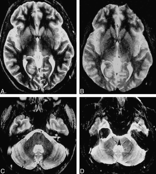
Comparison of BW-constant FIRMS and EP-FIRMS sequences (axial sections).
A and B, Basal ganglia level: FIRMS (A) and EP-FIRMS (B).
C and D, Brain stem level: FIRMS (C) and EP-FIRMS (D).
Although EP-FIRMS has a greater C/N value, subjectively the images are comparable in quality at superior levels (A, B) with minimal geometric distortion. Inferior levels show severe geometric distortion due to the proximity of sphenoidal sinus air, evident at the anterior margin of the pons in the EP-FIRMS image (arrowhead, D), with no artifact in the FIRMS image (C).
BW-Optimized Group
The FIRMS sequence in the BW-optimized group exhibited a superior C/N value in 100% of cases (n = 18), producing a mean C/N value of 26.7 as opposed to the mean C/N of 8.1 for the EP-FIRMS sequence. In 100% of cases, both interpreters judged the FIRMS sequence to have superior gray/white matter contrast, finding the EP-FIRMS images to be excessively noisy. The subjective readings thereby supported the poor C/N values of the EP-FIRMS sequence in the BW-optimized group (Figs 2 and 3). Furthermore, the superior gray/white conspicuity of FIRMS images in the BW-optimized group was maintained at the different ETLs tested. For the ETL = 16 group, the FIRMS sequence produced a mean C/N approximately three times that of the EP-FIRMS sequence. The larger ETL of 32 resulted in a mean C/N value that was more than four times greater for FIRMS.

A and B, Magnified axial MR images of the basal ganglia and thalamus obtained with BW-optimized FIRMS (A) and EP-FIRMS (B) sequences show superior contrast of FIRMS over EP-FIRMS. Note the insula (arrowheads): the gray/white matter is clearly delineated with FIRMS while poorly visualized with the much noisier EP-FIRMS
Discussion
In an earlier study (1) FIRMS was shown to yield improved gray/white contrast over conventional dual-echo (proton density–or T2-weighted), T2-weighted FSE, and T1-weighted radio-frequency spoiled gradient-recalled-echo imaging. Because of its improved gray/white contrast, FIRMS proved to be useful in imaging patients with seizures because of improved demonstration of migration disorders (including heterotopic gray matter, cortical dysplasia, and lissencephaly) and of the hippocampal alveus. The excellent depiction of the cortex obtained with FIRMS has also been useful in the setting of basal encephalocele (3). EP imaging is a technique with great potential because of its extreme speed. This has been used widely for diffusion-weighted (4) and functional (5) MR imaging. The imaging speed of the sequence also drastically decreases the prevalence of motion artifacts, a feature that has been extremely useful for body MR imaging (6), and holds promise for examining uncooperative patients. The short imaging time and decreased susceptibility to motion artifacts are compelling reasons for further study of this sequence.
EP imaging can be more rapid than FSE. The speed of FSE depends on obtaining a train of echoes with intervening 180° pulses. The speed of EP imaging lies in its ability to quickly fill Fourier space (K-space), using oscillating gradients with a negative and positive polarity, thus producing both odd and even echoes (7). In the case of EP-FIRMS, the initial IR portion (180°, 90°, 180°) of the sequence is followed by the EP oscillating gradient. As the echoes are sampled across the echo train, the spins accumulate a phase shift, since the signal is never again realigned by another 180° pulse, as is the case with FSE. As a result, EP imaging is often subject to geometric distortions, particularly at air-tissue interfaces (7). This artifact is further accentuated when scanning with a long effective echo spacing, as in the case of single-shot EP imaging (8). We found that even with multishot EP imaging, geometric distortion degraded the EP-FIRMS sequences when a narrow BW (31.3 kHz) was used (BW-constant group) (Fig 3), because the narrow BW also increased effective echo spacing, allowing increased accumulation of phase shift.

A and B, Axial MR images at the level of the midbrain obtained with BW-optimized FIRMS (A) and EP-FIRMS (B) sequences. FIRMS image has superior C/N values as compared with EP-FIRMS image. The EP-FIRMS image has no significant geometric distortion at this superior level
Geometric distortion can be reduced dramatically by widening the BW. This decreases the signal receive time, which reduces the time available for accumulation of phase shift. However, this introduces another problem. Widening the BW increases the range of frequencies to be sampled, resulting in a decreased signal-to-noise ratio, as the system is sampling more noise per unit of image signal (8). Thus, a wider BW decreases artifact occurrence at the cost of C/N levels, creating noisier images, as was observed subjectively by both our reviewers and reflected in our C/N values. In fact, with the BW-optimized sequences, FIRMS with a BW of 16 kHz had a mean C/N value more than three times that of EP-FIRMS using a BW of 111 kHz (see Table).
IR improves tissue contrast by suppressing a particular tissue, which, in the case of FIRMS and EP-FIRMS, is white matter. Maximal suppression of a tissue is achieved when the TI = ln2 × T1, the null point of the tissue (9–11). Consequently, by determining the null point of a tissue type, any tissue may be suppressed. Some examples of other IR techniques include short-inversion-time IR (STIR) and fluid-attenuated IR (FLAIR). STIR and fast-STIR are IR sequences that use a TI of 165 for suppression of adipose tissue (11, 12). Similarly, FLAIR and turbo-FLAIR, with a TI of approximately 2000, maximally suppress CSF, which is beneficial in regions of the CNS where CSF hyperintensity may obscure disease, such as in the cortex and periventricular region (13). FIRMS, with a TI of 250, suppresses white matter to optimize gray/white matter contrast (1, 2). (It is important to note that all the TIs mentioned above assume a 1.5-T field strength. The optimal TI needs to be shortened with lower B0 systems, since the T1 of a given tissue is field-strength dependent.)
A previous study using FIRMS, which is based on FSE IR, investigated optimal TI values for visualizing gray/white contrast by comparing C/N values of scans obtained with TIs ranging from 200 to 400 (2). The study determined that a TI of 250 to 300 is optimal for visualizing gray/white matter contrast. While a TI of 350 resulted in maximal white matter suppression, excessive gray matter suppression led to poorer overall C/N values. The preliminary validation study explored the effect on the C/N ratio of varying the TI between 250 and 300 for EP-FIRMS. Because the values obtained from images acquired with TIs of 250 and 300 showed no significant difference in contrast quality, a TI of 250 was used throughout the remainder of the study.
Another recently published study also examined the use of IR in EP imaging to improve gray/white contrast (14). The IR sequence used did not apply myelin suppression, which is the halllmark of FIRMS and EP-FIRMS, to achieve increased gray/white contrast. Rather, a TI of 400 was used, a value that was previously shown to cause breakthrough of white matter signal, even with a TR of 4000 (2). Since these authors used a shorter TR of 1500, the sequence would be expected to cause little white matter suppression (14). The observers found that their IR-EP imaging sequence led to increased gray/white contrast as compared with conventional T1-weighted SE sequences. A comparison study with the FSE-based sequence, FIRMS, may prove useful. Judging by our results, we speculate that an FSE-based sequence will be superior. Future studies might focus on comparisons of FIRMS or EP-FIRMS with true IR, short-tau FLAIR, and double IR (10) sequences.
Although this study found that EP-FIRMS imaging was inferior to the FIRMS sequence, because geometric distortion created a need for wide BW imaging with the former, further studies of the sequence as a single-shot technique may be warranted. Single-shot EP imaging greatly increases the speed of image acquisition as compared with multishot EP imaging, at the cost of increased effective echo spacing. The decision to use multishot EP-FIRMS imaging in this study, as opposed to the single-shot technique, was made because of the desire to compare the EP-FIRMS technique with a proved technique (FIRMS), controlling for ETL. In certain patients, in whom motion is a severe problem and speed is of utmost importance, we believe there could be a role for single-shot EP-FIRMS.
Conclusion
Our results indicate that FIRMS is superior to EP-FIRMS for visualizing gray/white contrast. When controlling the BW, we found that EP-FIRMS images had excessive geometric distortion. When optimizing the BW to avoid distortion, FIRMS images had far superior gray/white contrast, both objectively and subjectively, owing to increased noise with EP-FIRMS.
Footnotes
1 Presented at the annual meeting of the American Society of Neuroradiology, Philadelphia, May 1998.
↵2 Address reprint requests to Leo J. Wolansky, MD, MRI Section Chief, UMDNJ, Radiology/UH C-320, 150 Bergen St, Newark, NJ 07103.
References
- Received October 26, 1998.
- Accepted after revision June 1, 1999.
- Copyright © American Society of Neuroradiology
In this issue


Jump to section
Related Articles
Cited By...
- No citing articles found.