
Abstract
Summary: We describe our experience with an arteriotomy closure device that has become a routine tool for the management of most patients in our neurointerventional service. In our experience, this device contributes significantly to patient comfort by allowing mobilization within 2 hours of a procedure, even with anticoagulants. Efficacy and safety of this suture device requires proctoring during initial experience.
Arteriotomy-related complications represent a significant concern during neurointerventional practice. Reported arterial puncture-site complication rates during diagnostic and interventional transfemoral procedures in the radiologic and cardiologic literature range between 0.52% and 16% (1–6). Even in the absence of complications, puncture-site management accounts for a significant proportion of the time and vexation associated with some neurointerventional procedures. Compared with an average interventional cardiology practice, neurointerventional patients are more likely to require prolonged heparinization after a procedure, often mandating deferral of sheath removal. With the advent of carotid and vertebral artery angioplasty and stenting procedures requiring use of 8–10F sheaths, the potential for significant morbidity increases. Furthermore, sheaths of this size, in conjunction with heparinization and antiplatelet therapy, represent a challenge for safe hemostasis. Deferring sheath removal and sending the patient back to the intensive care unit with the sheath sutured in place has been a common practice in interventional neuroradiology. Nevertheless, thromboembolic complications, hematoma formation, retroperitoneal hemorrhage, or infection are serious complications that can be seen when arterial sheaths are left in place after neurointerventional procedures. Up to 78% of femoral arterial sheaths left in place overnight develop significant clots, even when the sheath is perfused with heparin (7). The ability to remove femoral sheaths from heparinized patients safely immediately after the procedure, without having to reverse anticoagulation, should help to eliminate the potential for many of these complications.
Percutaneous devices have been developed to reduce time to hemostasis. The Vasoseal (Datascope, Montvale, NJ) and Angioseal (Kensy Nash, Nexton, PA) closure devices deploy a collagen plug against the arterial wall forming a sealing coagulum. This report relates to the Techstar and Prostar Plus devices (Perclose Inc., Menlo Park, CA), which deploy 3–0 braided polyester sutures through the walls of the arteriotomy site to achieve hemostasis.
Device and Technique
The Perclose range of arteriotomy closure devices has been available in the United States since 1997. It is designed to close the arterial puncture site after removal of a standard sheath over a wire. The device is available in 6F, 8F, and 10F sheath denominations. The device consists of a sheath-like J design with hydrophilic coating for ease of advancement into the arteriotomy site (Figs 1, 2). It contains two (6F) or four (8F and 10F) nitinol needles within its shaft, to which one or two sutures of 3.0 braided polyester are attached. Because the device is intended for use in the common femoral artery only, an angiographic run of the femoral artery via the arterial sheath is required to demonstrate that the puncture site is not in a bifurcation or branch vessel.

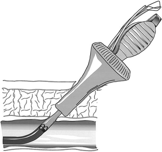
The device resembles a J-shaped 40-cm sheath with hydrophilic coating. It is inserted over a wire into the artery. Package also includes a knot pusher used for achieving tight closure of sutures under subcutaneous tissues
Insertion of the device takes approximately 5 to 10 minutes. Preparation of the subcutaneous tissues with 5–10 mL of 2% lidocaine containing 1/100,000 epinephrine may reduce subcutaneous bleeding and is used routinely. Extension of the skin and subcutaneous dissection site to approximately 1.0–1.5 cm in width then can be performed with a butterfly clamp and scalpel. A 0.035-in or 0.038-in wire is inserted, the sheath removed, and the device is advanced over the wire into the arterial site. Compression of the artery is not necessary after this point. The device has a “monorail” design and is inserted into the artery until the side-hole for the wire is advanced to the skin site. Then the wire is removed and the device is inserted all the way to the hub. A dissection process then is used to burrow the blunt edges of the hub through the subcutaneous tissues until they appose the wall of the artery. When the shaft of the device has been advanced fully into the arterial lumen, a flash of blood escapes through a portal in the device, indicating that the device is adequately advanced and that the needles can be deployed (Figs 2, 3). The needles are retracted from the device with a ring-handle, pulling the sutures through the edges of the arteriotomy site as they exit. Then the needles enter slots in the hub and emerge to external view on the proximal end of the handle, where they are withdrawn fully from the device (Figs 4A–B). The needles are cut from the sutures and the device is withdrawn partially, leaving the sutures in place. A specific series of alternating single hand knots or an “improved clinch knot” (8) is formed and cinched tightly to the artery as the device is withdrawn completely from the artery. Then the knot can be tightened against the arterial wall by use of a knot pusher (Figs 5A–B). The sutures are cut as short as possible. If there is persistent bleeding after the application of the suture knots, this can be controlled most often by manual compression of the groin for 5 to 10 minutes. Alternatively, the suture threads can be backloaded into a tamper device and used to apply pressure to the arteriotomy for a more prolonged period, after which the suture threads are cut.
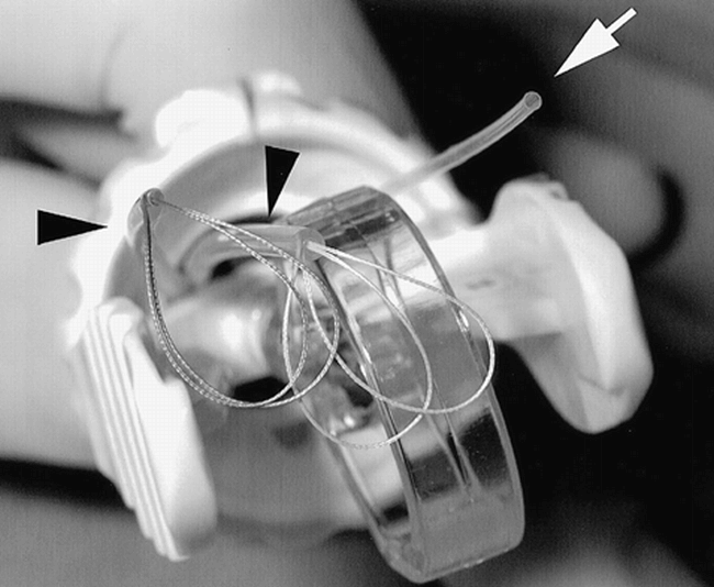
Hub of the device features a translucent ring handle for retracting the needles out of the shaft. A clear marker tubing (white arrow) emits pulsatile flow of blood when device is fully in position within vessel. Prior to deployment of the needles, slack from sutures is housed within two other clear tubings (black arrowheads) extending from the hub

After anesthetization of groin area with lidocaine and epinephrine, the device is dissected through the subcutaneous tissues until the narrow shaft is fully within the lumen of the artery. A hole near end of shaft allows escape of arterial flow into the clear tubing extending from hub (white arrow in 2), to indicate to the operator that the device is in place

A and B, Needles are retracted from shaft by pulling on ring handle. As they emerge from shaft (close-up view A), needles bevel away from shaft so as to pierce edges of the arteriotomy site, and then enter the hub. Then they emerge from hub with suture strings attached (black arrowhead in B). A forceps is used to withdraw needles and sutures from hub completely. Note that thread is attached to the end of needle, which emerges first from the hub (arrow)
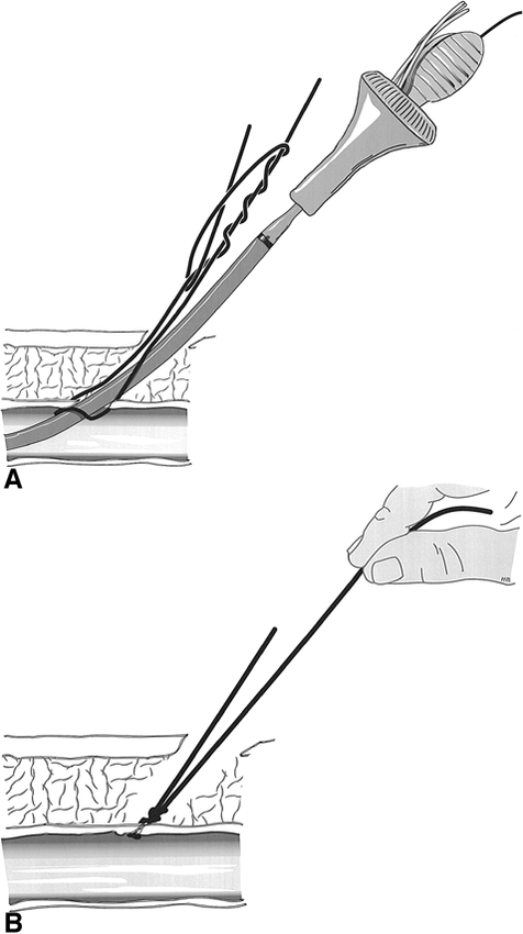
A and B, needles are cut from sutures and the device is withdrawn partially to give the operator control of sutures on the arterial side of hub. An improved clinch knot (A) or a series of surgeon's knots are applied loosely and tightened against the arterial wall as the device is withdrawn from artery (B). The knot pusher can be used then to tighten the knot
To date this device has been used on 69 occasions in our department on 65 neurointerventional patients (8F on 28 patients, 6F on 41 patients). Double-wall puncture technique was used to gain access to the common femoral artery in all cases. In two patients, a second device was placed in the same artery without difficulty or complication within a week of the first placement. All patients underwent heparinization at the time of sheath removal to an activated clotting time (ACT) level of at least twice that of the baseline. Failure to achieve hemostasis was encountered in two patients when the threads broke free of the artery during the suturing process. These vessels were compressed manually without complication to the patients. One significant groin hematoma occurred in this series of patients, requiring transfusion and surgical repair of the artery. This complication was related primarily to an inadvertently high puncture site in combination with the need to maintain full anticoagulation. During surgery, it was seen that the artery had been sutured to the inguinal ligament. The artery then had torn itself away from the ligament, leaving the suture behind, and developed a significant pseudoaneurysm. In the remaining (> 95%) patients of this series, the device has achieved full and immediate hemostasis without discomfort to the patients. No delayed complications or infections were seen. Use of this device virtually has eliminated the need for maintaining arterial sheaths in our patients after a procedure. This facilitates greater patient comfort and eliminates potential problems with sheath maintenance and pain related to prolonged compression with delayed removal.
Discussion
This initial experience with the Perclose device is similar to that reported by experienced operators who report a greater than 96% success rate and a 1% significant complication rate for retrograde and antegrade femoral artery punctures (9, 10). The process of accreditation for the Perclose device involves performing 20 cases under supervision. This is probably a reasonable requirement, considering the potential for arterial injury, or injury to the femoral nerve involved with its use. Furthermore, familiarity with the device is necessary to be able to recognize and correct technical problems with it. Ease and speed of insertion improve significantly during the learning process. In addition to those precepts involved in the proctoring process, a number of practical lessons have been learned with the increased use of this device in our department.
The device is certified for use only in the common femoral artery of adult patients. Inadvertent sheath location in an arterial bifurcation or branch precludes its use. Therefore, when starting an interventional case in which it is anticipated that a Perclose device will be of particular value (ie, an 8F sheath in an anticoagulated patient), it has become routine for us to perform an evaluation of the puncture site at the beginning of the case by using a 5F dilator. Thus, in two patients, we avoided dilating where the puncture site was undesirable. In those patients, we removed the 5F dilator, compressed for hemostasis and repunctured at a better site, allowing effective use of the device at the completion of the case.
The subcutaneous dissection is of pivotal importance for safe and effective deployment of the device. Unless the hub of the device has unimpeded access to the subfascial tissue of the groin, it is likely that the edges of the dermatotomy or the immediate subcutaneous tissue will be drawn into the wound and possibly become transfixed by the needles as they exit. This will create a wound site with poor apposition of tissue edges and a large skin dimple over the arterial site. Moreover, the presence of subcutaneous fascia in the knot or along its path will prevent the achievement of a tight knot on the artery, possibly leading to failure of the device. Therefore, after infiltrating the skin tissues with lidocaine and epinephrine, the dermatotomy site is extended on both sides with a scalpel. A suture clamp then is used to dissect on all four points of the compass around the sheath to mobilize the sheath completely. Care must be taken not to dissect all the way down to or into the artery itself.
The Perclose device is used commonly in the daily practice of our cardiology department, where satisfactory apposition of skin edges is obtained by application of adhesive strips. Patients generally are permitted or encouraged to ambulate within 2 hours after the cardiologic procedure, or when the ACT has normalized. With neurointerventional patients, immediate ambulation is not of such relevance, because most or all of our patients are confined to bed because of the nature of their illness and postprocedural care. Use of the device, however, permits patients to begin moving in bed an hour after the procedure, thus avoiding the considerable discomfort of prolonged immobilization. After returning to the neurointensive care unit, three of our patients experienced prolonged minor oozing from the groin site and responded to bandage or pneumatic compression over some hours. This problem was seen only in patients who underwent heparinization in whom partial thromboplastin time had drifted above the desired therapeutic range. In subsequent patients in whom heparinization was anticipated, this problem has been eliminated by the application of two or three skin sutures of 4.0 silk to the skin edges to assure good wound apposition. These 4.0 skin sutures generally are removed 2 to 3 days later.
The risk of infection needs to be considered because of the insertion of a foreign-body suture at the groin site. Although prophylactic antibiotic coverage is not routinely necessary for cardiologic procedures (11), neurointerventional procedures tend to be significantly longer. The increased length of our procedures suggests that the risk of bacteremia or site contamination might be greater with neurointerventional procedures. For this reason, we have administered a single dose of antibiotic for all patients in whom this device was used and who did not receive antibiotics for other reasons. This single dose usually consisted of 1 g of either cefuroxime or vancomycin (wholesale cost to the hospital less than $10). Sterile technique is practiced scrupulously, and members of the team change gloves after a long case in preparation for the insertion. A fresh J-wire is used for the same reason.
A number of problems can be encountered while the needles are being deployed from the device. To facilitate the correct withdrawal of the needles from the shaft, through the arteriotomy edges and then into the collection grooves on the hub, it is necessary for the hub to be aligned as coaxially as possible with the shaft. If this is not done, it is possible for the nitinol needles to miss the collecting groove and be deflected into the adjacent soft tissues. If this happens, the device may cause serious trauma to the artery if an attempt is made to remove it without first rehousing the needles completely in the shaft. This can be done by grasping the wire shaft of the withdrawal ring close to the hub and pushing back firmly in short increments into the hub. If the device still is not free to move within the wound at that point, it is sometimes the case that the last 1–2 millimeters of the needle have not retracted fully into the shaft. This can be accomplished by pulling on the threads firmly to retract the needles completely. A faulty device can be withdrawn then over a wire and replaced with a new one. There are no inherent restrictions or delays in puncturing the groin site again after insertion of the Perclose device.
Footnotes
↵1 Address reprint requests to Pearse Morris, MB, BCh, Department of Radiology, Wake Forest University School of Medicine, Winston-Salem NC 27157.
- Received February 17, 1999.
- Copyright © American Society of Neuroradiology
In this issue


Jump to section
Related Articles
Cited By...
- Safety and efficacy of percutaneous femoral artery access followed by Mynx closure in cerebral neurovascular procedures: a single center analysis
- Prospective Comparison of Angio-Seal versus Manual Compression for Hemostasis after Neurointerventional Procedures under Systemic Heparinization
- Procedural Safety and Short-Term Outcome of Ambulatory Carotid Stenting Editorial Comment