
Article Figures & Data
Figures
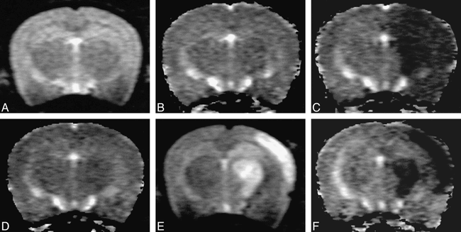
fig. 1. A–F, T2-weighted images (A and E) and ADC maps (B, C, D, F) of a representative rat (group C-1) that showed the transient ADC reduction pattern. T2-weighted images show no abnormal signal intensity at any time. A, before the hypoxic/ischemic insult; E, 48 hours after the insult. ADC maps show no ADC change before the hypoxic/ischemic insult (B), acute ADC reduction in the ipsilateral parietal cortex during the insult (C), ADC recovery 60 minutes after resuscitation (D), and no ADC change 48 hours after the insult (F)
fig. 2. A–F, T2-weighted images (A and E) and ADC maps (B, C, D, F) of a representative rat (group C-2) that showed the biphasic ADC reduction pattern. T2-weighted images show no abnormal signal intensity before the hypoxic/ischemic insult (A), during the insult, or 60 minutes after resuscitation; they do show high signal intensity in the ipsilateral parietal cortex 48 hours after the insult (E). ADC maps show no ADC change before the hypoxic/ischemic insult (B), acute ADC reduction in the ipsilateral parietal cortex during the insult (C), ADC recovery 60 minutes after resuscitation (D), and secondary ADC reduction in the ipsilateral parietal cortex 48 hours after the insult (F)
fig. 3. A–H, T2-weighted images (A–D) and ADC maps (E–H) of a representative rat (group C-3) that showed the persistent ADC reduction pattern.
A–D, T2-weighted images show no abnormal signal intensity before the hypoxic/ischemic insult (A), during the insult (B), or 60 minutes after resuscitation (C); they do show high signal intensity in the ipsilateral parietal cortex 6 hours after the insult (D).
E–H, ADC maps show no ADC change before the hypoxic/ischemic insult (E), acute ADC reduction in the ipsilateral parietal cortex during the insult (F), and sustained ADC decrease 60 minutes after resuscitation (G) and 6 hours after the insult (H).
fig. 4. Histopathologic findings of the ipsilateral parietal cortices of the control and experimental rats (H and E, original magnification ×400).
A, Light micrograph of a parietal cortex section from a control rat (group A) shows intact neurons (arrowheads) and well-preserved cell density 48 hours after CCA ligation.
B, Light micrograph of a parietal cortex section from a rat with the transient ADC reduction pattern (group C-1) shows neuronal loss with ischemic changes (cytoplasmic eosinophilia and pyknotic nuclei, arrows) 48 hours after the hypoxic/ischemic insult.
C, Light micrograph of a parietal cortex section from a rat with the biphasic ADC reduction pattern (group C-2) shows pan-cellular necrosis, indicative of cerebral infarction, 48 hours after the hypoxic/ischemic insult.
D, Light micrograph of a parietal cortex section from a rat with the persistent ADC reduction pattern (group C-3) shows cerebral infarction with extensive neuronal pyknosis (thick arrows) and severe neuropilar microvacuolation (thin arrow) 6 hours after the hypoxic/ischemic insult.


Tables

Time course of ADC changes and histopathologic findings in the ipsilateral parietal cortex in a rat model of hypoxia/ishemia

In this issue


Jump to section
Related Articles
Cited By...
- Motor Deficits Are Triggered by Reperfusion-Reoxygenation Injury as Diagnosed by MRI and by a Mechanism Involving Oxidants
- Transient Global Amnesia: Diffusion-Weighted Imaging Lesions and Cerebrovascular Disease
- Predictors of Hemorrhagic Transformation After Intravenous Recombinant Tissue Plasminogen Activator: Prognostic Value of the Initial Apparent Diffusion Coefficient and Diffusion-Weighted Lesion Volume
- Proton Spectroscopy and Diffusion Imaging on the First Day of Life after Perinatal Asphyxia: Preliminary Report
- Vasospasm After Subarachnoid Hemorrhage: Interest in Diffusion-Weighted MR Imaging
- Direct, Longitudinal Comparison of 1H and 23Na MRI After Transient Focal Cerebral Ischemia