Advertisement

Main menu

  • Home
  • Content
    • Current Issue
    • Accepted Manuscripts
    • Article Preview
    • Past Issue Archive
    • Video Articles
    • AJNR Case Collection
    • Case of the Week Archive
    • Case of the Month Archive
    • Classic Case Archive
  • Special Collections
    • AJNR Awards
    • ASNR Foundation Special Collection
    • Most Impactful AJNR Articles
    • Photon-Counting CT
    • Spinal CSF Leak Articles (Jan 2020-June 2024)
  • Multimedia
    • AJNR Podcasts
    • AJNR SCANtastic
    • Trainee Corner
    • MRI Safety Corner
    • Imaging Protocols
  • For Authors
    • Submit a Manuscript
    • Submit a Video Article
    • Submit an eLetter to the Editor/Response
    • Manuscript Submission Guidelines
    • Statistical Tips
    • Fast Publishing of Accepted Manuscripts
    • Graphical Abstract Preparation
    • Imaging Protocol Submission
    • Author Policies
  • About Us
    • About AJNR
    • Editorial Board
    • Editorial Board Alumni
  • More
    • Become a Reviewer/Academy of Reviewers
    • Subscribers
    • Permissions
    • Alerts
    • Feedback
    • Advertisers
    • ASNR Home

User menu

  • Alerts
  • Log in

Search

  • Advanced search
American Journal of Neuroradiology

American Journal of Neuroradiology

ASHNR American Society of Functional Neuroradiology ASHNR American Society of Pediatric Neuroradiology ASSR
  • Alerts
  • Log in

Advanced Search

  • Home
  • Content
    • Current Issue
    • Accepted Manuscripts
    • Article Preview
    • Past Issue Archive
    • Video Articles
    • AJNR Case Collection
    • Case of the Week Archive
    • Case of the Month Archive
    • Classic Case Archive
  • Special Collections
    • AJNR Awards
    • ASNR Foundation Special Collection
    • Most Impactful AJNR Articles
    • Photon-Counting CT
    • Spinal CSF Leak Articles (Jan 2020-June 2024)
  • Multimedia
    • AJNR Podcasts
    • AJNR SCANtastic
    • Trainee Corner
    • MRI Safety Corner
    • Imaging Protocols
  • For Authors
    • Submit a Manuscript
    • Submit a Video Article
    • Submit an eLetter to the Editor/Response
    • Manuscript Submission Guidelines
    • Statistical Tips
    • Fast Publishing of Accepted Manuscripts
    • Graphical Abstract Preparation
    • Imaging Protocol Submission
    • Author Policies
  • About Us
    • About AJNR
    • Editorial Board
    • Editorial Board Alumni
  • More
    • Become a Reviewer/Academy of Reviewers
    • Subscribers
    • Permissions
    • Alerts
    • Feedback
    • Advertisers
    • ASNR Home
  • Follow AJNR on Twitter
  • Visit AJNR on Facebook
  • Follow AJNR on Instagram
  • Join AJNR on LinkedIn
  • RSS Feeds

AJNR is seeking candidates for the AJNR Podcast Editor. Read the position description.

Research ArticleBRAIN

High-b-value Diffusion-weighted MR Imaging of Suspected Brain Infarction

Joel R. Meyer, Arturo Gutierrez, Bryan Mock, Delon Hebron, Jordan M. Prager, Michael T. Gorey and Daniel Homer
American Journal of Neuroradiology November 2000, 21 (10) 1821-1829;
Joel R. Meyer
  • Find this author on Google Scholar
  • Find this author on PubMed
  • Search for this author on this site
Arturo Gutierrez
  • Find this author on Google Scholar
  • Find this author on PubMed
  • Search for this author on this site
Bryan Mock
  • Find this author on Google Scholar
  • Find this author on PubMed
  • Search for this author on this site
Delon Hebron
  • Find this author on Google Scholar
  • Find this author on PubMed
  • Search for this author on this site
Jordan M. Prager
  • Find this author on Google Scholar
  • Find this author on PubMed
  • Search for this author on this site
Michael T. Gorey
  • Find this author on Google Scholar
  • Find this author on PubMed
  • Search for this author on this site
Daniel Homer
  • Find this author on Google Scholar
  • Find this author on PubMed
  • Search for this author on this site
  • Article
  • Figures & Data
  • Info & Metrics
  • Responses
  • References
  • PDF
Loading

Article Figures & Data

Figures

  • Tables
  • fig 1.
    • Download figure
    • Open in new tab
    • Download powerpoint
    fig 1.

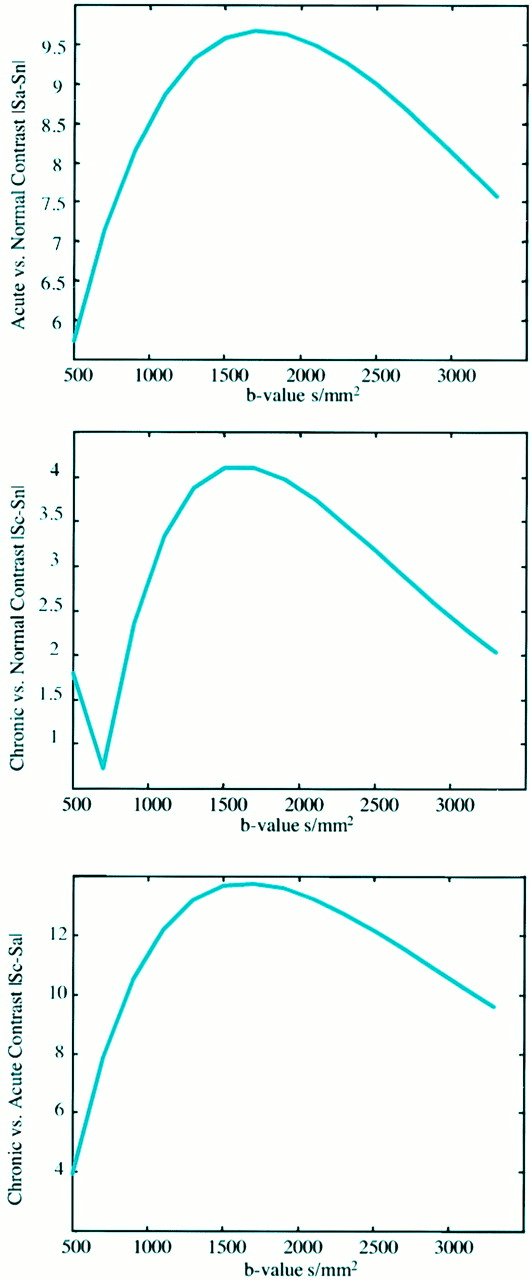
    The signal intensity difference between three tissue types (normal gray matter tissue [assumed ADC = 0.8 × 10⁻³ mm2/s, T2 = 100 ms], acute ischemic tissue [decreased ADC = 0.4 × 10⁻³ mm2/s, T2 = 100 ms], and chronic lesions [elevated ADC = 1.3 × 10⁻³ mm2/s, elevated T2 = 100 ms]) plotted as a function of the b value. In addition to the ADC and T2 assumptions for each tissue, the tissue T1s were assumed to be equal (1000 ms) and significantly smaller than the imaging TR (10 s) such that T1 effects could be ignored.

    A, The signal difference in normal gray matter tissue and acute lesion is shown.

    B, Normal tissue signal is compared with chronic lesion signal, assuming the T2 and ADC are elevated in the chronic lesion.

    C, The relative signal difference between acute and chronic lesion signal is demonstrated. In each case, the contrast reaches a maximum in the b-value range of 1500–2000 s/mm2. Note, however, that the largest contrast is seen in C (acute vs chronic), suggesting that higher b values may be beneficial in delineating chronically infarcted versus acutely ischemic regions.

  • fig 2.
    • Download figure
    • Open in new tab
    • Download powerpoint
    fig 2.

    Trace image of hyperacute infarct in an 89-year-old woman scanned 6 hours after developing right-sided weakness and aphasia.

    A, Diffusion-weighted image (b = 1000 s/mm2) demonstrates a high-signal area consistent with acute infarct in the left basal ganglia and posterior limb of the internal capsule.

    B, Diffusion-weighted image (b = 2500 s/mm2) demonstrates the increased contrast of the high-signal lesion.

    C, Diffusion-weighted image (b = 3000 s/mm2) also demonstrates this lesion more clearly than at b = 1000; anisotropic effects are evident in the posterior periventricular regions.

  • fig 3.
    • Download figure
    • Open in new tab
    • Download powerpoint
    fig 3.

    Acute infarct in an 82-year-old man scanned at 22 hours.

    A, Diffusion-weighted image with b = 1000 s/mm2 demonstrates a high-signal area consistent with acute infarct in the right basal ganglia along the posterior lenticular nucleus. The posterior horns of the lateral ventricles are prominent.

    B, Diffusion-weighted image at b = 2500 s/mm2 demonstrates the high-signal lesion with greater contrast.

    C, Diffusion-weighted image at b = 3000 s/mm2 also demonstrates this lesion more clearly than at b = 1000; anisotropic effects are evident in the contralateral internal capsule and corona radiata.

  • fig 4.
    • Download figure
    • Open in new tab
    • Download powerpoint
    fig 4.

    Acute infarct in a 94-year-old woman scanned 12 hours after developing left-sided hemiparesis.

    A, Diffusion-weighted image with b = 1000 s/mm2 demonstrates a high-signal area consistent with acute infarct in the right parietal lobe.

    B, Diffusion-weighted image at b = 2500 s/mm2 demonstrates the high-signal lesion with greater contrast.

    C, Diffusion-weighted image at b = 3000 s/mm2 also demonstrates this high-signal lesion more clearly than at b = 1000.

  • fig 5.
    • Download figure
    • Open in new tab
    • Download powerpoint
    fig 5.

    Transient ischemic attack and chronic infarct in a 90-year-old woman with a remote history of cerebral infarct, scanned 2 hours after developing right-sided weakness and left-sided facial droop. Her symptoms resolved spontaneously within 11 hours.

    A, Diffusion-weighted image with b = 1000 s/mm2 negative for acute infarcts, though a low-signal area of increased diffusion, consistent with chronic infarct, is noted in the right occipital lobe.

    B, Diffusion-weighted image at b = 2500 s/mm2 demonstrates the low-signal lesion with greater contrast.

    C, Diffusion-weighted image at b = 3000 s/mm2 also demonstrates this lesion more clearly than at b = 1000.

  • fig 6.
    • Download figure
    • Open in new tab
    • Download powerpoint
    fig 6.

    Acute and chronic infarcts.

    A, Diffusion-weighted image with b = 1000 s/mm2 demonstrates a high-signal lesion, consistent with acute infarct in the left corona radiata, and a low-signal area in the right periventricular white matter (arrow), consistent with a chronic infarct. A second low-signal lesion is seen anterior to the high-signal area in the left subcortical white matter.

    B, Diffusion-weighted image at b = 2500 s/mm2 shows increased contrast of the high-signal and two low-signal lesions.

    C, Diffusion-weighted image at b = 3000 s/mm2 also demonstrates the high- and low-signal areas more conspicuously than at b = 1000.

    D, FLAIR image demonstrating the encephalomalacia and reactive gliosis associated with the two chronic strokes seen as low-signal lesions on a diffusion-weighted image, and increased signal in the lesion seen as high signal on a diffusion-weighted image.

  • fig 7.
    • Download figure
    • Open in new tab
    • Download powerpoint
    fig 7.

    Primary brain tumor in a 72-year-old woman scanned 30 hours after developing aphasia and slurred speech.

    A, Diffusion-weighted image with b = 1000 s/mm2 is negative for acute infarcts, but an isointense irregularity of the cortical sulci is noted along the left sylvian fissure.

    B, Diffusion-weighted image at b = 2500 s/mm2 demonstrates a large low-signal lesion along the left sylvan fissure and in white matter posterior to the posterior horns of the lateral ventricles, representing increased diffusion that was not appreciated at b = 1000.

    C, Diffusion-weighted image at b = 3000 s/mm2 also demonstrates these low-signal lesions not seen at b = 1000.

    D, Fast spin-echo T2-weighted image reveals a high signal along the left sylvian fissure and posterior to the posterior lateral ventricles, corresponding to the low-signal lesions seen on the diffusion-weighted images at b = 2500 and b = 3000. Stereotactic biopsy confirmed the large lesion to be an oligostrocytoma.

Tables

  • Figures
  • TABLE 1:
    • View popup
    • Download powerpoint
    TABLE 1:

    Hyperintense lesions

  • TABLE 2:
    • View popup
    • Download powerpoint
    TABLE 2:

    Mean ADC of acute infarcts and normal tissue

  • TABLE 3:
    • View popup
    • Download powerpoint
    TABLE 3:

    Hypointense lesions

PreviousNext
Back to top

In this issue

American Journal of Neuroradiology
Vol. 21, Issue 10
1 Nov 2000
  • Table of Contents
  • Index by author
Advertisement
Print
Download PDF
Email Article

Thank you for your interest in spreading the word on American Journal of Neuroradiology.

NOTE: We only request your email address so that the person you are recommending the page to knows that you wanted them to see it, and that it is not junk mail. We do not capture any email address.

Enter multiple addresses on separate lines or separate them with commas.
High-b-value Diffusion-weighted MR Imaging of Suspected Brain Infarction
(Your Name) has sent you a message from American Journal of Neuroradiology
(Your Name) thought you would like to see the American Journal of Neuroradiology web site.
CAPTCHA
This question is for testing whether or not you are a human visitor and to prevent automated spam submissions.
Cite this article
Joel R. Meyer, Arturo Gutierrez, Bryan Mock, Delon Hebron, Jordan M. Prager, Michael T. Gorey, Daniel Homer
High-b-value Diffusion-weighted MR Imaging of Suspected Brain Infarction
American Journal of Neuroradiology Nov 2000, 21 (10) 1821-1829;

Citation Manager Formats

  • BibTeX
  • Bookends
  • EasyBib
  • EndNote (tagged)
  • EndNote 8 (xml)
  • Medlars
  • Mendeley
  • Papers
  • RefWorks Tagged
  • Ref Manager
  • RIS
  • Zotero
0 Responses
Respond to this article
Share
Bookmark this article
High-b-value Diffusion-weighted MR Imaging of Suspected Brain Infarction
Joel R. Meyer, Arturo Gutierrez, Bryan Mock, Delon Hebron, Jordan M. Prager, Michael T. Gorey, Daniel Homer
American Journal of Neuroradiology Nov 2000, 21 (10) 1821-1829;
del.icio.us logo Twitter logo Facebook logo Mendeley logo
  • Tweet Widget
  • Facebook Like
  • Google Plus One

Jump to section

  • Article
    • Abstract
    • Methods
    • Results
    • Discussion
    • Conclusion
    • References
  • Figures & Data
  • Info & Metrics
  • Responses
  • References
  • PDF

Related Articles

  • No related articles found.
  • PubMed
  • Google Scholar

Cited By...

  • Stroke Assessment With Diffusional Kurtosis Imaging
  • Value of diffusion-weighted imaging in the detection of viable tumour after neoadjuvant chemoradiation therapy in patients with locally advanced rectal cancer: comparison with T2 weighted and PET/CT imaging
  • Apparent Diffusion Coefficient with Higher b-Value Correlates Better with Viable Cell Count Quantified from the Cavity of Brain Abscess
  • Crossref
  • Google Scholar

This article has not yet been cited by articles in journals that are participating in Crossref Cited-by Linking.

More in this TOC Section

  • Usefulness of Quantitative Susceptibility Mapping for the Diagnosis of Parkinson Disease
  • White Matter Alterations in the Brains of Patients with Active, Remitted, and Cured Cushing Syndrome: A DTI Study
  • Qualitative and Quantitative Analysis of MR Imaging Findings in Patients with Middle Cerebral Artery Stroke Implanted with Mesenchymal Stem Cells
Show more BRAIN

Similar Articles

Advertisement

Indexed Content

  • Current Issue
  • Accepted Manuscripts
  • Article Preview
  • Past Issues
  • Editorials
  • Editor's Choice
  • Fellows' Journal Club
  • Letters to the Editor
  • Video Articles

Cases

  • Case Collection
  • Archive - Case of the Week
  • Archive - Case of the Month
  • Archive - Classic Case

Special Collections

  • AJNR Awards
  • ASNR Foundation Special Collection
  • Most Impactful AJNR Articles
  • Photon-Counting CT
  • Spinal CSF Leak Articles (Jan 2020-June 2024)

More from AJNR

  • Trainee Corner
  • Imaging Protocols
  • MRI Safety Corner

Multimedia

  • AJNR Podcasts
  • AJNR Scantastics

Resources

  • Turnaround Time
  • Submit a Manuscript
  • Submit a Video Article
  • Submit an eLetter to the Editor/Response
  • Manuscript Submission Guidelines
  • Statistical Tips
  • Fast Publishing of Accepted Manuscripts
  • Graphical Abstract Preparation
  • Imaging Protocol Submission
  • Evidence-Based Medicine Level Guide
  • Publishing Checklists
  • Author Policies
  • Become a Reviewer/Academy of Reviewers
  • News and Updates

About Us

  • About AJNR
  • Editorial Board
  • Editorial Board Alumni
  • Alerts
  • Permissions
  • Not an AJNR Subscriber? Join Now
  • Advertise with Us
  • Librarian Resources
  • Feedback
  • Terms and Conditions
  • AJNR Editorial Board Alumni

American Society of Neuroradiology

  • Not an ASNR Member? Join Now

© 2025 by the American Society of Neuroradiology All rights, including for text and data mining, AI training, and similar technologies, are reserved.
Print ISSN: 0195-6108 Online ISSN: 1936-959X

Powered by HighWire