
Abstract
BACKGROUND AND PURPOSE: The role of concurrent chemoradiation for treatment of head and neck squamous cell carcinoma is expanding. We sought to evaluate the CT appearance of diseased and normal cervical lymph nodes before and after concurrent chemoradiation and to correlate lymph node volume reduction as revealed by CT with histopathologic findings of resected nodes.
METHODS: Using concurrent chemoradiation, we treated seven patients with locally advanced head and neck squamous cell carcinoma. Our chemotherapeutic regimen consisted of cisplatin (100 mg/m2 body surface area administered on days 1 through 4 and 29 through 32) and 5-fluorouracil (1000 mg/m2 body surface area, administered on days 1 through 4 and 29 through 32). Radiotherapy was administered twice per day on dosing days 1 through 42 to a total dose of 7200 cGy to the primary tumor and 6000 cGy to the involved lymph nodes. Pre- and post-treatment CT scans were used to calculate lymph node volumes for all CT-positive (size criteria or extracapsular spread or both) diseased nodes (n = 19) and one normal node per patient (n = 7). Volume reduction was determined by CT results and correlated with the histopathologic findings of resected nodes.
RESULTS: Average volume reduction (± standard error of the mean) for the 19 diseased nodes was 91% ± 4% and for the seven normal nodes was 55% ± 21% (P < .02, two-sided t test). Fifteen of 19 of the diseased lymph nodes showed extracapsular spread before treatment and none of 19 after treatment. The histopathologic findings of resected nodes included persistent tumor in one of the 19 diseased lymph nodes. Six of seven patients remained alive and disease-free, with an average follow-up duration of 24 months.
CONCLUSION: Nodal volume reduction of greater than 90% was associated with eradication of tumor as assessed by histopathologic analysis of resected nodes. Serial CT scans obtained both before and after concurrent chemoradiation may be useful for predicting which patients will benefit from adjuvant surgical therapy.
Treatment of head and neck squamous cell carcinoma (HNSCC) is based on the stage of disease at time of presentation (1). Stage I and II disease, characterized by locally confined tumor without detectable lymph node involvement, is effectively treated with either surgical excision or radiation therapy. Stage III and IV disease, characterized by larger locally invasive tumor or lymph node metastases or both, is treated with combined surgery and radiation therapy. Unresectable Stage III or IV disease, deemed surgically unresectable because of invasion into vital structures (ie, carotid artery), has carried an extremely poor prognosis because of poor locoregional control (2).
Attempts at better locoregional control of HNSCC have led to the incorporation of chemotherapy into treatment protocols. This includes induction (neoadjuvant) chemotherapy in which chemotherapy is used to reduce tumor size in the hope that subsequent radiation therapy or surgical resection will eradicate remaining disease (3). More recently, chemotherapy has been used in combination with radiation therapy based on the premise that the two techniques are, at the least, additive if not synergistic. Recent reviews comparing concurrent chemoradiation versus radiation therapy with and without surgery conclude that chemoradiation is associated with an improved disease-free state as well as improved overall survival (2, 4).
With the efficacy of concurrent chemoradiation established, efforts are now focusing on monitoring the response to therapy to determine which patients would benefit from adjuvant surgical resection and which patients could be spared such additional morbidity (5). One noninvasive means of monitoring the response of lymph nodes to therapy is post-treatment radiographic studies. In this study, we describe CT-assessed lymph node volume reduction in response to concurrent chemoradiation. We further correlate this with the surgical pathologic findings of the imaged lymph nodes.
Methods
The current protocol for treatment of HNSCC with concurrent chemoradiation at our institution is open to all patients with newly diagnosed, previously untreated, locally advanced HNSCC. The chemotherapy protocol consists of IV administered cisplatin (100 mg/m2 body surface area infused over 1 hour on treatment days 1 through 4 and 29 through 32) and IV administered 5-fluorouracil (1000 mg/m2 body surface area infused over 24 hours on days 1 through 4 and 29 through 32). Concurrent radiation therapy is administered using a 6-Mev linear accelerator in doses of 150 cGy given twice daily on treatment days 1 through 12 and 20 through 32. The total projected dose of radiation therapy is 7200 cGy to the primary site and 6000 cGy to the involved lymph node regions.
From July 1996 until December 1997, 32 patients with HNSCC were enrolled in the concurrent chemoradiation protocol. We reviewed the charts of all 32 patients to identify those patients who underwent CT both before and after concurrent chemoradiation as well as surgical resections or biopsies of affected lymph nodes. We reviewed patient records to analyze the effects of chemoradiation on the radiographic appearance of lymph nodes and to correlate these findings with the surgical pathologic findings of the nodes. Seven of the 32 patients fit these criteria. Patient demographics and diagnoses for these seven patients are shown in the Table. The primary sites were the base of the tongue (three patients), hypopharynx (three patients), and tonsil (one patient). All patients presented with Stage IV disease.
HNSCC patient demographics, diagnosis, and disease-free state
CT was performed using a helical technique to acquire 3-mm-thick contiguous sections (field of view, 16 cm; matrix size, 512 × 512 mm) from the skull base to the thoracic inlet with IV administered contrast material (2 mL/sec; total load = 150 mL). Soft tissue algorithms were used to reconstruct images. Pretreatment CT scans were obtained an average of 20 days before beginning chemoradiation, and post-treatment scans were obtained an average of 40 days after completion of chemoradiation and 2 weeks before surgery. Analysis of the pre- and post-treatment CT scans revealed 19 CT-positive, diseased nodes in the seven patients. CT criteria for diseased nodes consisted of size (greatest diameter >1 cm [>1.5 cm in level II]) or extracapsular spread (thickening and irregularity of the capsule associated with obliteration of the surrounding fat) or both. In addition, for each patient, one normal node (greatest dimension <1 cm, no extracapsular spread, and homogeneous appearance) that fell within the radiation therapy field was chosen (n = 7).
All nodes were graded both subjectively and objectively. Subjective grading consisted of noting presence versus absence of extracapsular spread and heterogeneous versus homogeneous radiographic appearance. Objective grading consisted of pre- and post-treatment volume measurements (volpre, volpost) estimated by the ellipsoid approximation with which volume is approximated as half the product of the maximum dimension in each axis (vol ∼ ½ × x × y × z). Percentage volume reduction (%volred) was then calculated as 100 × (1 − volpost/volpre), such that a 100% volred corresponded to complete resolution of the lymph node and a 0% volred corresponded to no change in lymph node volume. Statistical comparison between the %volred of the diseased and normal lymph nodes was made using two-way Student's t test, with significance noted for P < .05.
After chemoradiation, biopsies of lymph nodes were obtained via either modified radical neck dissection (patients 1, 2, 3, 4, 5, and 7 [Table]) or open lymph node biopsy (patient 6). In addition, patient 7 underwent surgical treatment of the primary site (laryngopharyngectomy with jejunal interposition graph) and a biopsy from the primary site (tonsil) was taken from patient 2. Correlation between CT-identified lymph nodes and pathologic specimens was made based on neck level (levels I−VI) as well as surgical tags placed with reference to anatomic landmarks.
Specimens were prepared as per standard pathology protocols. Briefly, lymph nodes and surrounding tissue were bisected or quartered (based on size) and placed in tissue canisters for fixation, sectioning, and staining with hematoxylin and eosin. A single pathologic section thus consisted of two to four separate specimens from each node on a single slide. The number of slides made from a tissue block varied (typically two to three sections) according to the pathologist's subjective suspicion for malignancy in the tissue based on gross appearance of the specimen and initial microscopic examination. Using this technique, staff pathologists graded the nodal tissue regarding the presence or absence of malignant cells.
Results
Representative pre- and post-treatment radiographs are shown in Figure 1. These contrast-enhanced CT scans from the level of the hyoid bone show diseased (open arrows) and normal (curved arrows) lymph nodes. For this patient, the diseased node had a pretreatment volume of 5.94 mL and a post-treatment volume of 0.924 mL. The normal node had a pretreatment volume of 0.158 mL and a post-treatment volume of 0.003 mL. Regarding extracapsular spread, 15 of 19 diseased lymph nodes showed extracapsular spread before treatment and none of 19 showed extracapsular spread after treatment, as assessed by both CT and pathologic analysis.

Fig 1. Representative CT scans (pretreatment [A]; post-treatment [B]) show the response to chemoradiation of a pathologically abnormal node (open arrow) and a normal node (closed arrow). The pathologically abnormal node shows a volume reduction of 84%, and the normal node shows a volume reduction of 98%. The extracapsular spread seen in the pathologically abnormal node in the pretreatment scan is not seen in the post-treatment scan
Pre- and post-treatment lymph node measurements for all patients are shown in Figure 2. The left panel shows the effects of chemoradiation on diseased lymph node (n = 19) volume. The average pretreatment volume was 7.38 ± 2.26 mL, and the average post-treatment volume was 0.88 mL ± 0.46 mL (P < .01, two-way t test). The right panel of Figure 2 shows the effects of chemoradiation on normal lymph node (n = 7) volume. The average pretreatment volume was 0.18 mL ± 0.043 mL (± standard error of the mean), and the average post-treatment volume was 0.077 mL ± 0.037 mL (P > .1, two-way t test). Comparing lymph node volume in response to concurrent chemoradiation, the average volume reduction of the diseased lymph nodes, 91% ± 4%, was significantly greater than that for the normal lymph nodes, 55% ± 21% (P < .02, two-sided t test).

Fig 2. Volume reduction of lymph nodes in response to chemoradiation. A, A significant (P < .01) volume reduction of 91 ± 4% is seen for the pathologically abnormal nodes. B, Volume reduction in the normal nodes, 55 ± 21%, is not significant (P > .1). Note the different scales between the panels
Concurrent chemoradiation was associated with severe toxicities, including weight loss (average = 4.8 kg), neutropenia, and mucositis. No treatment-associated deaths occurred.
After completion of concurrent chemoradiation, six of seven patients underwent modified radical neck dissection and one patient underwent lymph node biopsy. Operations were performed an average of 55 days after completion of chemoradiation. Pathologic findings of resected tissue obtained from six of seven patients (18 lymph nodes) showed fibrosis, inflammation, but no evidence of malignancy (Fig 3A). In one patient (one lymph node), a small focus of squamous cell carcinoma was identified (Fig 3B). This single positive lymph node showed a volumetric decrease of 90% on the normal post-treatment CT scan (homogeneous, no extracapsular spread).
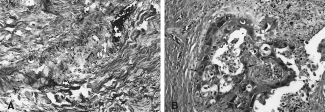
Fig 3. Histopathologic findings of resected lymph nodes.
A, Eighteen of 19 previously clinically positive lymph nodes show fibrosis and inflammation (original magnification, ×20).
B, In the other node (one of 19), a small focus of squamous cell carcinoma is apparent (arrows) (original magnification, ×20).
The primary site was surgically treated in two of seven patients. Patient 2 underwent biopsy of the primary site (tonsil), which was negative for tumor. Patient 7, who underwent laryngopharyngectomy with jejunal reconstruction for a hypopharyngeal primary, had a 2-mm-diameter focus of squamous cell carcinoma within the en bloc resection; the margins were free of tumor. In addition, this patient had one pathologically positive node in the left neck, which was not identified as radiographically positive on either the pre- or post-treatment chemoradiation CT scans.
Six of seven patients remained alive and six of six remained disease-free, with an average follow-up duration of 24 months. One patient experienced a recurrence at 17 months and ultimately succumbed to HNSCC at 19 months.
Discussion
The expanding role of combined chemoradiation therapy for organ preservation and unresectable HNSCC has brought to light a dilemma regarding when surgical therapy is necessary to remove lymph nodes that are grossly positive before medical therapy. In an effort to address this quandary, the current study was undertaken to analyze the effects of combined chemotherapy and radiotherapy on cervical lymph node volumes for both diseased and normal lymph nodes. In addition, we sought to correlate radiographic changes with the surgical pathologic findings of excised tissue.
Although chemoradiation did cause volume reduction in normal nodes (55% ± 21%), this reduction was not statistically significant. Normal nodes responded with a wide degree of variability; one node was no longer detectable on CT scans after chemoradiation, whereas another showed a small increase in size, presumably because of inflammation. In contrast, the volume reduction in diseased nodes (91% ± 4 %) was statistically significant; diseased nodes consistently showed large volume reduction, and chemoradiation resolved extracapsular spread as detected by CT and confirmed by surgical pathology. Surgical pathologic findings indicated that 18 (95%) of 19 lymph nodes were free of tumor after concurrent chemoradiation.
To analyze lymph node response, we advocate the use of volume estimation with measurements derived from the x, y, and z directions. This is in contrast to area estimations with which measurements are made only from the x and y axes (6). Such 2D analysis may result in inaccurate nodal size assessment, considering the z-axis variation between nodes. With the increasing use of contiguous scanning techniques, we believe the progression to 3D analysis is warranted because it provides more accurate determination of lymph node size.
Although there are a number of studies that quantify lymph node reduction for radiation therapy alone, none have done so for combined chemotherapy and radiation therapy. In necks irradiated with 6500 to 7500 cGy, Mukherji et al (7) reported shrinkage of 74% at 10.2 months and 65% at 12.1 months; in addition, they reported enlargement of a normal node in a therapeutic radiation field. Schafer et al (8) advocated sonographic monitoring and measurement of lymph node volume during radiation treatment; they reported volume reduction of 50% after 21 days of therapy. Liska et al (9) used CT to show cervical lymph node volume reduction between 60% and 72% for diseased nodes treated with 6600 cGy over 33 days. None of these investigators correlated radiographic changes with pathologic findings of affected tissue.
The present study shows that significant nodal volume reduction revealed by CT scanning correlates with a high degree of negative pathologic findings. Surgical specimens were labeled regarding neck level (levels I−VI) and were tagged in relation to anatomic structures to allow positive identification of nodes with those previously identified on CT scans. Eighteen (95%) of 19 previously clinically positive lymph nodes showed no histologic evidence of disease in surgical specimens. Of note, the single surgical specimen showing tumor occurred in a patient who intentionally received subtherapeutic radiation therapy (4500 cGy) in anticipation of the need for surgical resection of the primary disease site and free-flap reconstruction. Our results of tumor ablation in 95% of lymph nodes compare favorably with results from both chemotherapy alone and radiation therapy alone. Norris et al (10) reported 15% tumor ablation in cervical nodes after the administration of cisplatin-, bleomycin-, and methotrexate-based induction chemotherapy. Boyd et al (11) reported 68% tumor ablation in cervical nodes after the administration of high-dose radiation therapy. Our results of 95% tumor ablation suggest that the combination of chemotherapy and radiation therapy is synergistic.
The use of CT to follow clinical response offers certain advantages (including low cost and ease of availability) over other techniques. Such techniques proposed in the recent literature include [18F] fluorodeoxyglucose positron emission tomography (12) to follow metabolic activity of tumor and sonographically guided fine needle aspiration (13) to follow cytology. CT correlation with surgical pathologic findings, although high in our study in which nodes that showed 90% volume reduction had a 95% incidence of tumor ablation, is ultimately dependent on the reliability of pathologic analysis of resected nodes. Micrometastases may be missed, especially in larger surgical specimens in which limited numbers of sections are representative of the entire specimen. Although our preliminary results are promising, ultimate confirmation of CT results with histopathologic findings will depend on better techniques to determine tumor viability in surgical specimens (ie, molecular tumor markers).
Conclusion
In seven patients with stage IV HNSCC treated with concurrent chemoradiation, CT-assessed volume reduction of cervical lymph nodes showed that diseased nodes had a significantly greater volume reduction (91% ± 4%) compared with normal nodes (55% ± 21%) (P < .02, two-sided Student's t test). Chemoradiation resolved extracapsular spread of all diseased nodes. Surgical resection and pathologic analysis confirmed tumor ablation in 18 (95%) of 19 previously clinically positive nodes. Although these results are promising, suggesting that radiographic imaging may direct the need for adjuvant surgical therapy, our current recommendations are based on the conservative algorithm presented by Norris et al (10). Norris et al recommend adjuvant surgery for patients who show only partial response (ie, nodal volume reduction <90%) after therapeutic chemoradiation dosages.
Footnotes
↵1 Presented at the annual meeting of The American Society of Head and Neck Radiology, April 1998, Phoenix, AZ.
2 Address reprint requests to Robert F. Labadie, MD, PhD, CB #7070 Burnett-Womack Building, Division of Otolaryngology-Head and Neck Surgery, University of North Carolina at Chapel Hill, Chapel Hill, NC 27599.
References
- Received March 8, 1999.
- Copyright © American Society of Neuroradiology


