Advertisement

Main menu

  • Home
  • Content
    • Current Issue
    • Accepted Manuscripts
    • Article Preview
    • Past Issue Archive
    • Video Articles
    • AJNR Case Collection
    • Case of the Week Archive
    • Case of the Month Archive
    • Classic Case Archive
  • Special Collections
    • AJNR Awards
    • ASNR Foundation Special Collection
    • Most Impactful AJNR Articles
    • Photon-Counting CT
    • Spinal CSF Leak Articles (Jan 2020-June 2024)
  • Multimedia
    • AJNR Podcasts
    • AJNR SCANtastic
    • Trainee Corner
    • MRI Safety Corner
    • Imaging Protocols
  • For Authors
    • Submit a Manuscript
    • Submit a Video Article
    • Submit an eLetter to the Editor/Response
    • Manuscript Submission Guidelines
    • Statistical Tips
    • Fast Publishing of Accepted Manuscripts
    • Graphical Abstract Preparation
    • Imaging Protocol Submission
    • Author Policies
  • About Us
    • About AJNR
    • Editorial Board
    • Editorial Board Alumni
  • More
    • Become a Reviewer/Academy of Reviewers
    • Subscribers
    • Permissions
    • Alerts
    • Feedback
    • Advertisers
    • ASNR Home

User menu

  • Alerts
  • Log in

Search

  • Advanced search
American Journal of Neuroradiology

American Journal of Neuroradiology

ASHNR American Society of Functional Neuroradiology ASHNR American Society of Pediatric Neuroradiology ASSR
  • Alerts
  • Log in

Advanced Search

  • Home
  • Content
    • Current Issue
    • Accepted Manuscripts
    • Article Preview
    • Past Issue Archive
    • Video Articles
    • AJNR Case Collection
    • Case of the Week Archive
    • Case of the Month Archive
    • Classic Case Archive
  • Special Collections
    • AJNR Awards
    • ASNR Foundation Special Collection
    • Most Impactful AJNR Articles
    • Photon-Counting CT
    • Spinal CSF Leak Articles (Jan 2020-June 2024)
  • Multimedia
    • AJNR Podcasts
    • AJNR SCANtastic
    • Trainee Corner
    • MRI Safety Corner
    • Imaging Protocols
  • For Authors
    • Submit a Manuscript
    • Submit a Video Article
    • Submit an eLetter to the Editor/Response
    • Manuscript Submission Guidelines
    • Statistical Tips
    • Fast Publishing of Accepted Manuscripts
    • Graphical Abstract Preparation
    • Imaging Protocol Submission
    • Author Policies
  • About Us
    • About AJNR
    • Editorial Board
    • Editorial Board Alumni
  • More
    • Become a Reviewer/Academy of Reviewers
    • Subscribers
    • Permissions
    • Alerts
    • Feedback
    • Advertisers
    • ASNR Home
  • Follow AJNR on Twitter
  • Visit AJNR on Facebook
  • Follow AJNR on Instagram
  • Join AJNR on LinkedIn
  • RSS Feeds

AJNR is seeking candidates for the AJNR Podcast Editor. Read the position description.

Research ArticleHEAD AND NECK

The Persistent Stapedial Artery

Richard Silbergleit, Douglas J. Quint, Bharat A. Mehta, Suresh C. Patel, Joseph J. Metes and Samir E. Noujaim
American Journal of Neuroradiology March 2000, 21 (3) 572-577;
Richard Silbergleit
aFrom the Department of Diagnostic Radiology and Medical Imaging, Henry Ford Hospital, Detroit, MI (R.S., B.A.M., S.C.P.); the Department of Radiology, University of Michigan Hospitals, Ann Arbor (D.J.Q.); the Department of Radiology, St. John's Hospital and Medical Center, Detroit (J.J.M.); and the Department of Radiology, William Beaumont Hospital, Royal Oak, MI (S.E.N.).
  • Find this author on Google Scholar
  • Find this author on PubMed
  • Search for this author on this site
Douglas J. Quint
aFrom the Department of Diagnostic Radiology and Medical Imaging, Henry Ford Hospital, Detroit, MI (R.S., B.A.M., S.C.P.); the Department of Radiology, University of Michigan Hospitals, Ann Arbor (D.J.Q.); the Department of Radiology, St. John's Hospital and Medical Center, Detroit (J.J.M.); and the Department of Radiology, William Beaumont Hospital, Royal Oak, MI (S.E.N.).
  • Find this author on Google Scholar
  • Find this author on PubMed
  • Search for this author on this site
Bharat A. Mehta
aFrom the Department of Diagnostic Radiology and Medical Imaging, Henry Ford Hospital, Detroit, MI (R.S., B.A.M., S.C.P.); the Department of Radiology, University of Michigan Hospitals, Ann Arbor (D.J.Q.); the Department of Radiology, St. John's Hospital and Medical Center, Detroit (J.J.M.); and the Department of Radiology, William Beaumont Hospital, Royal Oak, MI (S.E.N.).
  • Find this author on Google Scholar
  • Find this author on PubMed
  • Search for this author on this site
Suresh C. Patel
aFrom the Department of Diagnostic Radiology and Medical Imaging, Henry Ford Hospital, Detroit, MI (R.S., B.A.M., S.C.P.); the Department of Radiology, University of Michigan Hospitals, Ann Arbor (D.J.Q.); the Department of Radiology, St. John's Hospital and Medical Center, Detroit (J.J.M.); and the Department of Radiology, William Beaumont Hospital, Royal Oak, MI (S.E.N.).
  • Find this author on Google Scholar
  • Find this author on PubMed
  • Search for this author on this site
Joseph J. Metes
aFrom the Department of Diagnostic Radiology and Medical Imaging, Henry Ford Hospital, Detroit, MI (R.S., B.A.M., S.C.P.); the Department of Radiology, University of Michigan Hospitals, Ann Arbor (D.J.Q.); the Department of Radiology, St. John's Hospital and Medical Center, Detroit (J.J.M.); and the Department of Radiology, William Beaumont Hospital, Royal Oak, MI (S.E.N.).
  • Find this author on Google Scholar
  • Find this author on PubMed
  • Search for this author on this site
Samir E. Noujaim
aFrom the Department of Diagnostic Radiology and Medical Imaging, Henry Ford Hospital, Detroit, MI (R.S., B.A.M., S.C.P.); the Department of Radiology, University of Michigan Hospitals, Ann Arbor (D.J.Q.); the Department of Radiology, St. John's Hospital and Medical Center, Detroit (J.J.M.); and the Department of Radiology, William Beaumont Hospital, Royal Oak, MI (S.E.N.).
  • Find this author on Google Scholar
  • Find this author on PubMed
  • Search for this author on this site
  • Article
  • Figures & Data
  • Info & Metrics
  • Responses
  • References
  • PDF
Loading

Article Figures & Data

Figures

  • Tables
  • fig 1.
    • Download figure
    • Open in new tab
    • Download powerpoint
    fig 1.

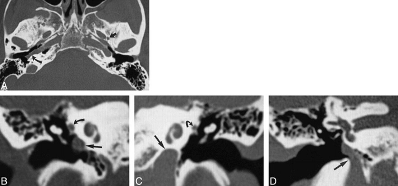
    Case 1: 52-year-old woman with buzzing in right ear.

    A, Axial CT scan of skull base shows a normal left foramen spinosum (straight arrow). The right foramen spinosum is absent. The right ICA is laterally displaced into the middle ear (curved arrow). The left ICA is in a normal position.

    B, Coronal CT scan through right middle ear shows the ICA in the middle ear cavity (straight arrow). The soft tissue at the tympanic segment of the facial nerve is prominent because of the presence of a stapedial artery (curved arrow).

    C, Coronal CT scan through left middle ear shows the ICA in a normal position below the cochlea (straight arrow). The tympanic facial nerve is normal in size (curved arrow).

    D, Coronal CT scan at level of vestibule shows the entrance of the aberrant ICA into the right middle ear (arrow).

  • fig 2.
    • Download figure
    • Open in new tab
    • Download powerpoint
    fig 2.

    Case 2: 14-year-old boy with headache, vomiting, dizziness, and ringing in the ear.

    A, Axial CT scan shows a normal right foramen spinosum (arrow) and absence of the left foramen spinosum.

    B, Coronal CT scan shows the aberrant ICA on the left (straight arrow) and the soft-tissue density of a PSA (curved arrow).

    C, Left carotid arteriogram, lateral view, shows a PSA arising from the aberrant ICA (arrow).

    D, Left carotid arteriogram, frontal view, shows a PSA arising from the aberrant ICA (arrow).

  • fig 3.
    • Download figure
    • Open in new tab
    • Download powerpoint
    fig 3.

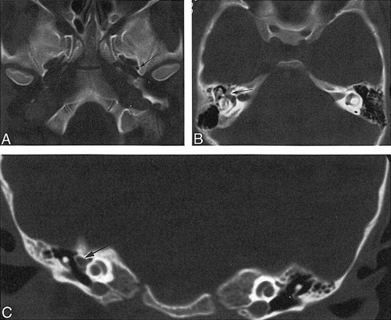
    Case 3: 6-year-old girl with vertigo.

    A, Axial CT scan shows a normal left foramen spinosum (arrow) and absence of the right foramen spinosum.

    B, Axial CT scan through middle ear shows prominent soft tissue, representing facial nerve and PSA (arrow).

    C, Coronal CT scan through middle ear shows prominent soft tissue, representing facial nerve and PSA (arrow).

  • fig 4.
    • Download figure
    • Open in new tab
    • Download powerpoint
    fig 4.

    Case 4: 25-year-old woman with pulsatile tinnitus.

    A, Axial CT scan shows a normal left foramen spinosum (arrow) and absence of the right foramen spinosum.

    B, Coronal CT scan through right middle ear shows the soft tissue at the tympanic segment of the facial nerve is prominent because of the presence of a stapedial artery (arrow).

    C, Coronal CT scan through left middle ear shows the tympanic facial nerve is normal in size (arrow).

  • fig 5.
    • Download figure
    • Open in new tab
    • Download powerpoint
    fig 5.

    Case 5: 5-year-old boy headache for 3 years. Carotid arteriogram, lateral view, shows a PSA (curved arrow) arising from the aberrant ICA, which represents the inferior tympanic branch of the ascending pharyngeal artery assuming the role of the ICA (straight arrow)

  • fig 6.
    • Download figure
    • Open in new tab
    • Download powerpoint
    fig 6.

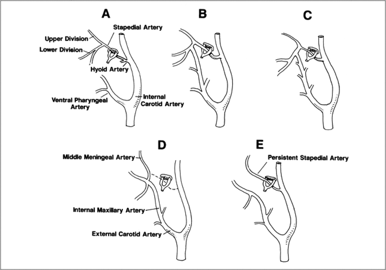
    Schematic representation of the developmental stages of the stapedial artery.

    A, The hyoid artery arises from the ICA. The stapedial artery arises from the hyoid artery near its origin. The stapedial artery branches into upper and lower divisions after passing through the stapes. The ventral pharyngeal arteries are the precursors of the definitive ECA. The stapedial artery is the only supply to the upper and lower divisions.

    B, Anastomosis forms between the ventral pharyngeal artery and the lower division branches.

    C, The stapedial artery decreases in size.

    D, Normal adult anatomy with involution of the stapedial artery.

    E, Anatomic configuration of typical PSA.

Tables

  • Figures
  • Table1

    Findings in five patients with persistent stapedial artery

PreviousNext
Back to top

In this issue

American Journal of Neuroradiology
Vol. 21, Issue 3
1 Mar 2000
  • Table of Contents
  • Index by author
Advertisement
Print
Download PDF
Email Article

Thank you for your interest in spreading the word on American Journal of Neuroradiology.

NOTE: We only request your email address so that the person you are recommending the page to knows that you wanted them to see it, and that it is not junk mail. We do not capture any email address.

Enter multiple addresses on separate lines or separate them with commas.
The Persistent Stapedial Artery
(Your Name) has sent you a message from American Journal of Neuroradiology
(Your Name) thought you would like to see the American Journal of Neuroradiology web site.
CAPTCHA
This question is for testing whether or not you are a human visitor and to prevent automated spam submissions.
Cite this article
Richard Silbergleit, Douglas J. Quint, Bharat A. Mehta, Suresh C. Patel, Joseph J. Metes, Samir E. Noujaim
The Persistent Stapedial Artery
American Journal of Neuroradiology Mar 2000, 21 (3) 572-577;

Citation Manager Formats

  • BibTeX
  • Bookends
  • EasyBib
  • EndNote (tagged)
  • EndNote 8 (xml)
  • Medlars
  • Mendeley
  • Papers
  • RefWorks Tagged
  • Ref Manager
  • RIS
  • Zotero
0 Responses
Respond to this article
Share
Bookmark this article
The Persistent Stapedial Artery
Richard Silbergleit, Douglas J. Quint, Bharat A. Mehta, Suresh C. Patel, Joseph J. Metes, Samir E. Noujaim
American Journal of Neuroradiology Mar 2000, 21 (3) 572-577;
del.icio.us logo Twitter logo Facebook logo Mendeley logo
  • Tweet Widget
  • Facebook Like
  • Google Plus One

Jump to section

  • Article
    • Abstract
    • Case Reports
    • Discussion
    • Conclusion
    • Footnotes
    • References
  • Figures & Data
  • Info & Metrics
  • Responses
  • References
  • PDF

Related Articles

  • No related articles found.
  • PubMed
  • Google Scholar

Cited By...

  • Cerebral neurovascular embryology, anatomic variations, and congenital brain arteriovenous lesions
  • The Many Faces of Persistent Stapedial Artery: CT Findings and Embryologic Explanations
  • Middle Meningeal Artery: Anatomy and Variations
  • Stapedial Artery: From Embryology to Different Possible Adult Configurations
  • Temporal Bone CT: Anatomy, Technique, and Associated Pathophysiology
  • A Venous Cause for Facial Canal Enlargement: Multidetector Row CT Findings and Histopathologic Correlation
  • Dangerous Extracranial-Intracranial Anastomoses and Supply to the Cranial Nerves: Vessels the Neurointerventionalist Needs to Know
  • Persistent Stapedial Artery: MR Angiographic and CT Findings
  • Bilateral Aberrant Internal Carotid Arteries with Bilateral Persistent Stapedial Arteries and Bilateral Duplicated Internal Carotid Arteries
  • Crossref
  • Google Scholar

This article has not yet been cited by articles in journals that are participating in Crossref Cited-by Linking.

More in this TOC Section

  • Correlation of Apparent Diffusion Coefficient at 3T with Prognostic Parameters of Retinoblastoma
  • Parathyroid Lesions: Characterization with Dual-Phase Arterial and Venous Enhanced CT of the Neck
  • Efficacy of Diffusion-Weighted Imaging for the Differentiation between Lymphomas and Carcinomas of the Nasopharynx and Oropharynx: Correlations of Apparent Diffusion Coefficients and Histologic Features
Show more HEAD AND NECK

Similar Articles

Advertisement

Indexed Content

  • Current Issue
  • Accepted Manuscripts
  • Article Preview
  • Past Issues
  • Editorials
  • Editor's Choice
  • Fellows' Journal Club
  • Letters to the Editor
  • Video Articles

Cases

  • Case Collection
  • Archive - Case of the Week
  • Archive - Case of the Month
  • Archive - Classic Case

Special Collections

  • AJNR Awards
  • ASNR Foundation Special Collection
  • Most Impactful AJNR Articles
  • Photon-Counting CT
  • Spinal CSF Leak Articles (Jan 2020-June 2024)

More from AJNR

  • Trainee Corner
  • Imaging Protocols
  • MRI Safety Corner

Multimedia

  • AJNR Podcasts
  • AJNR Scantastics

Resources

  • Turnaround Time
  • Submit a Manuscript
  • Submit a Video Article
  • Submit an eLetter to the Editor/Response
  • Manuscript Submission Guidelines
  • Statistical Tips
  • Fast Publishing of Accepted Manuscripts
  • Graphical Abstract Preparation
  • Imaging Protocol Submission
  • Evidence-Based Medicine Level Guide
  • Publishing Checklists
  • Author Policies
  • Become a Reviewer/Academy of Reviewers
  • News and Updates

About Us

  • About AJNR
  • Editorial Board
  • Editorial Board Alumni
  • Alerts
  • Permissions
  • Not an AJNR Subscriber? Join Now
  • Advertise with Us
  • Librarian Resources
  • Feedback
  • Terms and Conditions
  • AJNR Editorial Board Alumni

American Society of Neuroradiology

  • Not an ASNR Member? Join Now

© 2025 by the American Society of Neuroradiology All rights, including for text and data mining, AI training, and similar technologies, are reserved.
Print ISSN: 0195-6108 Online ISSN: 1936-959X

Powered by HighWire