
Abstract
BACKGROUND AND PURPOSE: Despite improvements in noninvasive imaging, some patients with contraindications to iodine-based contrast material still require angiography for the evaluation of carotid stenosis. Our aim was to assess the utility of gadopentetate dimeglumine as an intraarterial contrast agent in common carotid angiography.
METHODS: Twelve patients with suspected carotid artery stenosis were enrolled in the study. In addition to the standard injection sequences with iohexol, common carotid arteriograms were obtained after administration of gadopentetate dimeglumine. Neurologic status and vital signs were monitored during and for 6 hours after the examination. For each injection, five independent observers, blinded to the contrast agent used, measured the percentage of carotid stenosis and assessed their confidence in grading the stenosis, the overall quality of the examination, and, in cases of decreased quality, the reason(s) for it. Statistical analysis was done with paired and unpaired t-tests with equal variances.
RESULTS: No patient had an adverse clinical outcome, and measurements of carotid artery stenosis showed no statistically significant differences between the gadopentetate dimeglumine and iohexol examinations. Overall image quality and observer confidence in measurements of stenosis on the gadolinium-based studies were slightly but significantly lower than those of identical iodine-based studies.
CONCLUSION: Gadopentetate dimeglumine may be an alternative to iodine in selected patients undergoing carotid angiography. Although overall image quality of the gadolinium studies is slightly inferior to that of the iohexol studies, measurements of carotid artery stenosis are similar for the two examinations.
Despite recent advances in noninvasive imaging of the carotid arteries, catheter carotid arteriography remains the standard for evaluating patients with suspected carotid stenosis. Unfortunately, some patients with contraindications to iodinated contrast agents, such as azotemia or severe contrast allergy, still require carotid angiography. The purpose of this study was to assess the efficacy of gadopentetate dimeglumine as an alternative intraarterial contrast agent in the evaluation of the common carotid artery in patients with suspected carotid stenosis.
Methods
Approval from our human subjects committee was obtained before initiation of the study. Twelve patients who were referred for carotid angiography to either the University of Arizona Health Science Center or the Veteran's Affairs Hospital Tucson were enrolled in the study after giving informed consent. Patients who were pregnant or lactating, under the age of 21 years, had had a recent stroke or transient ischemic attack, or had a history of allergy to gadopentetate dimeglumine were ineligible.
Before the procedure, a brief neurologic examination (which consisted of motor strength, sensory symmetry, and cranial nerve assessment) was performed. The patient was then attached to both an EKG monitor and a pulse oximeter so that baseline vital signs could be obtained. Vital sign monitoring and neurologic assessments were continued throughout the examination.
Once the common femoral artery was accessed, an arch arteriogram was obtained. Subsequently, a 5F catheter was placed in either the right or left common carotid artery. Anteroposterior digital subtraction arteriography (DSA) of the cervical segment of the carotid artery was performed with a hand injection of 8 mL of undiluted gadopentetate dimeglumine, and two images per second were obtained. The identical procedure was repeated using iohexol 300, which was then followed by anteroposterior and lateral intracranial arteriograms obtained with the use of a power injection. Finally, lateral DSA of the common carotid artery in the neck was performed with 8 mL of hand-injected iohexol followed by 8 mL of hand-injected gadopentetate dimeglumine. At the discretion of the angiographer, three patients had only one injection with gadopentetate dimeglumine. After the procedure, the patients were observed for 6 hours, with continuous assessment of vital signs and neurologic status.
The 21 iodine-based and corresponding 21 gadolinium-based DSA images were then interpreted by five radiologists (four CAQ-certified or eligible neuroradiologists and one CAQ-certified angiographer) who were blinded to the contrast agent used in the study. The images were graded on a scale of 1 to 6 for overall image quality, with 1 representing an excellent study and 6 a nondiagnostic examination. Reasons for poor image quality were noted, such as inadequate contrast, patient motion, or patient positioning. The percentage of stenosis was assessed using the North American Symptomatic Carotid Endarterectomy Trial (NASCET) criteria, and confidence in each measurement was graded on a scale of 1 to 6.
The data collected were analyzed to test the null hypothesis, the observer confidence in contrast agents, the correlation of interobserver measurements, and the overall quality factors of the examination.
Results
Clinical Parameters
No neurologic complications were seen in any of the patients. Vital signs remained stable throughout the examination and during the recovery period for all patients. No subjective complaints were recorded as a result of the gadolinium injection.
Percentage of Stenosis
The null hypothesis of this study was that there is no statistical difference between iodine- and gadolinium-based contrast agents in the assessment of carotid artery stenosis when using the NASCET measurement criteria. For this data analysis, α was set at .001 for all statistical tests. Using an F-test, we determined that the data collected were of equal variances. A t-test was performed using unpaired and paired data, and both showed no statistically significant difference in measurements of stenosis between the iodine- and gadolinium-based studies (t = −1.27, df = 96, P = .21, two-tailed paired t-test). The mean measured stenosis on both the gadolinium- and iodine-based examinations was 45%, using the NASCET criteria.
Although the measurements of carotid artery stenosis were the same, there was a statistical difference in observer confidence in the measurements between the two studies (t = −3.42, df = 104, P = .0009, two-tailed paired t-test). The mean graded confidence in the iodine-based studies was 2.17, and the mean graded confidence in the gadolinium-based studies was 2.57. Both these values lie between 2 (confident) and 3 (somewhat confident). A receiver operating characteristic curve was not constructed owing to a lack of population values.
The interobserver correlation coefficient (determined by using a standard Pearson product moment correlation coefficient equation) was 0.77 for each observer's measurement of stenosis on each examination (Fig 1). This correlation is highly associative and shows that the measurement of stenosis (on all examinations) is reproducible from observer to observer.

Observer correlation in interpreting the degree of stenosis on studies obtained with gadolinium- and iodine-based contrast agents
Image Quality
The overall quality of the examination differed statistically between the iodine- and gadolinium-based studies. Iodine-based examinations were the better of the two, with a mean rating of 2.43 (2 = very good; 3 = good), whereas the overall quality of the gadolinium-based examinations had a mean rating of 2.95. An α setting of .001 and a paired t-test showed a statistical difference (t = −4.09, df = 104, P ≤ .00008, two-tailed paired t-test).
Quality ratings also included a reason for decreased overall quality. In both studies the quality was reduced most by inadequate contrast, then by patient positioning, and finally by other factors, such as filming technique and misregistration due to calcified plaque (Fig 2).
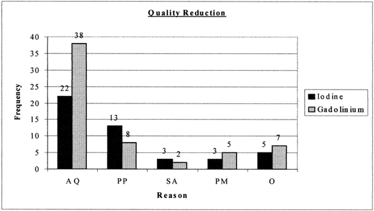
Histogram shows observers' reasons for reduced quality of iodine- and gadolinium-based studies. AQ indicates adequacy of contrast; PP, patient positioning; SA, saturation artifact; PM, patient motion; O, other factors, including filming technique and misregistration due to calcified plaque
Discussion
Exacerbation of chronic renal insufficiency with the use of iodinated contrast material is a well-documented phenomenon and may prevent patients with chronic renal insufficiency from undergoing carotid arteriography. Other patients are at increased risk owing to a history of anaphylactoid reaction to iodinated contrast agents. Therefore, there has been growing interest in the use of alternative, nonnephrotoxic contrast agents for use in arteriography.
Noninvasive imaging, such as sonography, CT angiography, and MR angiography, all have their limitations. Consequently, many vascular surgeons request carotid angiography for the preoperative assessment of patients with suspected carotid stenosis. Because of the continued need for carotid arteriography, and the prevalence of relative or absolute contraindications to iodinated contrast agents, an alternative to iodine-based contrast media would be valuable. There is growing experience with gadolinium-based DSA as an alternative to both iodine and CO2 in peripheral angiography (1–4). A recent case report (5) has demonstrated the possibility of using gadolinium-based contrast material in carotid angiography.
In this study, measurable carotid arterial stenosis was not significantly different between studies performed with gadopentetate dimeglumine and iohexol. However, the images obtained with the use of the gadolinium agent were judged to be of lower quality than those obtained with iohexol (Fig 3). Some of this difference may be attributed to the study design, in that if an iodine-based study was judged to be nondiagnostic, the examination was repeated. No repeat examinations were performed when the gadolinium-based contrast agent was used, even if the examination was clearly substandard. In practical application, cerebral arteriograms are reviewed as the examination is performed. If during the course of gadolinium-based carotid angiography there is doubt about the degree of stenosis, or if an arteriogram is judged to be nondiagnostic, additional injections can be performed. With our injection sequences, a total of 32 mL of gadopentetate dimeglumine was injected. Depending on the size of the patient, it may be possible to repeat all of the gadolinium injections without exceeding the commonly used dosage of 0.3 mmol/kg.

A and B, Gadolinium- (A) and iodine-based (B) frontal projections of common carotid arteriogram. Although the depiction of vascular anatomy is equivalent, there is relatively decreased intravascular contrast on the gadolinium examination
Gadolinium is less radiopaque than iodine (6), which undoubtedly contributed to the perception in this study that the gadolinium images were of poorer quality than the iodine images. In general, when filming the examinations, the image contrast had to be increased, which resulted in increased quantum mottle (Fig 4). None of the patients included in this study had near total carotid occlusion, so the utility of gadolinium in this circumstance cannot be assessed. Because of its relatively decreased radiodensity, it may be that an overestimate of a near total occlusion could occur when using a gadolinium-based contrast agent. If this were the case, a misdiagnosis could be made and a near total occlusion (string sign) could be mistaken for total carotid occlusion. Gadolinium should be used with caution if near total carotid occlusion is suspected.

A and B, Gadolinium- (A) and iodine-based (B) common carotid arteriograms. Lateral injections are nearly identical, except that the gadolinium examination has increased graininess due to film contrast
The osmolality of gadopentetate dimeglumine is greater than that of iodine. A prior report (5) described a burning sensation suffered by the patient during the gadolinium injection, an experience that none of our patients reported.
Previous investigators have reported that intraarterial gadolinium in doses up to 0.3 mmol/kg is less nephrotoxic than iodinated contrast injection in patients with azotemia. There is no evidence of nephrotoxicity to humans with impaired renal function with doses up to 0.3 mmol/kg. In fact, animal studies have shown no evidence of nephrotoxicity due to gadolinium in doses up to 10 mmol/kg (7, 8). For patients with compromised renal function who cannot undergo MR angiography, gadolinium cerebral angiography may be useful.
Conclusion
Gadolinium-based contrast agents may be used as an alternative to iodine-based agents in selected patients undergoing carotid angiography. Although, in our study, the overall image quality of the gadolinium-based examination was judged to be slightly inferior to that of the iohexol examination, measurements of carotid stenosis were similar when comparing gadolinium-based arteriograms with identical examinations obtained with iohexol.
Footnotes
↵1 Address reprint requests to William K. Erly, MD.
- Received August 2, 1999.
- Copyright © American Society of Neuroradiology
In this issue


Jump to section
Related Articles
Cited By...
- No citing articles found.