
Abstract
BACKGROUND AND PURPOSE: Recent experimental studies and a few case reports reveal that coiling may not lead to permanent occlusion of aneurysms by an organized thrombus. Therefore, biologic long-term prognosis seems to be doubtful, and the physical effect of coils may be important. The purpose of this study was to investigate the physical effect of coils on pressure and flow dynamics in aneurysms.
METHODS: Bifurcation aneurysms were created in eight rabbits, explanted after 3 weeks, and tested in a model with pulsatile perfusion with 0.9% saline and heparinized blood. Before and after densely packing with coils, systemic and intraaneurysmal pressure, aneurysmal pulsation, and impact measurements were recorded.
RESULTS: The peak and shape of the pressure waves in the aneurysm and in the delivery system were not significantly different before and after coiling. Under physiological intraaneurysmal pressure (while being perfused with saline), significant reduction (P = .022) of aneurysmal wall pulsation after coil embolization was noted. Overall, the aneurysmal impact on surrounding structures was statistically unchanged after coiling. However, in a few cases, after coil embolization, the observed increase of impact was more than doubled compared with the original values before coiling.
CONCLUSION: Coils do not physically affect intraaneurysmal pressure. After coiling, there is no significant reduction of flow rates through the aneurysm and no reduction of aneurysmal impact, but aneurysmal wall pulsation may be decreased.
Endovascular coil embolization seems to have become an established method in the treatment of saccular intracranial aneurysms. Although permanent occlusion rate, and therefore long-term prognosis, may be doubtful (1–4), the number of patients treated with coils increases each year. Radiologic and clinical reports of the results of coil embolization have been published during the last 6 years (5–10). Knowledge of the physical effects of coils on pressure and flow dynamics in saccular bifurcation aneurysms, however, is still lacking. Because intraaneurysmal pressure is considered to be an important factor in aneurysm growth and rupture (11), some experimental studies assess pressure inside saccular aneurysms in relation to the parent artery. In well-created bifurcation aneurysms, no pressure difference was observed under normal conditions (12–14). Byrne and Guglielmi (15) assumed that coils absorb parts of the energy of the systolic pulse to the walls of the aneurysm, buffering the transmitted blood pressure before the systolic pulse wave hits the wall of the aneurysm. Novak et al (14) measured intraaneurysmal pressure before and after incomplete coiling (there was only one coil inside the sac) but did not observe any pressure reduction. They did observe a decrease in aneurysmal pulsation. The purpose of the present experimental study was to investigate the physical effect of densely packed coils on intraaneurysmal pressure and on aneurysmal pulsation. Moreover, the aneurysmal impact (the pulse-dependent development of force between the aneurysmal sac and the surrounding structures) was determined. To avoid proximal arterial stenoses (eg, spasms) and the effects of thrombosis, an ex vivo model was chosen.
Methods
Surgical Procedure
In eight male New Zealand albino rabbits (weight, 3–4 kg), bifurcation aneurysms were microsurgically created according to the technique presented by Forrest and O'Reilly (16) and modified by Spetzger et al (1). The aneurysms were produced at a surgically created bifurcation of the common carotid artery by means of a venous graft pouch taken from the confluence of the right internal and external jugular vein. After 3 weeks, the aneurysms (final diameter, approximately 1 cm) were explanted.
Ex Vivo Model
Figure 1 shows a schematic drawing of the experimental system. An influx of 120 mL/min was obtained from a conventional peristaltic pump, and four air chambers and one check valve were used to produce a nearly constant, laminar, and unidirectional flow. This flow was then superimposed with a pulsatile flow generated by an electronically controlled valve (frequency generator PM 5139, Philips) and a resistance system. This led to a pump-independent aneurysmal systolic inflow and a passive diastolic outflow.

Schematic drawing of the experimental system. Systemic flow was obtained from a conventional peristaltic pump, and then four air chambers and one check valve were used to produce a nearly constant, laminar, and unidirectional flow. This flow was then superimposed with a pulsatile flow generated by an electronically controlled valve and a resistance system. Intraaneurysmal and systemic pressure were recorded online
The pressure in the delivery system (systemic pressure) was recorded online by a transducer (BPT 5326; World Precision Instruments, Sarasota, FL). The intraaneurysmal pressure was recorded online by a Camino probe (Camino Laboratories, San Diego, CA). Aneurysmal pulsation was measured without contact by a laser displacement sensor (No. LAA-20-2-670-1000; Di-el, Umbach, Germany).
Impact measurements were obtained by using a mechanical strain gauge dynamometer (Statham force transducer, ±30 g). We recorded the pulsatile force of the aneurysm sac on a round rigid rod. The rod had a diameter of 1 mm and was mounted at a right angle onto the surface of the aneurysm with a constant preload of 0.6 g. Before all experiments, the strain gauge dynamometer was calibrated to correlate rod displacement with force. The rod displacement necessary for the determination of force was small and well below wall displacement determined without contact by laser (<10%). Therefore, our impact/force measurement can be regarded as being independent of wall displacement.
In vitro tests were performed by pulsatile perfusion with (first) 0.9% saline and (second) heparinized rabbit blood. Pulse frequency was always 180/min, which is close to the physiological frequency (130–200/min) of the rabbit heart (17). At the beginning of each experiment (during the first run with saline), the systolic and diastolic intraaneurysmal pressures were adjusted close to the physiological values (systolic, 85–140 mm Hg; diastolic, 50–80 mm Hg) of the rabbit circulation (18). The selected valve and resistance adjustments then remained unchanged during the entire experiment. Beause of the higher viscosity of heparinized blood, the pressure values automatically increased and were therefore higher than those with saline.
Aneurysmal pulsation and impact were recorded in at least five different locations (the same locations before and after coiling) on the aneurysm surface for 10 s each (a total of >50 pulses each). After saline and heparinized blood measurement, the aneurysm sac was densely packed (as much as possible) with Guglielmi detachable coils under angiographic control during perfusion with 0.9% saline. In all cases, an obliteration of more than 90% was achieved. Pulsation, impact, and intraaneurysmal and systemic pressure were then recorded again, as described above. The rate of data collection for pressure, pulsation, and impact measurements was always 100/s. For statistic evaluation, paired t test or signed rank test was used.
Results
The calculated mean waves of intraaneurysmal and systemic pressure before and after coiling are shown in Figure 2A for saline and in Figure 2B for blood. The peak and shape of the pressure waves in the aneurysmal dome and in the delivery system were statistically not significantly different before and after coiling (signed rank test, P > .05%).
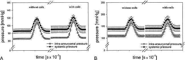
Intraaneurysmal and systemic pressure measurements (± standard error of mean) obtained before and after coiling. The amount and shape of the pressure waves were not significantly different before and after coil embolization. (x axis, each time interval = 10–1 s)
A, Circulating fluid, 0.9% saline.
B, Circulating fluid, heparinized blood.
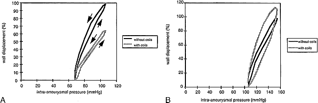
Because of technical reasons, laser displacement measurements obtained without contact were possible in only five experiments. Wall displacement (s) during pulsation (approximately 0.1–0.25 mm with 0.9% saline and 0.05–0.15 mm with blood) depends not only on intraaneurysmal pressure but also on the physical dimensions of the aneurysm (14). Because all specimens had different dimensions, relative values were used to define pulsation (maximal aneurysmal wall displacement without coils, smax = 100%). In Figure 3, the mean curves of aneurysmal pulsation are shown. Only for 0.9% saline (Fig 3A) did the statistical analysis (t test) yield a significant difference (P = .022) for wall displacement before and after coiling. Figure 4 shows the calculated wall displacement/pressure curves before and after coiling with 0.9% saline and blood, respectively. All curves describe hysteresis loops with pulsation as a function of pressure. Wall displacement follows variations in pressure with delay. The arrows in Figure 4A indicate the direction of the hysteresis loop. At the point of identical pressure, an aneurysm is smaller during the ascending versus the descending portion of the hysteresis curve. The area within each hysteresis loop was measured with a planimeter. Even though the statistical analysis yields a significant difference for wall displacement before and after coiling for 0.9% saline and even though the hysteresis loops in Figure 4A seem to be different, the differences in planimetered areas within the hysteresis loops did not reach statistical significance in either fluid used (t test, P = .134 for 0.9% saline). Even the quotient of area and maximal wall displacement did not reveal a significant difference (P > .05).

Aneurysmal pulsation measurements (± standard error of mean) obtained before and after coil embolization. (x axis, each time interval = 10–1 s)
A, After coiling and while circulating a 0.9% saline solution, a statistically significant (P = .022) reduction of wall displacement was observed
B, No statistically significant reduction was observed with blood.

Wall displacement/pressure curves measured before and after coil embolization describe hysteresis loops. Wall displacement follows changes in pressure with delay (arrows).
A, Circulating fluid, 0.9% saline.
B, Circulating fluid, heparinized blood.
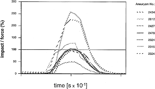
Aneurysmal impact was measured in seven specimens. Statistically, there was no significant difference (t test, P > .05) between the measured force before (100%) and after coiling and perfusion with 0.9% saline or blood, respectively. In two cases, however, a distinct increase of impact of more than twice the original values was measured after coil embolization. Figure 5 shows the results of impact measurements after coiling of all experiments with the circulating 0.9% saline fluid.

Results of impact/force measurements obtained for all experiments after coiling and while circulating a 0.9% saline solution. After embolization, in two cases, an increase of impact of more than twice the original values was measured. (x axis, each time interval = 10–1 s; y axis, impact/force [F]; diastolic value of impact = 0%; sytolic peak value of impact before coiling = 100%)
Discussion
Some experimental studies deal with the pressure inside saccular aneurysms in relation to its parent artery. In well-created bifurcation aneurysms, no pressure difference was observed under normal conditions (12–14). In our models, systemic and intraaneurysmal pressure differed with a nearly constant variation. This variation can easily be explained by the difference in the technique used for systemic and intraaneurysmal pressure measurement. There are only a few experimental studies dealing with experimental aneurysms and their flow dynamics after coiling (14, 19, 20). Gobin et al (20) and Novak et al (14) measured intraaneurysmal pressure in relation to the pressure in the parent artery after partial coil occlusion. They did not find any change, whether coils were present within the aneurysm. Our study confirms this finding in that we also did not observe any difference in intraaneurysmal pressure before and after coiling, even after densely packing the aneurysmal sac.
Novak et al (14) measured aneurysmal pulsation and intraaneurysmal pressure in sidewall and bifurcation aneurysms. In contrast to the present study, their aneurysms were only partially embolized with one coil. Moreover, they did not use a displacement sensor without contact. Similar to our study, the study by Novak et al (14) revealed a hysteresis in the relationship of intraaneurysmal pressure and pulsation. Already after partial coil embolization, they observed a reduction of aneurysmal pulsation and hysteresis loop. They assumed complete removal of hysteresis after further embolization.
The areas within the hysteresis loops are characterized by pressure, wall pulsation, and time lag between these components. In the series with 0.9% saline in the present study, significant reduction of aneurysmal pulsation was also observed, but the differences in hysteresis did not reach statistical significance (P = .134). The main reason may be the small number of experiments (n = 5). For statistical analysis, the area within the loop was measured. The area was distinctly decreased after coiling in three of five experiments. In only one aneurysm was the value somewhat increased; however, the t test did not reveal a statistically significant difference. Although more experiments may be necessary, it is important to note that the present study revealed that even in a completely coiled aneurysm, the hysteresis between pulsation and pressure does not dissapear. This is contrary to the assumption made by Novak et al (14).
We were unable to confirm the data regarding reduction of aneurysmal pulsation when blood was used to perfuse the aneurysms. In our models, aneurysm packing with Guglielmi detachable coils and contactless laser pulsation measurements were performed first during perfusion with 0.9% saline and “physiological” intraaneurysmal pressure values. Under these conditions, significant reduction of aneurysmal pulsation was observed. This finding is probably correct and can be explained by the higher tension of the aneurysmal wall due to the memory of the coil loops. Under “unphysiologically” elevated intraaneurysmal pressure values (such as in our series with blood perfusion), the tension of the aneurysmal wall was primarily high and the coil effect, therefore, may have been too small.
No significant difference could be determined between the calculated mean aneurysmal impact/force values to the surrounding structures before and after coiling. Because of the memory of the coils, the aneurysmal wall is under higher tension and thus stiffer than before. Therefore, impact may increase. However, in a few experiments, impact was distinctly higher after coiling. This finding may be important in patients with visual field defects that are due to aneurysms compressing the optic chiasma.
Coils do not affect the peak and shape of the pressure wave inside the aneurysmal dome, but at physiological pressures, aneurysmal pulsation seems to be smaller after coiling. Perhaps the reduced wall movement is an important factor in the development of thrombosis within the first hours after embolization (21). However, if there are really large spaces within the aneurysmal sac months after treatment (1–4), the physical effect of the presence of coils in the aneurysm may be unimportant and the mere presence does not prevent rupture. In addition to the physical effects of coils, there is a biologic reaction between the coils and the aneurysmal wall. Histologic investigations of experimental bifurcation aneurysms in rabbits 3 months after coiling (unpublished results) show a distinct increase of aneurysmal wall thickness due to chronic inflammation. Similar findings in aneurysms in humans have recently been described by Bavinzski et al (22). It may be that this biologic effect prevents aneurysmal rupture after partial recanalization.
Conclusion
Coils do not physically affect the peak and shape of the pressure wave at the aneurysmal wall. Under physiological intraaneurysmal pressure, aneurysmal pulsation seems to be reduced after coiling. The aneurysmal impact/force on the surrounding structures is not significantly different before and after coil embolization. However, in individual experiments, even a distinct increase was measured after coiling.
Footnotes
1 This work was presented in part at the ASNR/ASHNR/ASPNR/ASITN/ASSR Joint Meeting, San Diego, CA, May 1999.
This work was supported by a grant from the Friedhelm Frees Foundation, Wiesbaden, Germany.
↵2 Address reprint requests to Hans G Boecher-Schwarz, MD, Department of Neurosurgery, University Hospital, Langenbeckstr. 1, 55131 Mainz, Germany.
References
- Received June 7, 1999.
- Accepted after revision February 21, 2000.
- Copyright © American Society of Neuroradiology
In this issue


Jump to section
Related Articles
Cited By...
- Histopathological analysis of in vivo specimens of recurrent aneurysms after coil embolization
- The Distribution and Role of M1 and M2 Macrophages in Aneurysm Healing after Platinum Coil Embolization
- Contrast Enhancement of Intracranial Aneurysms on 3T 3D Black-Blood MRI and Its Relationship to Aneurysm Recurrence following Endovascular Treatment
- Intracranial Aneurysms: Wall Motion Analysis for Prediction of Rupture
- Mechanisms of Healing in Coiled Intracranial Aneurysms: A Review of the Literature
- In Vitro Study of Near-Wall Flow in a Cerebral Aneurysm Model with and without Coils
- Intracranial Aneurysms Treated With Endovascular Coils: Detection of Recurrences Using Unenhanced and Contrast-Enhanced Transcranial Color-Coded Duplex Sonography
- Basilar Trunk Occlusion during Endovascular Treatment of Giant and Fusiform Aneurysms of the Basilar Artery
- Image-Based Computational Simulation of Flow Dynamics in a Giant Intracranial Aneurysm
- Effect of Guglielmi Detachable Coil Placement on Intraaneurysmal Pressure: Experimental Study in Canines