Advertisement

Main menu

  • Home
  • Content
    • Current Issue
    • Accepted Manuscripts
    • Article Preview
    • Past Issue Archive
    • Video Articles
    • AJNR Case Collection
    • Case of the Week Archive
    • Case of the Month Archive
    • Classic Case Archive
  • Special Collections
    • AJNR Awards
    • ASNR Foundation Special Collection
    • Most Impactful AJNR Articles
    • Photon-Counting CT
    • Spinal CSF Leak Articles (Jan 2020-June 2024)
  • Multimedia
    • AJNR Podcasts
    • AJNR SCANtastic
    • Trainee Corner
    • MRI Safety Corner
    • Imaging Protocols
  • For Authors
    • Submit a Manuscript
    • Submit a Video Article
    • Submit an eLetter to the Editor/Response
    • Manuscript Submission Guidelines
    • Statistical Tips
    • Fast Publishing of Accepted Manuscripts
    • Graphical Abstract Preparation
    • Imaging Protocol Submission
    • Author Policies
  • About Us
    • About AJNR
    • Editorial Board
    • Editorial Board Alumni
  • More
    • Become a Reviewer/Academy of Reviewers
    • Subscribers
    • Permissions
    • Alerts
    • Feedback
    • Advertisers
    • ASNR Home

User menu

  • Alerts
  • Log in

Search

  • Advanced search
American Journal of Neuroradiology

American Journal of Neuroradiology

ASHNR American Society of Functional Neuroradiology ASHNR American Society of Pediatric Neuroradiology ASSR
  • Alerts
  • Log in

Advanced Search

  • Home
  • Content
    • Current Issue
    • Accepted Manuscripts
    • Article Preview
    • Past Issue Archive
    • Video Articles
    • AJNR Case Collection
    • Case of the Week Archive
    • Case of the Month Archive
    • Classic Case Archive
  • Special Collections
    • AJNR Awards
    • ASNR Foundation Special Collection
    • Most Impactful AJNR Articles
    • Photon-Counting CT
    • Spinal CSF Leak Articles (Jan 2020-June 2024)
  • Multimedia
    • AJNR Podcasts
    • AJNR SCANtastic
    • Trainee Corner
    • MRI Safety Corner
    • Imaging Protocols
  • For Authors
    • Submit a Manuscript
    • Submit a Video Article
    • Submit an eLetter to the Editor/Response
    • Manuscript Submission Guidelines
    • Statistical Tips
    • Fast Publishing of Accepted Manuscripts
    • Graphical Abstract Preparation
    • Imaging Protocol Submission
    • Author Policies
  • About Us
    • About AJNR
    • Editorial Board
    • Editorial Board Alumni
  • More
    • Become a Reviewer/Academy of Reviewers
    • Subscribers
    • Permissions
    • Alerts
    • Feedback
    • Advertisers
    • ASNR Home
  • Follow AJNR on Twitter
  • Visit AJNR on Facebook
  • Follow AJNR on Instagram
  • Join AJNR on LinkedIn
  • RSS Feeds

AJNR is seeking candidates for the AJNR Podcast Editor. Read the position description.

Research ArticlePEDIATRICS

Fetal Cerebral Cortex: Normal Gestational Landmarks Identified Using Prenatal MR Imaging

Catherine Garel, Emmanuel Chantrel, Hervé Brisse, Monique Elmaleh, Dominique Luton, Jean-François Oury, Guy Sebag and Max Hassan
American Journal of Neuroradiology January 2001, 22 (1) 184-189;
Catherine Garel
  • Find this author on Google Scholar
  • Find this author on PubMed
  • Search for this author on this site
Emmanuel Chantrel
  • Find this author on Google Scholar
  • Find this author on PubMed
  • Search for this author on this site
Hervé Brisse
  • Find this author on Google Scholar
  • Find this author on PubMed
  • Search for this author on this site
Monique Elmaleh
  • Find this author on Google Scholar
  • Find this author on PubMed
  • Search for this author on this site
Dominique Luton
  • Find this author on Google Scholar
  • Find this author on PubMed
  • Search for this author on this site
Jean-François Oury
  • Find this author on Google Scholar
  • Find this author on PubMed
  • Search for this author on this site
Guy Sebag
  • Find this author on Google Scholar
  • Find this author on PubMed
  • Search for this author on this site
Max Hassan
  • Find this author on Google Scholar
  • Find this author on PubMed
  • Search for this author on this site
  • Article
  • Figures & Data
  • Info & Metrics
  • Responses
  • References
  • PDF
Loading

Article Figures & Data

Figures

  • Tables
  • fig 1.
    • Download figure
    • Open in new tab
    • Download powerpoint
    fig 1.

    Distribution of fetuses by gestational age (GA)

  • fig 2.
    • Download figure
    • Open in new tab
    • Download powerpoint
    fig 2.

    Slice no. 1. T2-weighted midline sagittal image (20/9.2/12) (31 weeks' gestation). 1, parietooccipital fissure; 2, calcarine fissure; small arrow, marginal sulcus; large arrow, cingular sulcus.fig 3. Slice no. 2. T2-weighted parasagittal image (20/9.2/12) at the level of the sylvian fissure (28 weeks' gestation). Opercularization of the insula is completed posteriorly (arrow). Anteriorly, the insula (star) is wide open
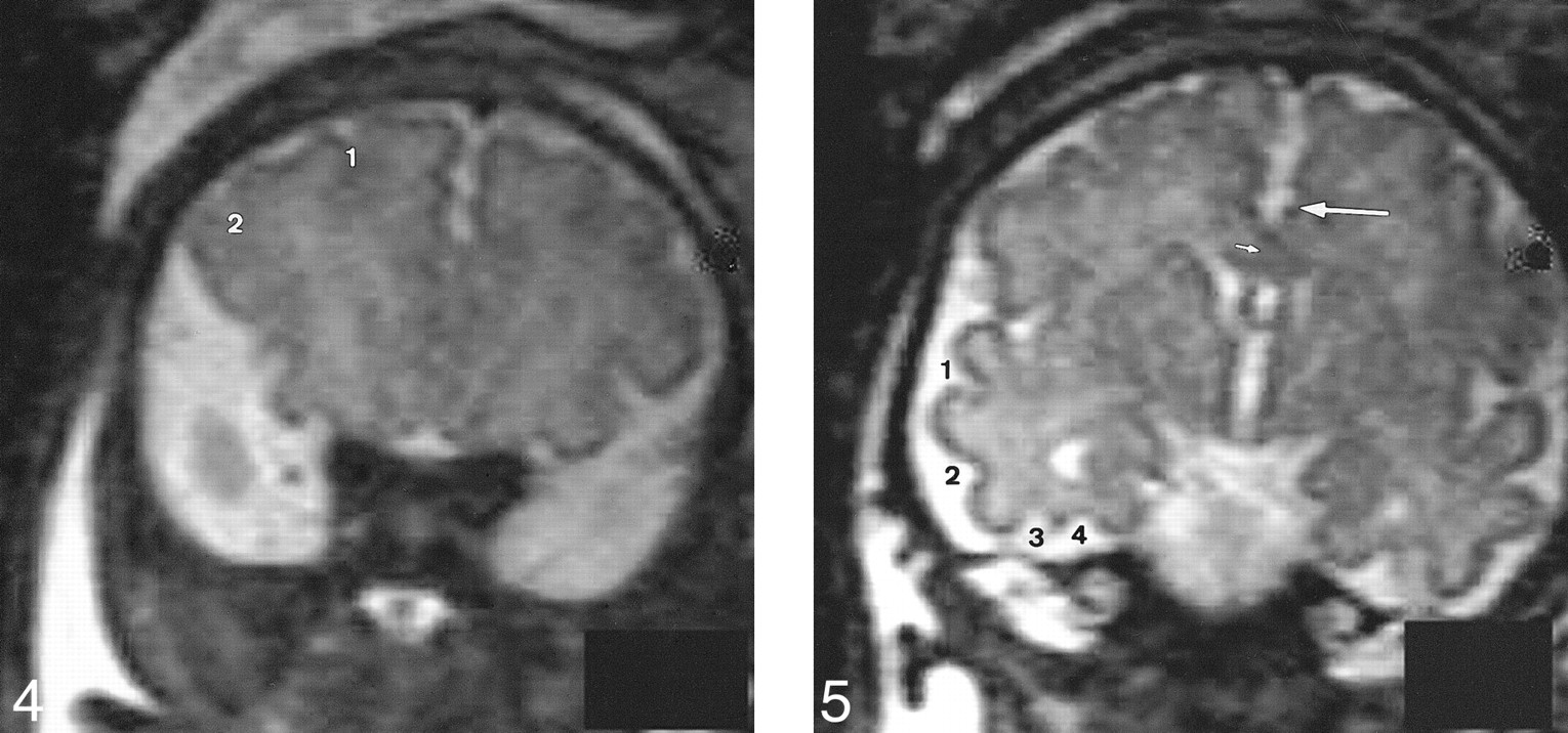
  • fig 4.
    • Download figure
    • Open in new tab
    • Download powerpoint
    fig 4.

    Slice no. 3. T2-weighted anterior coronal image (20/9.2/12) at the anterior part of the frontal lobes (35 weeks' gestation). 1, superior frontal sulcus; 2, inferior frontal sulcus.fig 5. Slice no. 4. T2-weighted coronal image (20/9.2/12) at the level of the third ventricle (35 weeks' gestation). Large arrow, cingulate sulcus; small arrow, callosal sulcus (barely visible); 1, superior temporal sulcus (anterior part); 2, inferior temporal sulcus; 3, external occipital temporal sulcus; 4, collateral sulcus.

  • fig 6.
    • Download figure
    • Open in new tab
    • Download powerpoint
    fig 6.

    Slice no. 5. T2-weighted coronal image (20/9.2/12) at the level of the temporal horns (33 weeks' gestation). Large arrow, cingulate sulcus; small arrow, callosal sulcus (barely visible); 1, superior temporal sulcus (posterior part); 2, collateral sulcus; 3, hippocampal fissure.fig 7. Slice no. 6. T2-weighted coronal image (20/9.2/12) at the level of the ventricular atria (33 weeks' gestation). 1, collateral sulcus; 2, intraparietal sulcus

  • fig 8.
    • Download figure
    • Open in new tab
    • Download powerpoint
    fig 8.

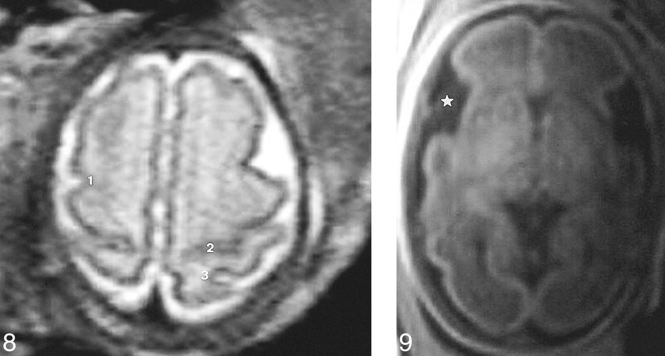
    Slice no. 7. T2-weighted transverse image (20/9.2/12) at the level of the vertex (31 weeks' gestation). 1, precentral sulcus; 2, central sulcus (almost abutting the hemispheric fissure); 3, postcentral sulcus.fig 9. Slice no. 8. T1-weighted transverse image (300/15/4) at the level of the third ventricle (29 weeks' gestation). The insula (star) is wide open

Tables

  • Figures
  • Table1

    Chronology of sulcation in our MR imaging series and in neuropathologic studies

PreviousNext
Back to top

In this issue

American Journal of Neuroradiology
Vol. 22, Issue 1
1 Jan 2001
  • Table of Contents
  • Index by author
Advertisement
Print
Download PDF
Email Article

Thank you for your interest in spreading the word on American Journal of Neuroradiology.

NOTE: We only request your email address so that the person you are recommending the page to knows that you wanted them to see it, and that it is not junk mail. We do not capture any email address.

Enter multiple addresses on separate lines or separate them with commas.
Fetal Cerebral Cortex: Normal Gestational Landmarks Identified Using Prenatal MR Imaging
(Your Name) has sent you a message from American Journal of Neuroradiology
(Your Name) thought you would like to see the American Journal of Neuroradiology web site.
CAPTCHA
This question is for testing whether or not you are a human visitor and to prevent automated spam submissions.
Cite this article
Catherine Garel, Emmanuel Chantrel, Hervé Brisse, Monique Elmaleh, Dominique Luton, Jean-François Oury, Guy Sebag, Max Hassan
Fetal Cerebral Cortex: Normal Gestational Landmarks Identified Using Prenatal MR Imaging
American Journal of Neuroradiology Jan 2001, 22 (1) 184-189;

Citation Manager Formats

  • BibTeX
  • Bookends
  • EasyBib
  • EndNote (tagged)
  • EndNote 8 (xml)
  • Medlars
  • Mendeley
  • Papers
  • RefWorks Tagged
  • Ref Manager
  • RIS
  • Zotero
0 Responses
Respond to this article
Share
Bookmark this article
Fetal Cerebral Cortex: Normal Gestational Landmarks Identified Using Prenatal MR Imaging
Catherine Garel, Emmanuel Chantrel, Hervé Brisse, Monique Elmaleh, Dominique Luton, Jean-François Oury, Guy Sebag, Max Hassan
American Journal of Neuroradiology Jan 2001, 22 (1) 184-189;
del.icio.us logo Twitter logo Facebook logo Mendeley logo
  • Tweet Widget
  • Facebook Like
  • Google Plus One

Jump to section

  • Article
    • Abstract
    • Methods
    • Results
    • Discussion
    • Conclusion
    • Acknowledgments
    • Footnotes
    • References
  • Figures & Data
  • Info & Metrics
  • Responses
  • References
  • PDF

Related Articles

  • No related articles found.
  • PubMed
  • Google Scholar

Cited By...

  • How infant brains fold: Sulcal deepening is linked to development of sulcal span, thickness, curvature, and microstructure
  • Mechanics of the Spatiotemporal Evolution of Sulcal Pits in the Folding Brain
  • Unique longitudinal contributions of sulcal interruptions to reading acquisition in children
  • Dynamic changes in subplate and cortical plate microstructure at the onset of cortical folding in vivo
  • Automatic Quantification of Normal Brain Gyrification Patterns and Changes in Fetuses with Polymicrogyria and Lissencephaly Based on MRI
  • Fetal MR Imaging Anatomy of the Transverse Temporal Gyrus (Heschl Gyrus)
  • Spatiotemporal atlas of the fetal brain depicts cortical developmental gradient in Chinese population
  • Mapping Human Fetal Brain Maturation In Vivo Using Quantitative MRI
  • Using MR Imaging to Assess Fetal Neurological Pathologies
  • Association of Isolated Congenital Heart Disease with Fetal Brain Maturation
  • Mapping fetal brain development based on automated segmentation and 4D brain atlasing
  • Imaging local genetic influences on cortical folding
  • Deviant cortical sulcation related to schizophrenia, but not cognitive deficits, likely predate brain development in the second trimester
  • Does 3T Fetal MRI Improve Image Resolution of Normal Brain Structures between 20 and 24 Weeks' Gestational Age?
  • Quantitative Folding Pattern Analysis of Early Primary Sulci in Human Fetuses with Brain Abnormalities
  • Second-Trimester Sonographic Diagnosis of Polymicrogyria
  • Brain Injury in Neonates with Complex Congenital Heart Disease: What Is the Predictive Value of MRI in the Fetal Period?
  • Assessment of MRI-Based Automated Fetal Cerebral Cortical Folding Measures in Prediction of Gestational Age in the Third Trimester
  • Development of the Fetal Cerebral Cortex in the Second Trimester: Assessment with 7T Postmortem MR Imaging
  • Assessment of Sulcation of the Fetal Brain in Cases of Isolated Agenesis of the Corpus Callosum Using In Utero MR Imaging
  • Agenesis of the Corpus Callosum: An MR Imaging Analysis of Associated Abnormalities in the Fetus
  • Objective Evaluation of Sylvian Fissure Development by Multiplanar 3-Dimensional Ultrasonography
  • Abnormal orbitofrontal development due to prematurity
  • Fetal Magnetic Resonance Imaging in the Evaluation of Fetuses Referred for Sonographically Suspected Abnormalities of the Corpus Callosum
  • Prenatal Diagnosis of Polymicrogyria by Fetal Magnetic Resonance Imaging in Monochorionic Cotwin Death
  • Maternal-Fetal In Vivo Imaging: A Combined PET and MRI Study
  • Crossref
  • Google Scholar

This article has not yet been cited by articles in journals that are participating in Crossref Cited-by Linking.

More in this TOC Section

  • SyMRI & MR Fingerprinting in Brainstem Myelination
  • Comparison of Image Quality and Radiation Dose in Pediatric Temporal Bone CT Using Photon-Counting Detector CT and Energy-Integrating Detector CT
  • Neonatal Hypocalcemia: Cerebral MRI Abnormalities
Show more Pediatrics

Similar Articles

Advertisement

Indexed Content

  • Current Issue
  • Accepted Manuscripts
  • Article Preview
  • Past Issues
  • Editorials
  • Editor's Choice
  • Fellows' Journal Club
  • Letters to the Editor
  • Video Articles

Cases

  • Case Collection
  • Archive - Case of the Week
  • Archive - Case of the Month
  • Archive - Classic Case

Special Collections

  • AJNR Awards
  • ASNR Foundation Special Collection
  • Most Impactful AJNR Articles
  • Photon-Counting CT
  • Spinal CSF Leak Articles (Jan 2020-June 2024)

More from AJNR

  • Trainee Corner
  • Imaging Protocols
  • MRI Safety Corner

Multimedia

  • AJNR Podcasts
  • AJNR Scantastics

Resources

  • Turnaround Time
  • Submit a Manuscript
  • Submit a Video Article
  • Submit an eLetter to the Editor/Response
  • Manuscript Submission Guidelines
  • Statistical Tips
  • Fast Publishing of Accepted Manuscripts
  • Graphical Abstract Preparation
  • Imaging Protocol Submission
  • Evidence-Based Medicine Level Guide
  • Publishing Checklists
  • Author Policies
  • Become a Reviewer/Academy of Reviewers
  • News and Updates

About Us

  • About AJNR
  • Editorial Board
  • Editorial Board Alumni
  • Alerts
  • Permissions
  • Not an AJNR Subscriber? Join Now
  • Advertise with Us
  • Librarian Resources
  • Feedback
  • Terms and Conditions
  • AJNR Editorial Board Alumni

American Society of Neuroradiology

  • Not an ASNR Member? Join Now

© 2025 by the American Society of Neuroradiology All rights, including for text and data mining, AI training, and similar technologies, are reserved.
Print ISSN: 0195-6108 Online ISSN: 1936-959X

Powered by HighWire