Advertisement

Main menu

  • Home
  • Content
    • Current Issue
    • Accepted Manuscripts
    • Article Preview
    • Past Issue Archive
    • Video Articles
    • AJNR Case Collection
    • Case of the Week Archive
    • Case of the Month Archive
    • Classic Case Archive
  • Special Collections
    • AJNR Awards
    • ASNR Foundation Special Collection
    • Most Impactful AJNR Articles
    • Photon-Counting CT
    • Spinal CSF Leak Articles (Jan 2020-June 2024)
  • Multimedia
    • AJNR Podcasts
    • AJNR SCANtastic
    • Trainee Corner
    • MRI Safety Corner
    • Imaging Protocols
  • For Authors
    • Submit a Manuscript
    • Submit a Video Article
    • Submit an eLetter to the Editor/Response
    • Manuscript Submission Guidelines
    • Statistical Tips
    • Fast Publishing of Accepted Manuscripts
    • Graphical Abstract Preparation
    • Imaging Protocol Submission
    • Author Policies
  • About Us
    • About AJNR
    • Editorial Board
    • Editorial Board Alumni
  • More
    • Become a Reviewer/Academy of Reviewers
    • Subscribers
    • Permissions
    • Alerts
    • Feedback
    • Advertisers
    • ASNR Home

User menu

  • Alerts
  • Log in

Search

  • Advanced search
American Journal of Neuroradiology

American Journal of Neuroradiology

ASHNR American Society of Functional Neuroradiology ASHNR American Society of Pediatric Neuroradiology ASSR
  • Alerts
  • Log in

Advanced Search

  • Home
  • Content
    • Current Issue
    • Accepted Manuscripts
    • Article Preview
    • Past Issue Archive
    • Video Articles
    • AJNR Case Collection
    • Case of the Week Archive
    • Case of the Month Archive
    • Classic Case Archive
  • Special Collections
    • AJNR Awards
    • ASNR Foundation Special Collection
    • Most Impactful AJNR Articles
    • Photon-Counting CT
    • Spinal CSF Leak Articles (Jan 2020-June 2024)
  • Multimedia
    • AJNR Podcasts
    • AJNR SCANtastic
    • Trainee Corner
    • MRI Safety Corner
    • Imaging Protocols
  • For Authors
    • Submit a Manuscript
    • Submit a Video Article
    • Submit an eLetter to the Editor/Response
    • Manuscript Submission Guidelines
    • Statistical Tips
    • Fast Publishing of Accepted Manuscripts
    • Graphical Abstract Preparation
    • Imaging Protocol Submission
    • Author Policies
  • About Us
    • About AJNR
    • Editorial Board
    • Editorial Board Alumni
  • More
    • Become a Reviewer/Academy of Reviewers
    • Subscribers
    • Permissions
    • Alerts
    • Feedback
    • Advertisers
    • ASNR Home
  • Follow AJNR on Twitter
  • Visit AJNR on Facebook
  • Follow AJNR on Instagram
  • Join AJNR on LinkedIn
  • RSS Feeds

AJNR is seeking candidates for the AJNR Podcast Editor. Read the position description.

Research ArticleBRAIN

Preoperative Proton MR Spectroscopic Imaging of Brain Tumors: Correlation with Histopathologic Analysis of Resection Specimens

Chris Dowling, Andrew W. Bollen, Susan M. Noworolski, Michael W. McDermott, Nicholas M. Barbaro, Mark R. Day, Roland G. Henry, Susan M. Chang, William P. Dillon, Sarah J. Nelson and Daniel B. Vigneron
American Journal of Neuroradiology April 2001, 22 (4) 604-612;
Chris Dowling
  • Find this author on Google Scholar
  • Find this author on PubMed
  • Search for this author on this site
Andrew W. Bollen
  • Find this author on Google Scholar
  • Find this author on PubMed
  • Search for this author on this site
Susan M. Noworolski
  • Find this author on Google Scholar
  • Find this author on PubMed
  • Search for this author on this site
Michael W. McDermott
  • Find this author on Google Scholar
  • Find this author on PubMed
  • Search for this author on this site
Nicholas M. Barbaro
  • Find this author on Google Scholar
  • Find this author on PubMed
  • Search for this author on this site
Mark R. Day
  • Find this author on Google Scholar
  • Find this author on PubMed
  • Search for this author on this site
Roland G. Henry
  • Find this author on Google Scholar
  • Find this author on PubMed
  • Search for this author on this site
Susan M. Chang
  • Find this author on Google Scholar
  • Find this author on PubMed
  • Search for this author on this site
William P. Dillon
  • Find this author on Google Scholar
  • Find this author on PubMed
  • Search for this author on this site
Sarah J. Nelson
  • Find this author on Google Scholar
  • Find this author on PubMed
  • Search for this author on this site
Daniel B. Vigneron
  • Find this author on Google Scholar
  • Find this author on PubMed
  • Search for this author on this site
  • Article
  • Figures & Data
  • Info & Metrics
  • Responses
  • References
  • PDF
Loading

Article Figures & Data

Figures

  • Tables
  • fig 1.
    • Download figure
    • Open in new tab
    • Download powerpoint
    fig 1.

    Screen saver on the ISG workstation correlating to position of biopsy for a patient with oligoastrocytoma. The spectra and images for this biopsy are shown in figure 3A. MR images are from a 3D spoiled gradient (SPGR) (34/3/1 [TR/TE/excitations]) with 1.5-mm slice thicknesses

  • fig 2.
    • Download figure
    • Open in new tab
    • Download powerpoint
    fig 2.

    A, Axial image from a contrast-enhanced SPGR 3D volume dataset (34/3/1) of a patient with an oligoastrocytoma. The locations for which spectra are reconstructed are shown by the grid.

    B, Spectral array (16 × 8 × 8 [phase encodes], 1000/144/1) corresponding to the grid shown in the image of A. Note the high NAA levels and moderate choline and creatine levels in the spectra corresponding to the normal MR regions (right side of spectral array). Note the very high choline and low NAA in portions of the contrast enhancement and surrounding tissue (dashed lines) and the lower metabolites in adjacent tissue (double lines).
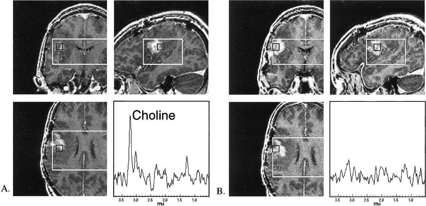
  • fig 3.
    • Download figure
    • Open in new tab
    • Download powerpoint
    fig 3.

    Contrast-enhanced SPGR (34/3/1) MR and MRS (16 × 8 × 8, 1000/144/1) data from a patient with oligoastrocytoma.

    A, MR location and the spectrum for a biopsy that yielded 75% oligoastrocytoma. Note the elevated choline (1.8 times normal) and absent NAA levels.

    B, MR location and the spectrum for a biopsy that correlated with the pathologic finding of necrosis, astrogliosis, and white matter. Note the minimal choline, creatine and NAA resonances in this spectrum. Although the image intensity is similar to the region in A, significant spectral differences were observed with elevated choline/normal choline levels correlated with the presence of tumor.

  • fig 4.
    • Download figure
    • Open in new tab
    • Download powerpoint
    fig 4.

    Multiple MR spectra (16 × 8 × 8, 1000/144/1) and corresponding images (contrast-enhanced SPGR [34/3/1]) centered on biopsy locations for a patient with a newly diagnosed GBM. Higher normalized choline levels were observed in the location of the biopsy with 90% tumor compared with others of lower percentages. (WM = white matter)

  • fig 5.
    • Download figure
    • Open in new tab
    • Download powerpoint
    fig 5.

    Metabolite levels in different tissues. The ellipses show the mean ± SD. for each tissue type. Abnormal regions have significantly lower normalized NAA, whereas tumor regions also have significantly increased normalized choline levels

Tables

  • Figures
  • Table1

    Normalized metabolite levels in different tissues

PreviousNext
Back to top

In this issue

American Journal of Neuroradiology
Vol. 22, Issue 4
1 Apr 2001
  • Table of Contents
  • Index by author
Advertisement
Print
Download PDF
Email Article

Thank you for your interest in spreading the word on American Journal of Neuroradiology.

NOTE: We only request your email address so that the person you are recommending the page to knows that you wanted them to see it, and that it is not junk mail. We do not capture any email address.

Enter multiple addresses on separate lines or separate them with commas.
Preoperative Proton MR Spectroscopic Imaging of Brain Tumors: Correlation with Histopathologic Analysis of Resection Specimens
(Your Name) has sent you a message from American Journal of Neuroradiology
(Your Name) thought you would like to see the American Journal of Neuroradiology web site.
CAPTCHA
This question is for testing whether or not you are a human visitor and to prevent automated spam submissions.
Cite this article
Chris Dowling, Andrew W. Bollen, Susan M. Noworolski, Michael W. McDermott, Nicholas M. Barbaro, Mark R. Day, Roland G. Henry, Susan M. Chang, William P. Dillon, Sarah J. Nelson, Daniel B. Vigneron
Preoperative Proton MR Spectroscopic Imaging of Brain Tumors: Correlation with Histopathologic Analysis of Resection Specimens
American Journal of Neuroradiology Apr 2001, 22 (4) 604-612;

Citation Manager Formats

  • BibTeX
  • Bookends
  • EasyBib
  • EndNote (tagged)
  • EndNote 8 (xml)
  • Medlars
  • Mendeley
  • Papers
  • RefWorks Tagged
  • Ref Manager
  • RIS
  • Zotero
0 Responses
Respond to this article
Share
Bookmark this article
Preoperative Proton MR Spectroscopic Imaging of Brain Tumors: Correlation with Histopathologic Analysis of Resection Specimens
Chris Dowling, Andrew W. Bollen, Susan M. Noworolski, Michael W. McDermott, Nicholas M. Barbaro, Mark R. Day, Roland G. Henry, Susan M. Chang, William P. Dillon, Sarah J. Nelson, Daniel B. Vigneron
American Journal of Neuroradiology Apr 2001, 22 (4) 604-612;
del.icio.us logo Twitter logo Facebook logo Mendeley logo
  • Tweet Widget
  • Facebook Like
  • Google Plus One

Jump to section

  • Article
    • Abstract
    • Methods
    • Results
    • Discussion
    • Conclusion
    • Acknowledgments
    • Footnotes
    • References
  • Figures & Data
  • Info & Metrics
  • Responses
  • References
  • PDF

Related Articles

  • No related articles found.
  • PubMed
  • Google Scholar

Cited By...

  • Diagnostic Accuracy of MR Spectroscopic Imaging and 18F-FET PET for Identifying Glioma: A Biopsy-Controlled Hybrid PET/MRI Study
  • Early Metabolic Changes in 1H-MRSI Predictive for Survival in Patients With Newly Diagnosed High-grade Glioma
  • Role of MRI in Primary Brain Tumor Evaluation
  • Advanced Magnetic Resonance Imaging of the Physical Processes in Human Glioblastoma
  • Clinical applications of imaging biomarkers. Part 1. The neuroradiologist's perspective
  • Proton MR Spectroscopy Improves Discrimination between Tumor and Pseudotumoral Lesion in Solid Brain Masses
  • Relative Cerebral Blood Volume Values to Differentiate High-Grade Glioma Recurrence from Posttreatment Radiation Effect: Direct Correlation between Image-Guided Tissue Histopathology and Localized Dynamic Susceptibility-Weighted Contrast-Enhanced Perfusion MR Imaging Measurements
  • Clinical Applications of Metabolomics in Oncology: A Review
  • The value of magnetic resonance spectroscopy in tumour imaging
  • Latest Advances in Molecular Imaging Instrumentation
  • Tumor Cell Metabolism Imaging
  • Diagnostic performance of spectroscopic and perfusion MRI for distinction of brain tumors
  • Diagnosis and Treatment of Recurrent High-Grade Astrocytoma
  • Physiologic and Metabolic Magnetic Resonance Imaging in Gliomas
  • NMR Spectroscopy and Pediatric Brain Tumors
  • Multivoxel Magnetic Resonance Spectroscopy of Brain Tumors
  • Crossref
  • Google Scholar

This article has not yet been cited by articles in journals that are participating in Crossref Cited-by Linking.

More in this TOC Section

  • Optimal MRI Sequence for Identifying Occlusion Location in Acute Stroke: Which Value of Time-Resolved Contrast-Enhanced MRA?
  • Evaluating the Effects of White Matter Multiple Sclerosis Lesions on the Volume Estimation of 6 Brain Tissue Segmentation Methods
  • Quiet PROPELLER MRI Techniques Match the Quality of Conventional PROPELLER Brain Imaging Techniques
Show more BRAIN

Similar Articles

Advertisement

Indexed Content

  • Current Issue
  • Accepted Manuscripts
  • Article Preview
  • Past Issues
  • Editorials
  • Editor's Choice
  • Fellows' Journal Club
  • Letters to the Editor
  • Video Articles

Cases

  • Case Collection
  • Archive - Case of the Week
  • Archive - Case of the Month
  • Archive - Classic Case

Special Collections

  • AJNR Awards
  • ASNR Foundation Special Collection
  • Most Impactful AJNR Articles
  • Photon-Counting CT
  • Spinal CSF Leak Articles (Jan 2020-June 2024)

More from AJNR

  • Trainee Corner
  • Imaging Protocols
  • MRI Safety Corner

Multimedia

  • AJNR Podcasts
  • AJNR Scantastics

Resources

  • Turnaround Time
  • Submit a Manuscript
  • Submit a Video Article
  • Submit an eLetter to the Editor/Response
  • Manuscript Submission Guidelines
  • Statistical Tips
  • Fast Publishing of Accepted Manuscripts
  • Graphical Abstract Preparation
  • Imaging Protocol Submission
  • Evidence-Based Medicine Level Guide
  • Publishing Checklists
  • Author Policies
  • Become a Reviewer/Academy of Reviewers
  • News and Updates

About Us

  • About AJNR
  • Editorial Board
  • Editorial Board Alumni
  • Alerts
  • Permissions
  • Not an AJNR Subscriber? Join Now
  • Advertise with Us
  • Librarian Resources
  • Feedback
  • Terms and Conditions
  • AJNR Editorial Board Alumni

American Society of Neuroradiology

  • Not an ASNR Member? Join Now

© 2025 by the American Society of Neuroradiology All rights, including for text and data mining, AI training, and similar technologies, are reserved.
Print ISSN: 0195-6108 Online ISSN: 1936-959X

Powered by HighWire