Advertisement

Main menu

  • Home
  • Content
    • Current Issue
    • Accepted Manuscripts
    • Article Preview
    • Past Issue Archive
    • Video Articles
    • AJNR Case Collection
    • Case of the Week Archive
    • Case of the Month Archive
    • Classic Case Archive
  • Special Collections
    • AJNR Awards
    • ASNR Foundation Special Collection
    • Most Impactful AJNR Articles
    • Photon-Counting CT
    • Spinal CSF Leak Articles (Jan 2020-June 2024)
  • Multimedia
    • AJNR Podcasts
    • AJNR SCANtastic
    • Trainee Corner
    • MRI Safety Corner
    • Imaging Protocols
  • For Authors
    • Submit a Manuscript
    • Submit a Video Article
    • Submit an eLetter to the Editor/Response
    • Manuscript Submission Guidelines
    • Statistical Tips
    • Fast Publishing of Accepted Manuscripts
    • Graphical Abstract Preparation
    • Imaging Protocol Submission
    • Author Policies
  • About Us
    • About AJNR
    • Editorial Board
    • Editorial Board Alumni
  • More
    • Become a Reviewer/Academy of Reviewers
    • Subscribers
    • Permissions
    • Alerts
    • Feedback
    • Advertisers
    • ASNR Home

User menu

  • Alerts
  • Log in

Search

  • Advanced search
American Journal of Neuroradiology

American Journal of Neuroradiology

ASHNR American Society of Functional Neuroradiology ASHNR American Society of Pediatric Neuroradiology ASSR
  • Alerts
  • Log in

Advanced Search

  • Home
  • Content
    • Current Issue
    • Accepted Manuscripts
    • Article Preview
    • Past Issue Archive
    • Video Articles
    • AJNR Case Collection
    • Case of the Week Archive
    • Case of the Month Archive
    • Classic Case Archive
  • Special Collections
    • AJNR Awards
    • ASNR Foundation Special Collection
    • Most Impactful AJNR Articles
    • Photon-Counting CT
    • Spinal CSF Leak Articles (Jan 2020-June 2024)
  • Multimedia
    • AJNR Podcasts
    • AJNR SCANtastic
    • Trainee Corner
    • MRI Safety Corner
    • Imaging Protocols
  • For Authors
    • Submit a Manuscript
    • Submit a Video Article
    • Submit an eLetter to the Editor/Response
    • Manuscript Submission Guidelines
    • Statistical Tips
    • Fast Publishing of Accepted Manuscripts
    • Graphical Abstract Preparation
    • Imaging Protocol Submission
    • Author Policies
  • About Us
    • About AJNR
    • Editorial Board
    • Editorial Board Alumni
  • More
    • Become a Reviewer/Academy of Reviewers
    • Subscribers
    • Permissions
    • Alerts
    • Feedback
    • Advertisers
    • ASNR Home
  • Follow AJNR on Twitter
  • Visit AJNR on Facebook
  • Follow AJNR on Instagram
  • Join AJNR on LinkedIn
  • RSS Feeds

AJNR is seeking candidates for the AJNR Podcast Editor. Read the position description.

Research ArticleBRAIN

Arterial Hyperintensity on Fast Fluid-attenuated Inversion Recovery Images: A Subtle Finding for Hyperacute Stroke Undetected by Diffusion-weighted MR Imaging

Masayuki Maeda, Tatsuya Yamamoto, Shouichiro Daimon, Hajime Sakuma and Kan Takeda
American Journal of Neuroradiology April 2001, 22 (4) 632-636;
Masayuki Maeda
  • Find this author on Google Scholar
  • Find this author on PubMed
  • Search for this author on this site
Tatsuya Yamamoto
  • Find this author on Google Scholar
  • Find this author on PubMed
  • Search for this author on this site
Shouichiro Daimon
  • Find this author on Google Scholar
  • Find this author on PubMed
  • Search for this author on this site
Hajime Sakuma
  • Find this author on Google Scholar
  • Find this author on PubMed
  • Search for this author on this site
Kan Takeda
  • Find this author on Google Scholar
  • Find this author on PubMed
  • Search for this author on this site
  • Article
  • Figures & Data
  • Info & Metrics
  • Responses
  • References
  • PDF
Loading

Article Figures & Data

Figures

  • Tables
  • fig 1.
    • Download figure
    • Open in new tab
    • Download powerpoint
    fig 1.

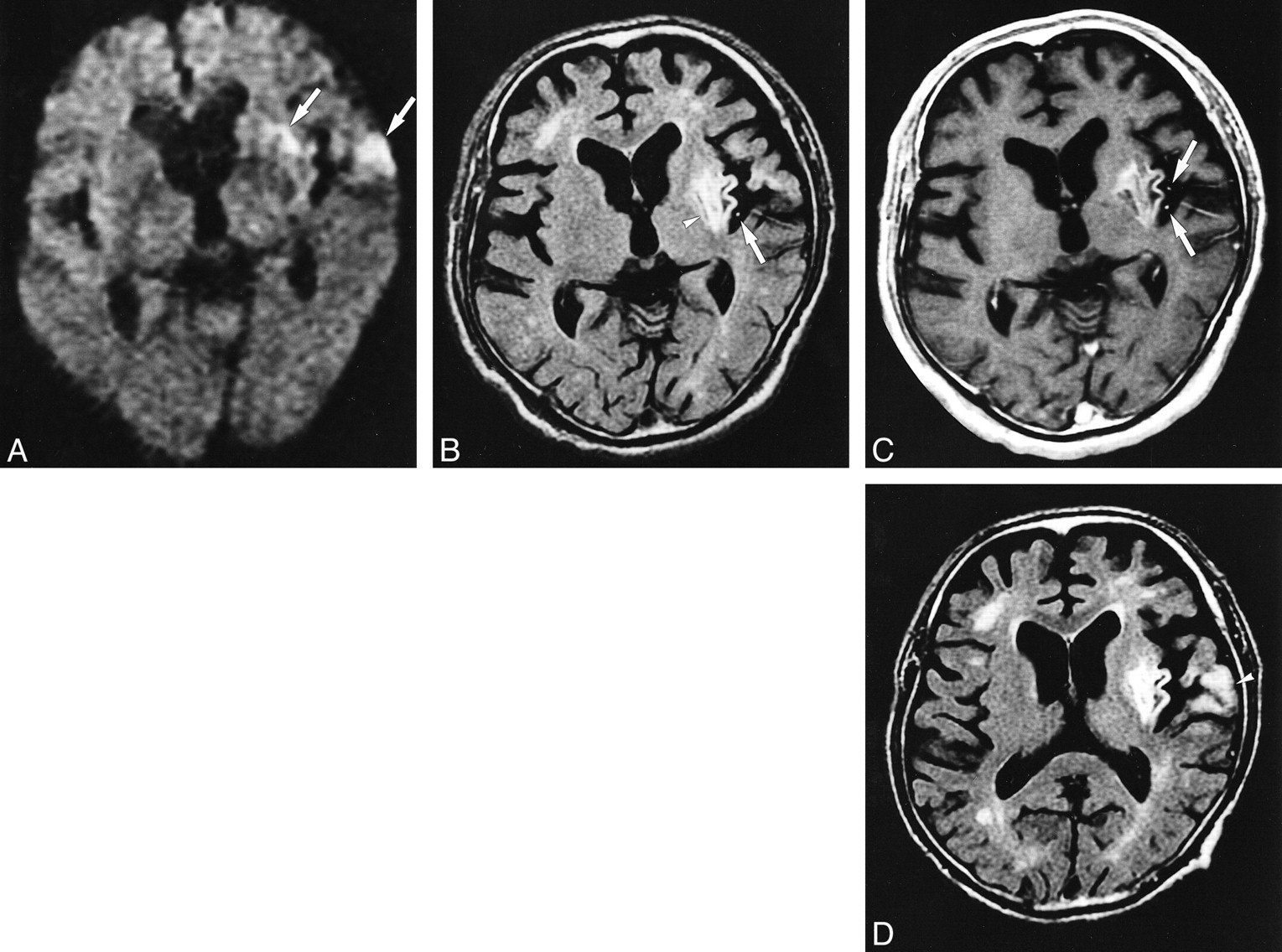
    Case 10. MR images of 75-year-old man, with right hemiparesis, scanned 3 hours (A–C) and 3 days (D) after symptom onset.

    A, Diffusion-weighted (4999/126/1, b = 1000) MR image shows high signal intensity in the left MCA territory (arrows), suggesting the infarction pertinent to this episode.

    B, Fast-FLAIR (8000/110/1, TI = 1800) image shows arterial hyperintensity in the left sylvian fissure (arrow). Note that there is chronic infarction that occurred 7 weeks before this stroke (arrowhead).

    C, Contrast-enhanced T1-weighted (500/12/2) image shows arterial enhancement more extensively than does the arterial hyperintensity exhibited in B (arrows).

    D, Fast-FLAIR (8000/110/1, TI = 1800) image shows a hyperintense area of acute infarction at 3 days after onset (arrowhead). Note that the arterial hyperintensity seen at 3 hours disappears at this time.

  • fig 2.
    • Download figure
    • Open in new tab
    • Download powerpoint
    fig 2.

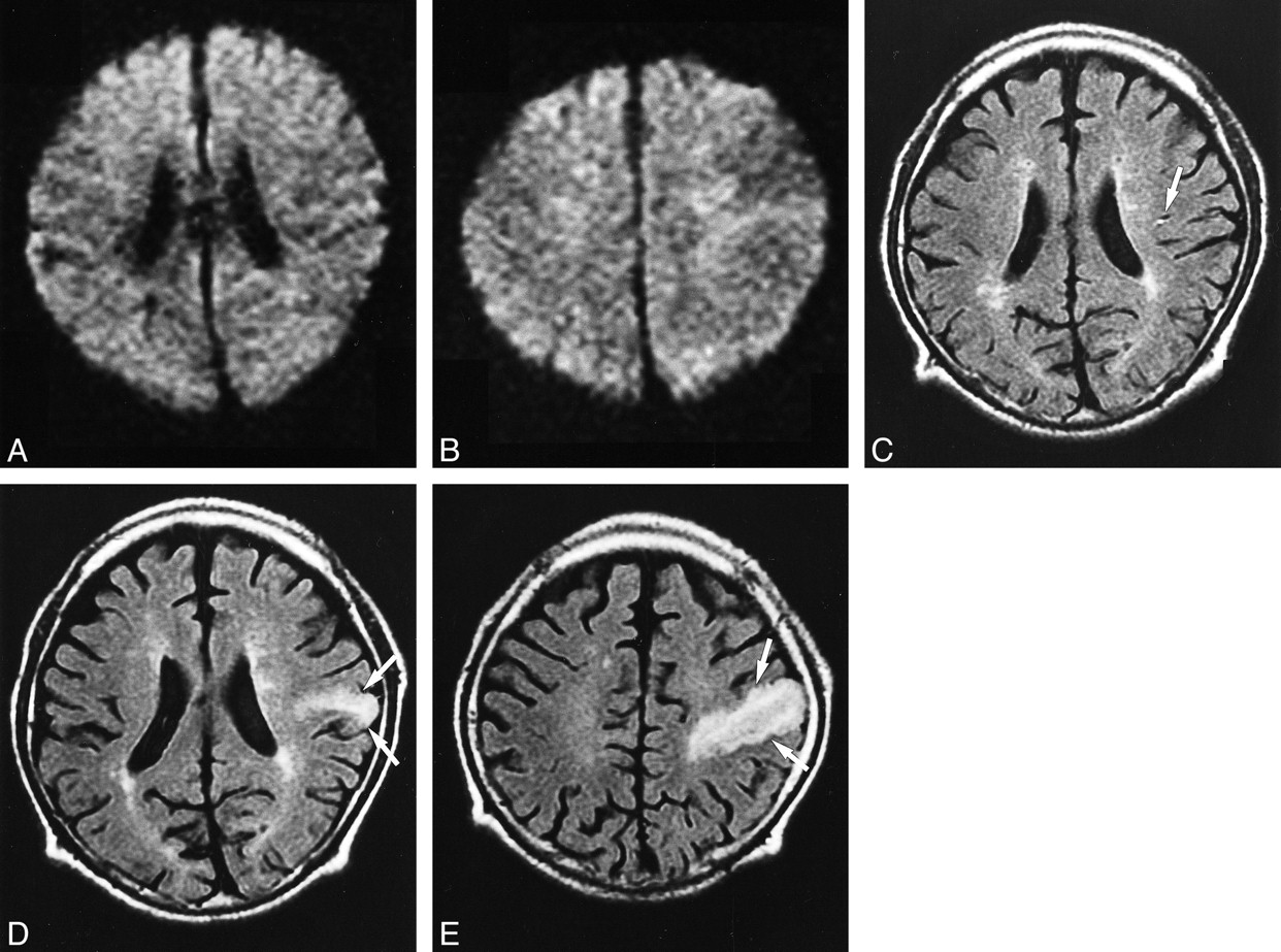
    Case 11. MR images of a 65-year-old man, with right-arm weakness, scanned 3 hours (A–C) and 3 days (D and E) after symptom onset.

    A and B, Diffusion-weighted MR images (4999/126/1, b = 1000) are negative for stroke 3 hours after onset.

    C, Fast-FLAIR (8000/110/1, TI = 1800) image shows arterial hyperintensity in the left MCA territory (arrow). This is the only abnormal finding pertinent to the patient's symptoms at the initial MR examination.

    D and E, Fast-FLAIR (8000/110/1, TI = 1800) images show extensive acute infarction in the left MCA territory 3 days after onset (arrows).

Tables

  • Figures
  • Table1

    Clinical data and MR findings of patients with stroke

PreviousNext
Back to top

In this issue

American Journal of Neuroradiology
Vol. 22, Issue 4
1 Apr 2001
  • Table of Contents
  • Index by author
Advertisement
Print
Download PDF
Email Article

Thank you for your interest in spreading the word on American Journal of Neuroradiology.

NOTE: We only request your email address so that the person you are recommending the page to knows that you wanted them to see it, and that it is not junk mail. We do not capture any email address.

Enter multiple addresses on separate lines or separate them with commas.
Arterial Hyperintensity on Fast Fluid-attenuated Inversion Recovery Images: A Subtle Finding for Hyperacute Stroke Undetected by Diffusion-weighted MR Imaging
(Your Name) has sent you a message from American Journal of Neuroradiology
(Your Name) thought you would like to see the American Journal of Neuroradiology web site.
CAPTCHA
This question is for testing whether or not you are a human visitor and to prevent automated spam submissions.
Cite this article
Masayuki Maeda, Tatsuya Yamamoto, Shouichiro Daimon, Hajime Sakuma, Kan Takeda
Arterial Hyperintensity on Fast Fluid-attenuated Inversion Recovery Images: A Subtle Finding for Hyperacute Stroke Undetected by Diffusion-weighted MR Imaging
American Journal of Neuroradiology Apr 2001, 22 (4) 632-636;

Citation Manager Formats

  • BibTeX
  • Bookends
  • EasyBib
  • EndNote (tagged)
  • EndNote 8 (xml)
  • Medlars
  • Mendeley
  • Papers
  • RefWorks Tagged
  • Ref Manager
  • RIS
  • Zotero
0 Responses
Respond to this article
Share
Bookmark this article
Arterial Hyperintensity on Fast Fluid-attenuated Inversion Recovery Images: A Subtle Finding for Hyperacute Stroke Undetected by Diffusion-weighted MR Imaging
Masayuki Maeda, Tatsuya Yamamoto, Shouichiro Daimon, Hajime Sakuma, Kan Takeda
American Journal of Neuroradiology Apr 2001, 22 (4) 632-636;
del.icio.us logo Twitter logo Facebook logo Mendeley logo
  • Tweet Widget
  • Facebook Like
  • Google Plus One

Jump to section

  • Article
    • Abstract
    • Methods
    • Results
    • Discussion
    • Conclusion
    • Footnotes
    • References
  • Figures & Data
  • Info & Metrics
  • Responses
  • References
  • PDF

Related Articles

  • No related articles found.
  • PubMed
  • Google Scholar

Cited By...

  • Hyperintense Basilar Artery on FLAIR MR Imaging: Diagnostic Accuracy and Clinical Impact in Patients with Acute Brain Stem Stroke
  • Fluid-Attenuated Inversion Recovery Vascular Hyperintensities: An Important Imaging Marker for Cerebrovascular Disease
  • Decrease in Leptomeningeal Ivy Sign on Fluid-Attenuated Inversion Recovery Images after Cerebral Revascularization in Patients with Moyamoya Disease
  • Angiography Reveals That Fluid-Attenuated Inversion Recovery Vascular Hyperintensities Are Due to Slow Flow, Not Thrombus
  • Exclusion of brain lesions: is MR contrast medium required after a negative fluid-attenuated inversion recovery sequence?
  • Evaluation of Hyperintense Vessels on FLAIR MRI for the Diagnosis of Multiple Intracerebral Arterial Stenoses
  • Crossref
  • Google Scholar

This article has not yet been cited by articles in journals that are participating in Crossref Cited-by Linking.

More in this TOC Section

  • Usefulness of Quantitative Susceptibility Mapping for the Diagnosis of Parkinson Disease
  • White Matter Alterations in the Brains of Patients with Active, Remitted, and Cured Cushing Syndrome: A DTI Study
  • Qualitative and Quantitative Analysis of MR Imaging Findings in Patients with Middle Cerebral Artery Stroke Implanted with Mesenchymal Stem Cells
Show more BRAIN

Similar Articles

Advertisement

Indexed Content

  • Current Issue
  • Accepted Manuscripts
  • Article Preview
  • Past Issues
  • Editorials
  • Editor's Choice
  • Fellows' Journal Club
  • Letters to the Editor
  • Video Articles

Cases

  • Case Collection
  • Archive - Case of the Week
  • Archive - Case of the Month
  • Archive - Classic Case

Special Collections

  • AJNR Awards
  • ASNR Foundation Special Collection
  • Most Impactful AJNR Articles
  • Photon-Counting CT
  • Spinal CSF Leak Articles (Jan 2020-June 2024)

More from AJNR

  • Trainee Corner
  • Imaging Protocols
  • MRI Safety Corner

Multimedia

  • AJNR Podcasts
  • AJNR Scantastics

Resources

  • Turnaround Time
  • Submit a Manuscript
  • Submit a Video Article
  • Submit an eLetter to the Editor/Response
  • Manuscript Submission Guidelines
  • Statistical Tips
  • Fast Publishing of Accepted Manuscripts
  • Graphical Abstract Preparation
  • Imaging Protocol Submission
  • Evidence-Based Medicine Level Guide
  • Publishing Checklists
  • Author Policies
  • Become a Reviewer/Academy of Reviewers
  • News and Updates

About Us

  • About AJNR
  • Editorial Board
  • Editorial Board Alumni
  • Alerts
  • Permissions
  • Not an AJNR Subscriber? Join Now
  • Advertise with Us
  • Librarian Resources
  • Feedback
  • Terms and Conditions
  • AJNR Editorial Board Alumni

American Society of Neuroradiology

  • Not an ASNR Member? Join Now

© 2025 by the American Society of Neuroradiology All rights, including for text and data mining, AI training, and similar technologies, are reserved.
Print ISSN: 0195-6108 Online ISSN: 1936-959X

Powered by HighWire