
Article Figures & Data
Figures
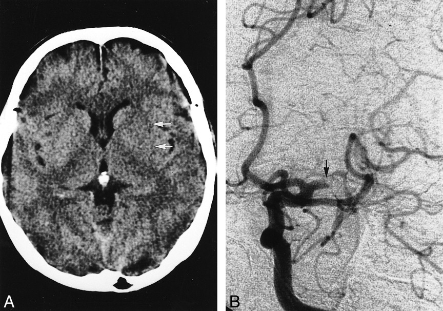
- fig 1.
Representative early CT sign of grade I occlusion.
A, Nonenhanced CT scan in a 71-year-old man obtained 1.5 hours after left MCA stroke. Note loss of the left insular ribbon (arrows) versus normal right insular ribbon (arrowheads). The left lentiform nucleus appears normal.
B, Left carotid angiogram, obtained immediately after initial CT scanning, shows an embolic occlusion of the left M2 segment (arrow). Lateral lenticulostriate arteries (arrowheads) were well opacified from their origins.
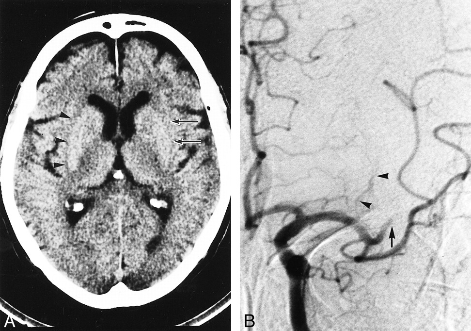
- fig 2.
Representative early CT sign of grade III occlusion.
A, Nonenhanced CT scan in a 60-year-old woman obtained 2 hours after left MCA stroke. Note obscuration of the left entire lentiform nucleus (arrows).
B, Left carotid angiogram, obtained immediately after initial CT scanning, shows left MCA trunk occlusion at it origin (arrow). None of the lenticulostriate arteries were opacified.
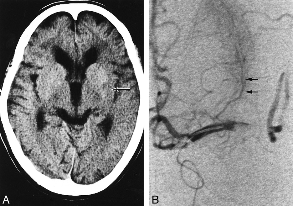
- fig 3.
Representative early CT sign of grade II occlusion.
A, Nonenhanced CT scan in a 57-year-old woman obtained 3 hours after left MCA stroke. Note obscuration of posterolateral part of the left lentiform nucleus (arrow).
B, Left local angiogram, obtained using a microcatheter, shows left MCA trunk occlusion with partial opacification of the lenticulostriate arteries (arrows).


Tables

- TABLE 2:
Early CT signs in the deep MCA territories at varying sites of MCA occlusion
- TABLE 3:
Sensitivity and specificity of early CT sign as an indicator of LSA involvement
- TABLE 4:
Rate of therapeutic recanalization and the incidence of symptomatic hemorrhagic complications in each grade of early CT signs


In this issue


Jump to section
Related Articles
Cited By...
- Ask the consultant: Stroke
- Perfusion patterns as a tool for emergency stroke diagnosis: differentiating proximal and distal MCA occlusions
- A case of large right MCA stroke with hyperdense MCA sign in CT imaging
- Visualization of Lenticulostriate Arteries by Flow-Sensitive Black-Blood MR Angiography on a 1.5T MRI System: A Comparative Study between Subjects with and without Stroke
- Presence of Early Ischemic Changes on Computed Tomography Depends on Severity and the Duration of Hypoperfusion: A Single Photon Emission-Computed Tomographic Study
- Appearance of Early Venous Filling During Intra-Arterial Reperfusion Therapy for Acute Middle Cerebral Artery Occlusion: A Predictive Sign for Hemorrhagic Complications
- Direct Percutaneous Transluminal Angioplasty for Acute Middle Cerebral Artery Trunk Occlusion: An Alternative Option to Intra-arterial Thrombolysis
- Parenchymal Hyperdensity on Computed Tomography After Intra-Arterial Reperfusion Therapy for Acute Middle Cerebral Artery Occlusion: Incidence and Clinical Significance