
Article Figures & Data
Figures
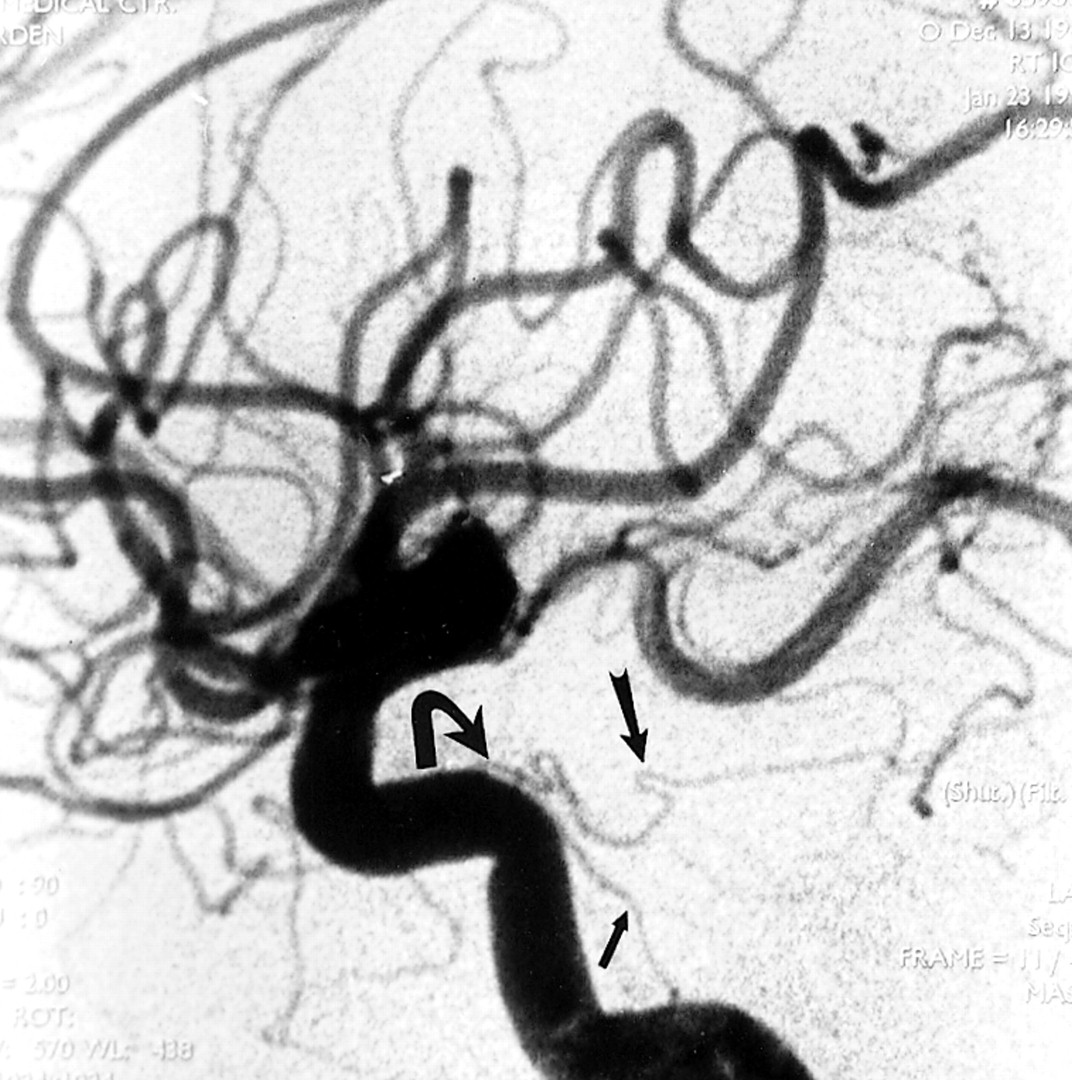
- fig 1.
Lateral DSA, arterial phase, shows normal caliber dorsal clival (small arrow) and tentorial (large arrow) branches of the MHT in a different patient with no disease in this region. Note the difficult geometry of the MHT relative to the C4–C5 segments of the ICA if an anterograde approach to microcatheterization is attempted (curved arrow)
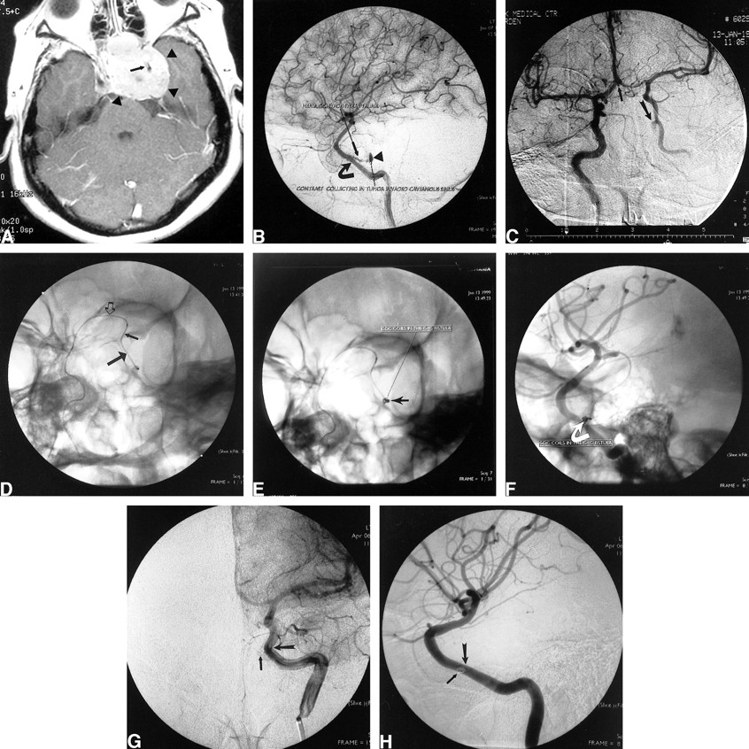
- fig 2.
Case 1: 33-year-old right-handed woman with a recurrent pituitary macroadenoma approximately 20 months after initial surgery. A, Contrast-enhanced T1-weighted axial MR image shows a densely enhancing pituitary macroadenoma (arrowheads) invading the left cavernous sinus and encasing the intracavernous left ICA (arrow). B, Lateral DSA, late arterial phase, shows contrast material pooling in the tumor-invaded cavernous sinus (arrowhead). The avulsed branch of the MHT (small arrow) arises from the tumor-encased intracavernous ICA (large arrow). The tumor diffusely narrows and uncoils the intracavernous ICA. C, Anteroposterior DSA from right ICA injection with left carotid compression shows good cross filling through the ACoM (small arrow) with retrograde filling of the left carotid-cavernous fistula (curved arrow). D, Left anterior oblique (LAO) fluoroscopic image shows the microcatheter crossing from right to left through the ACoM (open arrow), then down the supraclinoid left ICA (small arrow) into the horizontal (C4) intracavernous left ICA (large arrow). E, LAO fluoroscopic image shows two 2-mm × 2-cm GDC-10 coils (arrow) after deployment into the venous side of the carotid-cavernous fistula. F, Steep LAO nonsubtracted DSA shows two 2-mm × 2-cm GDC-10 coils in the venous side of the carotid-cavernous fistula (arrow). G, Anteroposterior DSA from left ICA injection in the late arterial, early capillary phase shows complete obliteration of the carotid-cavernous fistula. The GDCs (small arrow) are faintly seen just medial to the C4–C5 junction of the left ICA (large arrow). H, Steep LAO DSA from the left ICA injection in the arterial phase shows complete obliteration of the carotid-cavernous fistula. The GDCs (small arrow) overlap the C4–C5 junction of the left ICA (large arrow) in this projection

In this issue


Jump to section
Related Articles
Cited By...
- No citing articles found.