
Abstract
Summary: Transnasal needle access for sampling was used in two patients with posterior nasopharyngeal lesions. The procedure was performed under CT guidance. This new technique is simple and appears suitable for selected cases. The two cases and the details of the procedure are described.
For lesions affecting the skull base, tissue diagnosis before surgery or treatment planning poses a difficult problem. We describe a simple transnasal approach for tissue sampling that may be helpful in selected cases.
Case Reports
Case 1
A 35-year-old male farmer presented with a history of slight limitation of neck movement and intermittent neck pain of 4 years' duration, which was especially noticeable when carrying weight on his head. Five months earlier, a slight hoarseness of voice had developed, and for 1 month he had been experiencing regurgitation of liquids through the nose, inability to swallow, and breathlessness. For the preceding 15 days, he had been unable to walk or even to sit without support. There was no history of diabetes or hypertension, and no indication that the patient was immunocompromised. His ELISA was negative for HIV. On examination, multiple left-sided lower cranial nerve palsies were present from the fifth to the 12th nerve, except for the sixth nerve.
CT studies showed extensive destruction of the skull base along with the clivus, atlas, and sphenoidal sinus by a large soft tissue mass with nodular extensions that had a large nasopharyngeal component. The skull base destruction also led to a secondary basilar invagination. The soft tissue mass involved both cavernous sinuses and extended to both cerebellopontine angles. MR imaging showed the mass to be predominantly hypointense on both T2- and T1-weighted sequences with areas of T2 hyperintensity, suggestive of necrosis. Petrous and cavernous internal carotid arteries were of normal caliber and coursed adjacent to the soft tissue mass on both sides. There was a severe kink at the pontomedullary junction over the odontoid tip due to skull base destruction and invagination. The maxillary, frontal, and ethmoidal sinuses were well aerated, with no evidence of mucosal involvement. A contrast-enhanced MR study revealed intense enhancement of the mass lesion with central nonenhancing areas that were T2 hyperintense. The imaging findings, especially the T2 hypointense areas, suggested fungal granuloma with extensive skull base destruction.
Tissue sampling via the oblique retromaxillary or mandibular notch approach was considered to carry a risk of injury to the cavernous part of the internal carotid artery. A midline approach through the nasal cavity under CT guidance was undertaken, and 40 mL of thick, brownish pus was aspirated. A postprocedural CT scan showed the presence of an air-fluid level within the nasopharyngeal soft tissue. The specimen was sent for bacterial and fungal culture and cytologic examination. On microscopy, typical septate hyphae with dichotomous branching suggestive of Aspergillus were seen. On cultures, extensive growth of A. flavus was observed; the species was identified on the basis of colonial morphology and microscopic examination of lactophenol cotton blue preparation.
Case 2
A 64-year-diabetic man presented with a history of pain in the left ear of 3 months' duration followed by ear discharge. A few days later, motor facial weakness developed on the left side, followed by deafness, dysphonia, dysphagia, and dysarthria, which had increased progressively over the preceding 2 months. A history of mild headache was present, but there was no history of fever, weight loss, bleeding through the ear, change in consciousness, or vomiting.
Neurologic examination showed evidence of left seventh, ninth, 11th, and 12th nerve palsy without any cerebellar or long tract signs. ENT examination revealed no obvious nasopharyngeal mass; there was no evidence of pus discharge in the left external auditory canal, and the eardrum was intact. Conductive deafness was noted on the left side.
MR imaging performed on a 1.5-T unit showed an enhancing soft tissue mass extending to the left parapharyngeal space associated with T2 hyperintensity involving the left petrous apex. CT studies showed an enhancing left-sided nasopharyngeal lesion with central hypodensity and bone destruction of the inferomedial aspect of the left petrous bone. A few calcific densities, apparently fragments of destroyed bone, were also identified. A transnasal CT-guided needle biopsy of the mass was performed, and 5 mL of pus was aspirated and sent for culture and sensitivity examination. Cultures showed extensive growth of Pseudomonas aeruginosa, sensitive to amikacin and ceftazidime. The patient was started on appropriate antibiotics and was discharged. At the 3-month follow-up, the symptoms had nearly completely resolved.
Technique
The procedure was performed by radiologists. The patient was placed in a supine position on a CT table, and a preliminary noncontrast axial CT scan was obtained in the region of interest with 5-mm contiguous sections. The nasal cavity was anesthetized using 2% lidocaine spray. An open-end nasogastric tube (16F) was passed through the nasal cavity, and a 145-cm metal guidewire was placed through it. In the first case, the other end of the guidewire was brought out through the oral cavity to afford better anchorage for passing the metal canula over it. However, in the second case, the floppy end of the guidewire was left in the stomach. The nasogastric tube was withdrawn over the guidewire. A long 20-cm 18-gauge metal canula with a blunt distal end (from a trocar canula set) was passed over the snugly fitting guidewire after applying lidocaine jelly in the nasal cavity. A lateral scout view was obtained to confirm that the canula had reached the nasopharyngeal soft tissue (Fig 1A). The guidewire was slowly withdrawn, with the canula kept in situ (Fig 1B). Limited 5mmthick CT sections were obtained to confirm the position of the tip of the canula against the soft tissue. The trocar was then inserted in the canula and the canula with the trocar was advanced in 1.5-cm increments (Fig 1C). Aspiration after removal of the trocar yielded pus. The procedure was uneventful, and no complication was noticed in either patient.
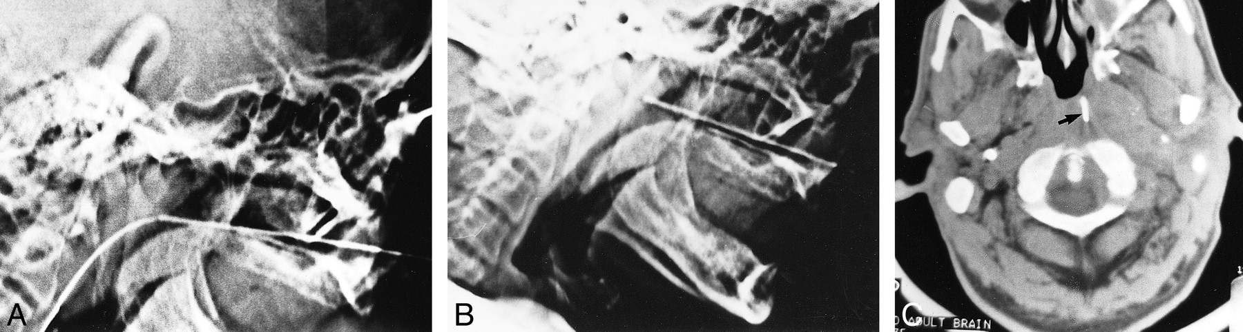
Case 2: 64-year-old man undergoing transnasal access for sampling a skull base lesion.
A and B, Lateral CT scout images. Metal canula was advanced over a guidewire placed transnasally (A). After removal of the guidewire, the canula was directed slightly superiorly to the nasopharyngeal mass (B).
C, Axial CT scan shows the tip of the metal canula in the nasopharyngeal lesion (arrow).
Discussion
Standard approaches for biopsy of mass lesions involving the skull base include a CT-guided lateral approach through the mandibular notch and a retromandibular transparotid or oblique retromaxillary approach (1). Various alternative techniques have been described, such as a mandibular sigmoid notch approach (2), an anterior approach (3), transoral and transnasal stereotactic procedures (4) and various modifications of these approaches (5), a transnasal stereotactic approach, for clival lesions (6, 7), and transoral fine-needle aspiration, for oropharyngeal masses (8). All the procedures have some advantages as well as some limitations, and all have been used with varying degrees of success. Most of them require good technical expertise and careful surgical planning and preparation; the stereotactic procedures also require special instruments, such as the Patil stereotactic frame (4, 5) or the Cosman-Roberts Wells frame (6).
In contrast, our method of transnasal aspiration is quite simple. The procedure is performed by radiologists and carried out under CT guidance. It is well tolerated by patients, and there is no real danger of transgressing major neural or vascular tissue. The CT scan helps to accurately identify the needle entry point as well as the placement of the needle in the lesion. A metal guidewire helps in the placement of the blunt distal end of the metal canula past the turbinates or conchi. A snugly fitting guidewire/metal canula combination avoids injury to the nasal mucosa. The procedure allows some movement of the canula after removal of the guidewire, which may be used to place the canula accurately against a paramidline lesion under CT guidance, as in our second case. Placement of the canula, per se, does not pose any danger. The possibility of abrasion of and bleeding from the nasal mucosa is avoided by adhering to the technique of placing the canula over the guidewire. Although we did not encounter any complications in our cases, postprocedural bleeding from the puncture site at the nasopharyngeal mucosa may be a problem in patients with highly vascular lesions and needs to be watched for. In paramidline lesions, the advancement of the canula needs to be done via CT guidance, and intravenous iodinated contrast material may be necessary to locate the internal carotid artery and internal jugular vein so as to avoid any injury to these vascular structures. In our patients, aspiration of liquified contents was sufficient to identify the infective nature of the lesion and the pathogen involved. However, the technique can be used for needle biopsy of a solid soft tissue mass involving the skull base through the metal canula coaxially once the latter is placed in position under CT guidance.
Conclusion
The technique of accessing a nasopharyngeal mass transnasally with a needle is simple, avoids injury to nasal mucosa, and appears inherently safe, as the path of the needle does not traverse any important soft tissue structures. As such, the procedure may be suitable for diagnostic tissue sampling in selected cases.
Footnotes
↵1 Address reprint requests to Dr. Rajendra Phadke, Additional Professor, Department of Radiodiagnosis, Sanjay Gandhi Postgraduate Institute of Medical Sciences, Raebareli Rd, Lucknow-226014 UP, India.
- Received August 28, 2000.
- Copyright © American Society of Neuroradiology
In this issue


Jump to section
Related Articles
Cited By...
- No citing articles found.