Advertisement

Main menu

  • Home
  • Content
    • Current Issue
    • Accepted Manuscripts
    • Article Preview
    • Past Issue Archive
    • Video Articles
    • AJNR Case Collection
    • Case of the Week Archive
    • Case of the Month Archive
    • Classic Case Archive
  • Special Collections
    • AJNR Awards
    • ASNR Foundation Special Collection
    • Most Impactful AJNR Articles
    • Photon-Counting CT
    • Spinal CSF Leak Articles (Jan 2020-June 2024)
  • Multimedia
    • AJNR Podcasts
    • AJNR SCANtastic
    • Trainee Corner
    • MRI Safety Corner
    • Imaging Protocols
  • For Authors
    • Submit a Manuscript
    • Submit a Video Article
    • Submit an eLetter to the Editor/Response
    • Manuscript Submission Guidelines
    • Statistical Tips
    • Fast Publishing of Accepted Manuscripts
    • Graphical Abstract Preparation
    • Imaging Protocol Submission
    • Author Policies
  • About Us
    • About AJNR
    • Editorial Board
    • Editorial Board Alumni
  • More
    • Become a Reviewer/Academy of Reviewers
    • Subscribers
    • Permissions
    • Alerts
    • Feedback
    • Advertisers
    • ASNR Home

User menu

  • Alerts
  • Log in

Search

  • Advanced search
American Journal of Neuroradiology

American Journal of Neuroradiology

ASHNR American Society of Functional Neuroradiology ASHNR American Society of Pediatric Neuroradiology ASSR
  • Alerts
  • Log in

Advanced Search

  • Home
  • Content
    • Current Issue
    • Accepted Manuscripts
    • Article Preview
    • Past Issue Archive
    • Video Articles
    • AJNR Case Collection
    • Case of the Week Archive
    • Case of the Month Archive
    • Classic Case Archive
  • Special Collections
    • AJNR Awards
    • ASNR Foundation Special Collection
    • Most Impactful AJNR Articles
    • Photon-Counting CT
    • Spinal CSF Leak Articles (Jan 2020-June 2024)
  • Multimedia
    • AJNR Podcasts
    • AJNR SCANtastic
    • Trainee Corner
    • MRI Safety Corner
    • Imaging Protocols
  • For Authors
    • Submit a Manuscript
    • Submit a Video Article
    • Submit an eLetter to the Editor/Response
    • Manuscript Submission Guidelines
    • Statistical Tips
    • Fast Publishing of Accepted Manuscripts
    • Graphical Abstract Preparation
    • Imaging Protocol Submission
    • Author Policies
  • About Us
    • About AJNR
    • Editorial Board
    • Editorial Board Alumni
  • More
    • Become a Reviewer/Academy of Reviewers
    • Subscribers
    • Permissions
    • Alerts
    • Feedback
    • Advertisers
    • ASNR Home
  • Follow AJNR on Twitter
  • Visit AJNR on Facebook
  • Follow AJNR on Instagram
  • Join AJNR on LinkedIn
  • RSS Feeds

AJNR is seeking candidates for the AJNR Podcast Editor. Read the position description.

Research ArticlePEDIATRICS

Lhermitte-Duclos Disease: Assessment with MR Imaging, Positron Emission Tomography, Single-photon Emission CT, and MR Spectroscopy

Joachim Klisch, Freimut Juengling, Joachim Spreer, Donatus Koch, Torsten Thiel, Martin Büchert, Sebastian Arnold, Friedrich Feuerhake and Martin Schumacher
American Journal of Neuroradiology May 2001, 22 (5) 824-830;
Joachim Klisch
  • Find this author on Google Scholar
  • Find this author on PubMed
  • Search for this author on this site
Freimut Juengling
  • Find this author on Google Scholar
  • Find this author on PubMed
  • Search for this author on this site
Joachim Spreer
  • Find this author on Google Scholar
  • Find this author on PubMed
  • Search for this author on this site
Donatus Koch
  • Find this author on Google Scholar
  • Find this author on PubMed
  • Search for this author on this site
Torsten Thiel
  • Find this author on Google Scholar
  • Find this author on PubMed
  • Search for this author on this site
Martin Büchert
  • Find this author on Google Scholar
  • Find this author on PubMed
  • Search for this author on this site
Sebastian Arnold
  • Find this author on Google Scholar
  • Find this author on PubMed
  • Search for this author on this site
Friedrich Feuerhake
  • Find this author on Google Scholar
  • Find this author on PubMed
  • Search for this author on this site
Martin Schumacher
  • Find this author on Google Scholar
  • Find this author on PubMed
  • Search for this author on this site
  • Article
  • Figures & Data
  • Info & Metrics
  • Responses
  • References
  • PDF
Loading

Article Figures & Data

Figures

  • fig 1.
    • Download figure
    • Open in new tab
    • Download powerpoint
    fig 1.

    A, Coronal multiplanar reconstruction (MPR) of the MPR sequence (9.7/4/12/160/1 [TR/TE/flip angle/partitions/slice thickness in mm]), control 1 year after surgery, showing regredient hydrocephalus and solid and more “tiger-striped” components of the mass. Note periventricular nodular heterotopia. A 1.5-T scanner (Magnetom Vision; Siemens, Erlangen, Germany) with a standard gradient system (25mT/m, 600 μs risetime) and a circular polarized head coil was used.

    B, Apparent diffusion coefficient (ADC) map of the Lhermitte-Duclos disease (LDD) after surgery shows no disturbance of diffusion within the lesion. The resection margin is detectable. High-speed multislice diffusion imaging was performed using echo-planar diffusion-weighted imaging (0.8/123 [TR/TE]) with b values of 50, 500, and 1000. ADC maps were calculated to quantify areas of low diffusion.

    C, Histologic analysis of LDD (hematoxylin and eosin staining, magnification ×430) shows high density of small vessels within the lesion and dysplastic Purkinje cells

  • fig 2.
    • Download figure
    • Open in new tab
    • Download powerpoint
    fig 2.

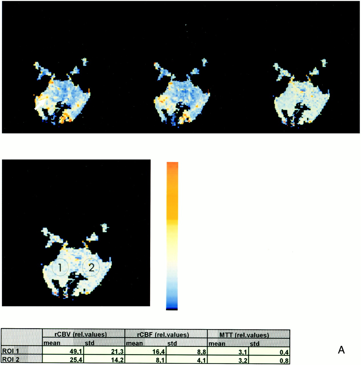
    A (above), Perfusion MR shows marked increase of regional cerebral blood volume (rCBV) and regional cerebral blood flow (rCBF) within the solid component of the lesion (arrow). Note that there is no interhemispheric difference in mean transit time (MTT). A total of 12 mL of gadopentate dimeglumine was administered (4 mL/s) and a T2*-weighted gradient-echo sequence with multishot echo-planar sequences 0.8/10/90 (TR/TE/flip angle) was obtained, followed by image postprocessing with maps of rCBV, MTT, rCBF, and time to peak.

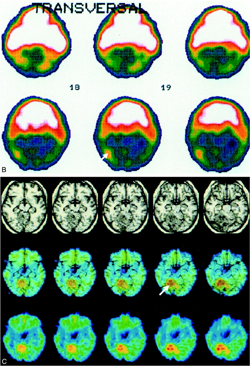
    B (facing page), Single-photon emission CT images obtained 120 minutes after administration of 94 MBq 201-thallium (TI) shows ncreased 201-TI uptake within the solid part of the residual LDD (arrow). This area corresponds to the zone of increased rCBV and rCBF shown in A. Images were aquired with a rotating gamma camera (Siemens ZLC 370/750, Germany) 5 minutes and 120 minutes after intravenous administration of 97 MBq 201-TI. Data acquisition was completed after 32 minutes by using a 128 × 128 matrix. Butterworth and Ramp filters were used to reconstruct images. Ratios of regions of interest compared with contralateral cerebellum were calculated.

    C (facing page), 18-FDG-PET with 3D MR imaging overlay shows the increased 18-FDG uptake is within the solid component of the LDD (arrow) and corresponds to the zone of increased rCBV and increased 201-TI uptake (see A and B). A Siemens CTI ECAT EXACT tomograph scanner (Germany) was used with a 10.8-cm field of veiw and 6.8-mm full width half maximum. The images were acquired 30 minutes after injection of 185 MBq 18-FDG. Images with attenuation correction were reconstructed using the standard mathematical algorithm implemented in ECAT software and using filtered back-projection by Shepp-Logan filter (cut-off 0.35 cycles/pixel). Automated spatial normalization to the stereotactic Talairach space was performed using SPM96. ROIs confined to the LDD were defined in all realigned slices covering the mass lesion, and identical regions were defined for the contralateral unaffected cerebellar hemisphere. Mean values for semiquantitative analysis of each region of interest (ECAT counts per pixel per second) were normalized to individual global brain glucose metabolism to give a glucose metabolic index as region of interest over global cerebral metabolic rate of glucose

  • fig 2.
    • Download figure
    • Open in new tab
    • Download powerpoint
    fig 2.

    Continued

  • fig 3.
    • Download figure
    • Open in new tab
    • Download powerpoint
    fig 3.

    (Case 1 and 2). MR spectroscopy shows increased lactate levels and reduced levels of choline and myo-inositol. For comparison, we added the data from Verheggen's patient (14). 1H MR spectra 1.5-T whole-body system (Sonata; Siemens, Erlangen, Germany) or 2-T whole-body system (Medsped S200 Avance; Bruker, Karlsruhe, Germany) with a gradient insert of 30mT/m stength and 190 ms ramp time were localized with a point-resolved spectroscopy select sequence (3000/30 or 128/88 or 20mm3 [TR/TE/repetitions/volume of interest]). A water suppression preparation period using three optimized chemical shift selective cycles was applied before each measurement. For each localization, a reference scan without water suppression was acquired. Spectra were analyzed using the LCModel software with the appropriate reference spectra

PreviousNext
Back to top

In this issue

American Journal of Neuroradiology
Vol. 22, Issue 5
1 May 2001
  • Table of Contents
  • Index by author
Advertisement
Print
Download PDF
Email Article

Thank you for your interest in spreading the word on American Journal of Neuroradiology.

NOTE: We only request your email address so that the person you are recommending the page to knows that you wanted them to see it, and that it is not junk mail. We do not capture any email address.

Enter multiple addresses on separate lines or separate them with commas.
Lhermitte-Duclos Disease: Assessment with MR Imaging, Positron Emission Tomography, Single-photon Emission CT, and MR Spectroscopy
(Your Name) has sent you a message from American Journal of Neuroradiology
(Your Name) thought you would like to see the American Journal of Neuroradiology web site.
CAPTCHA
This question is for testing whether or not you are a human visitor and to prevent automated spam submissions.
Cite this article
Joachim Klisch, Freimut Juengling, Joachim Spreer, Donatus Koch, Torsten Thiel, Martin Büchert, Sebastian Arnold, Friedrich Feuerhake, Martin Schumacher
Lhermitte-Duclos Disease: Assessment with MR Imaging, Positron Emission Tomography, Single-photon Emission CT, and MR Spectroscopy
American Journal of Neuroradiology May 2001, 22 (5) 824-830;

Citation Manager Formats

  • BibTeX
  • Bookends
  • EasyBib
  • EndNote (tagged)
  • EndNote 8 (xml)
  • Medlars
  • Mendeley
  • Papers
  • RefWorks Tagged
  • Ref Manager
  • RIS
  • Zotero
0 Responses
Respond to this article
Share
Bookmark this article
Lhermitte-Duclos Disease: Assessment with MR Imaging, Positron Emission Tomography, Single-photon Emission CT, and MR Spectroscopy
Joachim Klisch, Freimut Juengling, Joachim Spreer, Donatus Koch, Torsten Thiel, Martin Büchert, Sebastian Arnold, Friedrich Feuerhake, Martin Schumacher
American Journal of Neuroradiology May 2001, 22 (5) 824-830;
del.icio.us logo Twitter logo Facebook logo Mendeley logo
  • Tweet Widget
  • Facebook Like
  • Google Plus One

Jump to section

  • Article
    • Abstract
    • Case Reports
    • Discussion
    • Conclusion
    • Footnotes
    • References
  • Figures & Data
  • Info & Metrics
  • Responses
  • References
  • PDF

Related Articles

  • No related articles found.
  • PubMed
  • Google Scholar

Cited By...

  • Neuroimaging abnormalities in patients with Cowden syndrome: Retrospective single-center study
  • Crossref
  • Google Scholar

This article has not yet been cited by articles in journals that are participating in Crossref Cited-by Linking.

More in this TOC Section

  • SyMRI & MR Fingerprinting in Brainstem Myelination
  • Comparison of Image Quality and Radiation Dose in Pediatric Temporal Bone CT Using Photon-Counting Detector CT and Energy-Integrating Detector CT
  • Dandy-Walker Phenotype with Brainstem Involvement
Show more Pediatrics

Similar Articles

Advertisement

Indexed Content

  • Current Issue
  • Accepted Manuscripts
  • Article Preview
  • Past Issues
  • Editorials
  • Editor's Choice
  • Fellows' Journal Club
  • Letters to the Editor
  • Video Articles

Cases

  • Case Collection
  • Archive - Case of the Week
  • Archive - Case of the Month
  • Archive - Classic Case

Special Collections

  • AJNR Awards
  • ASNR Foundation Special Collection
  • Most Impactful AJNR Articles
  • Photon-Counting CT
  • Spinal CSF Leak Articles (Jan 2020-June 2024)

More from AJNR

  • Trainee Corner
  • Imaging Protocols
  • MRI Safety Corner

Multimedia

  • AJNR Podcasts
  • AJNR Scantastics

Resources

  • Turnaround Time
  • Submit a Manuscript
  • Submit a Video Article
  • Submit an eLetter to the Editor/Response
  • Manuscript Submission Guidelines
  • Statistical Tips
  • Fast Publishing of Accepted Manuscripts
  • Graphical Abstract Preparation
  • Imaging Protocol Submission
  • Evidence-Based Medicine Level Guide
  • Publishing Checklists
  • Author Policies
  • Become a Reviewer/Academy of Reviewers
  • News and Updates

About Us

  • About AJNR
  • Editorial Board
  • Editorial Board Alumni
  • Alerts
  • Permissions
  • Not an AJNR Subscriber? Join Now
  • Advertise with Us
  • Librarian Resources
  • Feedback
  • Terms and Conditions
  • AJNR Editorial Board Alumni

American Society of Neuroradiology

  • Not an ASNR Member? Join Now

© 2025 by the American Society of Neuroradiology All rights, including for text and data mining, AI training, and similar technologies, are reserved.
Print ISSN: 0195-6108 Online ISSN: 1936-959X

Powered by HighWire