
Article Figures & Data
Figures
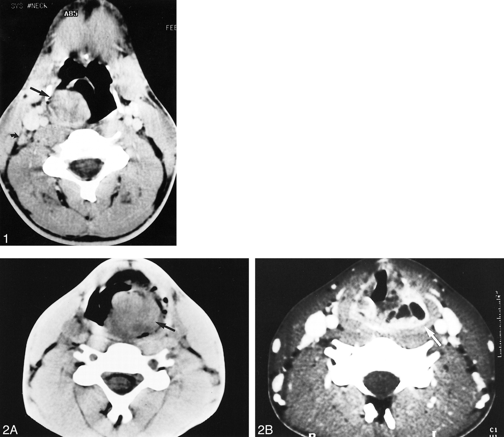
- fig 1.
Axial view contrast-enhanced CT scan of a 21-year-old man who consulted for dysphagia associated with dyspnea that worsened when lying supine. A homogeneous, well-demarcated mass can be seen, seemingly arising from the right lateral wall of the oro- and hypopharynx (arrow), associated with a homolateral deep cervical node (curved arrowhead). No recurrence had occurred 5 years after surgery, chemotherapy, and radiotherapy.
fig 2. Images from the case of a 17-year-old male patient who consulted for a painless progressively enlarging neck mass and complained of dyspnea.
A, Initial non-contrast-enhanced axial view CT scan reveals a left hypopharyngeal mass involving the left piriform sinus (black arrow).
B, Axial view contrast-enhanced CT scan, obtained 3 months after partial resection under endoscopic guidance and carbon dioxide laser therapy, shows a heterogeneous, multilocular recurrence of the tumor (white arrow)
- fig 3.
Initial axial view contrast-enhanced CT scan of a 61-year-old woman who noted a slowly growing mass of the left external fossa that was associated with intermittent pain in the left temporomandibular joint. A tumor of the left infratemporal fossa, with intra-tumoral calcifications (arrowheads), can be seen. The lesion is invading the left temporal muscle and the left greater wing of the sphenoid bone (arrow). Histologically, it is a spindle cell monophasic synovial sarcoma. No recurrence had occurred 8 years after surgery and radiotherapy.
fig 4. Images from the case of an 18-year-old female patient who presented with a synovial sarcoma arising from the right infratemporal fossa.
A, Axial view T1-weighted MR image (600/20/2 [TR/TE/ excitations]) shows a voluminous, homogeneous mass involving the right lateral pterygoid and temporal muscles and displacing the posterior wall of the right maxillary sinus anteriorly. The lesion exhibits an isointense signal (arrow).
B, Axial view T2-weighted MR image (2000/100/1) shows a voluminous, homogeneous mass involving the right lateral pterygoid and temporal muscles and displacing the posterior wall of the right maxillary sinus anteriorly. The lesion exhibits a hypointense signal (arrow)
- fig 5.
Images from the case of a 12-year-old male patient who complained of dental pain associated with nasal obstruction and progressive exophthalmos.
A, Initial contrast-enhanced CT scan shows a heterogeneous, necrotic mass involving the right maxillary sinus (arrows), invading the wall of the maxillary sinus, the right nasal fossa, and the right lateral pterygoid muscle.
B, T2-weighted MR image (3695/99/2) shows that the lesion is heterogeneous.
C, The lesion is isointense on the T1-weighted MR image (440/12/2). No recurrence had occurred 2 years after surgery, chemotherapy, and radiotherapy
- fig 6.
Histologic analysis of spindle cell monophasic synovial sarcoma (case 4).
A, Tumor cells are stained brown antibody anti-vimentin.
B, Only a few cells show positivity for cytokeratin
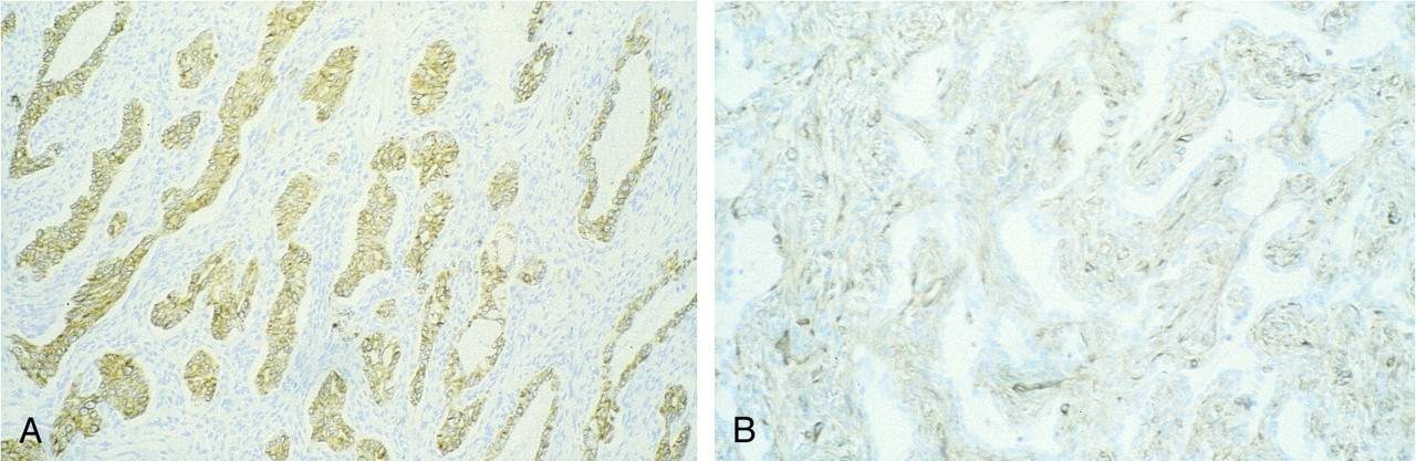
- fig 7.
Biphasic synovial sarcoma (case 8).
A, Prominent keratin-immunoreactive epithelial cells can be seen among keratin-negative spindle cells.
B, Spindle cells are stained brown antibody anti-vimentin


