Advertisement

Main menu

  • Home
  • Content
    • Current Issue
    • Accepted Manuscripts
    • Article Preview
    • Past Issue Archive
    • Video Articles
    • AJNR Case Collection
    • Case of the Week Archive
    • Case of the Month Archive
    • Classic Case Archive
  • Special Collections
    • AJNR Awards
    • ASNR Foundation Special Collection
    • Most Impactful AJNR Articles
    • Photon-Counting CT
    • Spinal CSF Leak Articles (Jan 2020-June 2024)
  • Multimedia
    • AJNR Podcasts
    • AJNR SCANtastic
    • Trainee Corner
    • MRI Safety Corner
    • Imaging Protocols
  • For Authors
    • Submit a Manuscript
    • Submit a Video Article
    • Submit an eLetter to the Editor/Response
    • Manuscript Submission Guidelines
    • Statistical Tips
    • Fast Publishing of Accepted Manuscripts
    • Graphical Abstract Preparation
    • Imaging Protocol Submission
    • Author Policies
  • About Us
    • About AJNR
    • Editorial Board
    • Editorial Board Alumni
  • More
    • Become a Reviewer/Academy of Reviewers
    • Subscribers
    • Permissions
    • Alerts
    • Feedback
    • Advertisers
    • ASNR Home

User menu

  • Alerts
  • Log in

Search

  • Advanced search
American Journal of Neuroradiology

American Journal of Neuroradiology

ASHNR American Society of Functional Neuroradiology ASHNR American Society of Pediatric Neuroradiology ASSR
  • Alerts
  • Log in

Advanced Search

  • Home
  • Content
    • Current Issue
    • Accepted Manuscripts
    • Article Preview
    • Past Issue Archive
    • Video Articles
    • AJNR Case Collection
    • Case of the Week Archive
    • Case of the Month Archive
    • Classic Case Archive
  • Special Collections
    • AJNR Awards
    • ASNR Foundation Special Collection
    • Most Impactful AJNR Articles
    • Photon-Counting CT
    • Spinal CSF Leak Articles (Jan 2020-June 2024)
  • Multimedia
    • AJNR Podcasts
    • AJNR SCANtastic
    • Trainee Corner
    • MRI Safety Corner
    • Imaging Protocols
  • For Authors
    • Submit a Manuscript
    • Submit a Video Article
    • Submit an eLetter to the Editor/Response
    • Manuscript Submission Guidelines
    • Statistical Tips
    • Fast Publishing of Accepted Manuscripts
    • Graphical Abstract Preparation
    • Imaging Protocol Submission
    • Author Policies
  • About Us
    • About AJNR
    • Editorial Board
    • Editorial Board Alumni
  • More
    • Become a Reviewer/Academy of Reviewers
    • Subscribers
    • Permissions
    • Alerts
    • Feedback
    • Advertisers
    • ASNR Home
  • Follow AJNR on Twitter
  • Visit AJNR on Facebook
  • Follow AJNR on Instagram
  • Join AJNR on LinkedIn
  • RSS Feeds

AJNR is seeking candidates for the AJNR Podcast Editor. Read the position description.

Research ArticleBRAIN

Perfusion-sensitive MR Imaging of Gliomas: Comparison between Gradient-echo and Spin-echo Echo-planar Imaging Techniques

Takeshi Sugahara, Yukunori Korogi, Masato Kochi, Yukitaka Ushio and Mutsumasa Takahashi
American Journal of Neuroradiology August 2001, 22 (7) 1306-1315;
Takeshi Sugahara
  • Find this author on Google Scholar
  • Find this author on PubMed
  • Search for this author on this site
Yukunori Korogi
  • Find this author on Google Scholar
  • Find this author on PubMed
  • Search for this author on this site
Masato Kochi
  • Find this author on Google Scholar
  • Find this author on PubMed
  • Search for this author on this site
Yukitaka Ushio
  • Find this author on Google Scholar
  • Find this author on PubMed
  • Search for this author on this site
Mutsumasa Takahashi
  • Find this author on Google Scholar
  • Find this author on PubMed
  • Search for this author on this site
  • Article
  • Figures & Data
  • Info & Metrics
  • Responses
  • References
  • PDF
Loading

Article Figures & Data

Figures

  • fig 1.
    • Download figure
    • Open in new tab
    • Download powerpoint
    fig 1.

    Relationship of the maximum rCBV of the low- and high-grade gliomas between the GE- (2000/46/1 [TR/TE/excitation]) and the SE-EPI (2000/97/1) techniques. For high-grade gliomas, the maximum rCBV ratios obtained with GE-EPI are significantly higher than those acquired with SE-EPI, whereas there is no significant difference for low-grade gliomas

  • fig 2.
    • Download figure
    • Open in new tab
    • Download powerpoint
    fig 2.

    A 35-year-old man with low-grade astrocytoma.

    A, T2-weighted image at 3700/96/1 (TR/TE/excitation). Left frontoparietal tumor with hyperintensity is not enhanced after contrast medium administration.

    B, Contrast-enhanced T1-weighted image at 690/14/1.

    C and D, SE-EPI at 2000/97/1 (C) and GE-EPI (D,) at 2000/46/1. On the perfusion-sensitive MR imaging, tumor CBV (arrow) is lower than that of gray matter obtained with the two techniques.

  • fig 3.
    • Download figure
    • Open in new tab
    • Download powerpoint
    fig 3.

    A 55-year-old man with glioblastoma.

    A, Contrast-enhanced T1-weighted image at 690/14/1 (TR/TE/excitation). B and C, SE-EPI at 2000/97/1 (B) and GE-EPI at 2000/46/1 (C). The area of high tumor rCBV (arrow) is clearly visualized with both SE- and GE-EPI.

  • fig 4.
    • Download figure
    • Open in new tab
    • Download powerpoint
    fig 4.

    A 33-year-old man with glioblastoma.

    A, Contrast-enhanced T1-weighted image at 3700/96/1 (TR/TE/excitation).

    B and C, SE-EPI at 2000/97/1 (B) and GE-EPI (C) at 2000/46/1. High rCBV of the tumor rim (arrow) is shown with SE-EPI but is more conspicuous with GE-EPI (arrow).

  • fig 5.
    • Download figure
    • Open in new tab
    • Download powerpoint
    fig 5.

    The maximum rCBV ratios calculated with the GE-EPI technique at 2000/46/1 (TR/TE/excitation) versus those obtained with SE-EPI at 2000/97/1

  • fig 6.
    • Download figure
    • Open in new tab
    • Download powerpoint
    fig 6.

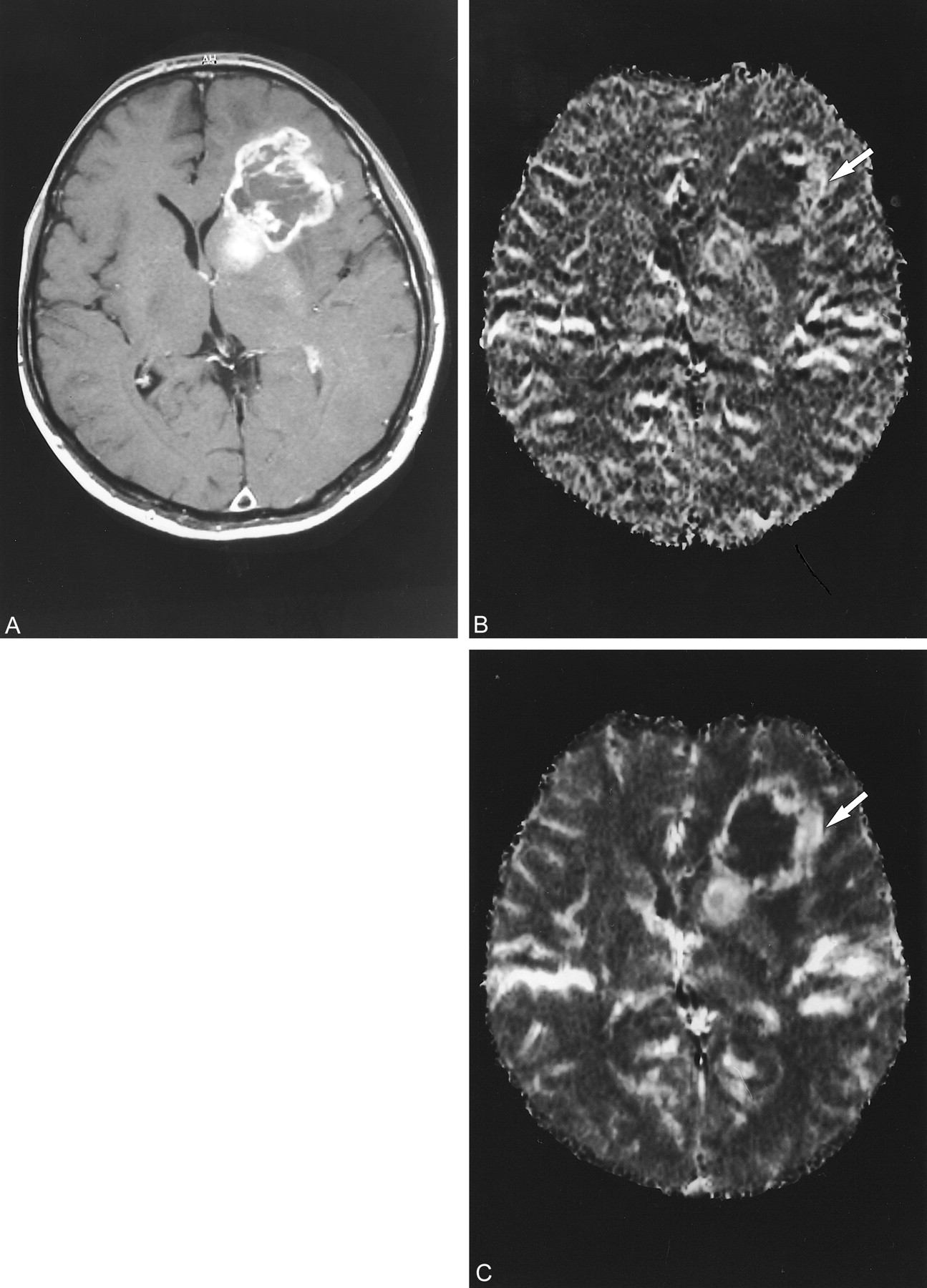
    A 47-year-old man with glioblastoma.

    A, Contrast-enhanced T1-weighted image at 3700/96/1 (TR/TE/excitation).

    B and C, SE-EPI at 2000/97/1 (B) and GE-EPI at 2000/46/1 (C). Tumor rCBV is higher than that of gray and white matter obtained with the two techniques, but the hypervascular area of the tumor (B and C, arrow) is more conspicuous with GE-EPI.

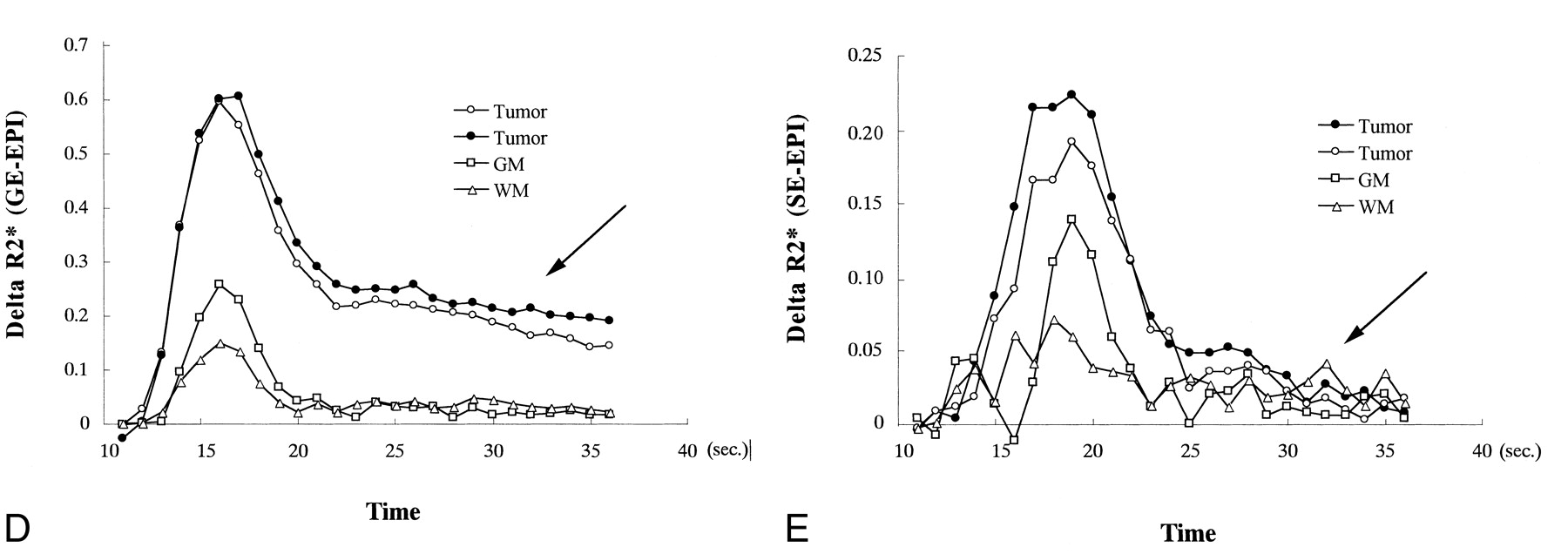
    D and E, The ΔR2* and ΔR2 curves during the transit of the contrast material through the gray and white matter regions of the brain and the two areas within the tumor that were measured using each of the SE- and GE-EPI techniques, respectively. The curves with white and black circles represent the tumor; the curves with squares and triangles represent the gray and white matter, respectively. Interestingly, the susceptibility effects within the tumor did not completely disappear after the first pass of the contrast medium on the GE-EPI image (D, arrow), whereas they almost completely disappeared on the SE-EPI image (E, arrow).

  • fig 6.
    • Download figure
    • Open in new tab
    • Download powerpoint
    fig 6.

    Continued

PreviousNext
Back to top

In this issue

American Journal of Neuroradiology: 22 (7)
American Journal of Neuroradiology
Vol. 22, Issue 7
1 Aug 2001
  • Table of Contents
  • Index by author
Advertisement
Print
Download PDF
Email Article

Thank you for your interest in spreading the word on American Journal of Neuroradiology.

NOTE: We only request your email address so that the person you are recommending the page to knows that you wanted them to see it, and that it is not junk mail. We do not capture any email address.

Enter multiple addresses on separate lines or separate them with commas.
Perfusion-sensitive MR Imaging of Gliomas: Comparison between Gradient-echo and Spin-echo Echo-planar Imaging Techniques
(Your Name) has sent you a message from American Journal of Neuroradiology
(Your Name) thought you would like to see the American Journal of Neuroradiology web site.
CAPTCHA
This question is for testing whether or not you are a human visitor and to prevent automated spam submissions.
Cite this article
Takeshi Sugahara, Yukunori Korogi, Masato Kochi, Yukitaka Ushio, Mutsumasa Takahashi
Perfusion-sensitive MR Imaging of Gliomas: Comparison between Gradient-echo and Spin-echo Echo-planar Imaging Techniques
American Journal of Neuroradiology Aug 2001, 22 (7) 1306-1315;

Citation Manager Formats

  • BibTeX
  • Bookends
  • EasyBib
  • EndNote (tagged)
  • EndNote 8 (xml)
  • Medlars
  • Mendeley
  • Papers
  • RefWorks Tagged
  • Ref Manager
  • RIS
  • Zotero
0 Responses
Respond to this article
Share
Bookmark this article
Perfusion-sensitive MR Imaging of Gliomas: Comparison between Gradient-echo and Spin-echo Echo-planar Imaging Techniques
Takeshi Sugahara, Yukunori Korogi, Masato Kochi, Yukitaka Ushio, Mutsumasa Takahashi
American Journal of Neuroradiology Aug 2001, 22 (7) 1306-1315;
del.icio.us logo Twitter logo Facebook logo Mendeley logo
  • Tweet Widget
  • Facebook Like
  • Google Plus One

Jump to section

  • Article
    • Abstract
    • Methods
    • Results
    • Discussion
    • Conclusion
    • Footnotes
    • References
  • Figures & Data
  • Info & Metrics
  • Responses
  • References
  • PDF

Related Articles

  • No related articles found.
  • PubMed
  • Google Scholar

Cited By...

  • Impact of Software Modeling on the Accuracy of Perfusion MRI in Glioma
  • Arterial Spin-Labeled Perfusion of Pediatric Brain Tumors
  • Semi-automated and automated glioma grading using dynamic susceptibility-weighted contrast-enhanced perfusion MRI relative cerebral blood volume measurements
  • Quantitative Blood Flow Measurements in Gliomas Using Arterial Spin-Labeling at 3T: Intermodality Agreement and Inter- and Intraobserver Reproducibility Study
  • Optimized Preload Leakage-Correction Methods to Improve the Diagnostic Accuracy of Dynamic Susceptibility-Weighted Contrast-Enhanced Perfusion MR Imaging in Posttreatment Gliomas
  • Spin-Echo Echo-Planar Perfusion MR Imaging in the Differential Diagnosis of Solitary Enhancing Brain Lesions: Distinguishing Solitary Metastases from Primary Glioma
  • Usefulness of diffusion/perfusion-weighted MRI in patients with non-enhancing supratentorial brain gliomas: a valuable tool to predict tumour grading?
  • Analysis of dynamic contrast enhanced MRI
  • Dynamic Magnetic Resonance Perfusion Imaging of Brain Tumors
  • Second harmonic imaging: a new ultrasound technique to assess human brain tumour perfusion
  • Crossref
  • Google Scholar

This article has not yet been cited by articles in journals that are participating in Crossref Cited-by Linking.

More in this TOC Section

  • White Matter Alterations in the Brains of Patients with Active, Remitted, and Cured Cushing Syndrome: A DTI Study
  • Qualitative and Quantitative Analysis of MR Imaging Findings in Patients with Middle Cerebral Artery Stroke Implanted with Mesenchymal Stem Cells
  • Fast Contrast-Enhanced 4D MRA and 4D Flow MRI Using Constrained Reconstruction (HYPRFlow): Potential Applications for Brain Arteriovenous Malformations
Show more BRAIN

Similar Articles

Advertisement

Indexed Content

  • Current Issue
  • Accepted Manuscripts
  • Article Preview
  • Past Issues
  • Editorials
  • Editor's Choice
  • Fellows' Journal Club
  • Letters to the Editor
  • Video Articles

Cases

  • Case Collection
  • Archive - Case of the Week
  • Archive - Case of the Month
  • Archive - Classic Case

Special Collections

  • AJNR Awards
  • ASNR Foundation Special Collection
  • Most Impactful AJNR Articles
  • Photon-Counting CT
  • Spinal CSF Leak Articles (Jan 2020-June 2024)

More from AJNR

  • Trainee Corner
  • Imaging Protocols
  • MRI Safety Corner

Multimedia

  • AJNR Podcasts
  • AJNR Scantastics

Resources

  • Turnaround Time
  • Submit a Manuscript
  • Submit a Video Article
  • Submit an eLetter to the Editor/Response
  • Manuscript Submission Guidelines
  • Statistical Tips
  • Fast Publishing of Accepted Manuscripts
  • Graphical Abstract Preparation
  • Imaging Protocol Submission
  • Evidence-Based Medicine Level Guide
  • Publishing Checklists
  • Author Policies
  • Become a Reviewer/Academy of Reviewers
  • News and Updates

About Us

  • About AJNR
  • Editorial Board
  • Editorial Board Alumni
  • Alerts
  • Permissions
  • Not an AJNR Subscriber? Join Now
  • Advertise with Us
  • Librarian Resources
  • Feedback
  • Terms and Conditions
  • AJNR Editorial Board Alumni

American Society of Neuroradiology

  • Not an ASNR Member? Join Now

© 2025 by the American Society of Neuroradiology All rights, including for text and data mining, AI training, and similar technologies, are reserved.
Print ISSN: 0195-6108 Online ISSN: 1936-959X

Powered by HighWire