
Article Figures & Data
Figures
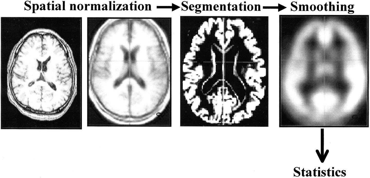
- fig 1.
Outline summary of voxel-based morphometry. Spatial normalization fitted each individual brain to a standard template brain in 3D space. Normalized MR images were then segmented into gray matter, white matter, cerebrospinal fluid, and other compartments. The gray matter images were smoothed with a 12-mm, full-width half-maximum isotropic gaussian kernel to use the partial volume effect to create a spectrum of gray matter intensities
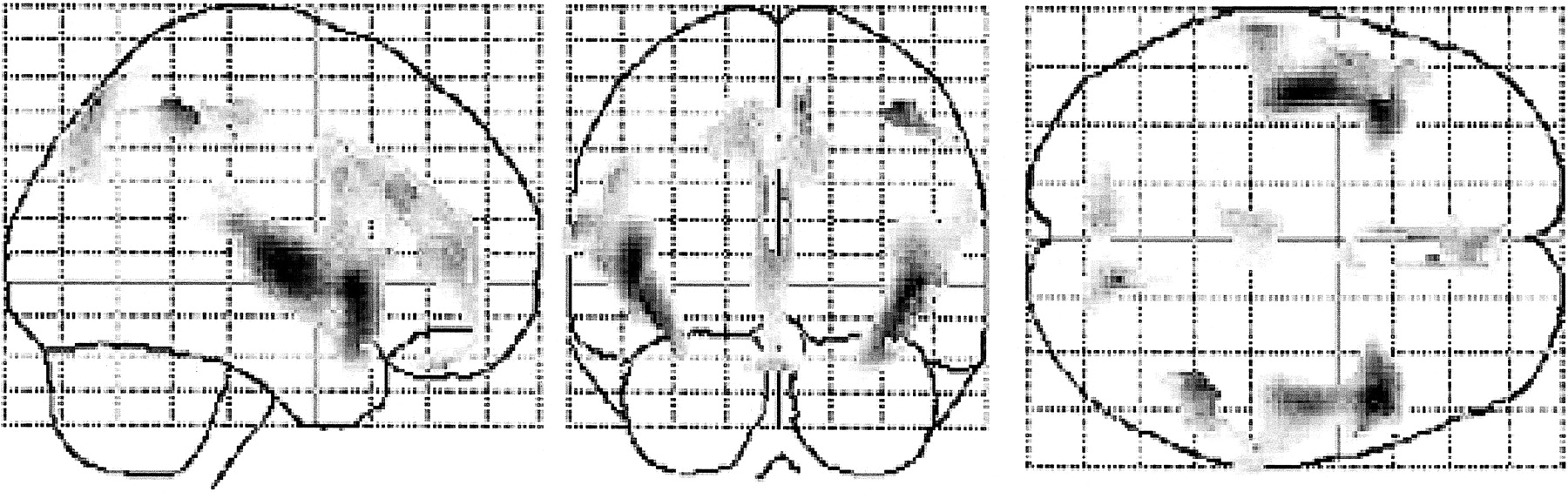
- fig 2.
Negative correlations between age and regional gray matter volume in healthy volunteers. The SPM of the t statistics (after transformation to a SPM [Z] for this contrast) is displayed in a standard format as a maximum intensity projection viewed from the right-hand side and from the back and the top of the brain. The anatomic space corresponds to the atlas of Talairach and Tournoux (18). Significant reduction of regional gray matter volume is noted in the inferior frontal gyrus, the insular cortex, cingulate gyrus, superior temporal gyrus, precuneus, left dorsolateral prefrontal cortex, right inferior parietal lobule, and orbitofrontal cortex
- fig 3.
Significant reduction of regional gray matter volumes in AD, which was noted in the bilateral hippocampal formation, entorhinal cortex, and parahippocampal cortex

Tables

Location and peak of significant declines in gray matter volume with age

In this issue


Jump to section
Related Articles
Cited By...
- Structural Degeneration of the Nucleus basalis of Meynert in Mild Cognitive Impairment and Alzheimers Disease - Evidence from an MRI-based Meta-Analysis
- Relationship between finger movement characteristics and voxel-based specific regional analysis systems for Alzheimers disease
- Distinct changes in morphometric networks in aging versus Alzheimers disease dementia
- Network Curvature as a Hallmark of Brain Structural Connectivity
- Initial Experience in Using Continuous Arterial Spin-Labeled MR Imaging for Early Detection of Alzheimer Disease
- Age, Alzheimer disease, and brain structure
- A voxel based morphometry study on mild cognitive impairment
- Correction for Partial-Volume Effects on Brain Perfusion SPECT in Healthy Men
- Detection of grey matter loss in mild Alzheimer's disease with voxel based morphometry
- Longitudinal Evaluation of Both Morphologic and Functional Changes in the Same Individuals with Alzheimer's Disease