Advertisement

Main menu

  • Home
  • Content
    • Current Issue
    • Accepted Manuscripts
    • Article Preview
    • Past Issue Archive
    • Video Articles
    • AJNR Case Collection
    • Case of the Week Archive
    • Case of the Month Archive
    • Classic Case Archive
  • Special Collections
    • AJNR Awards
    • ASNR Foundation Special Collection
    • Most Impactful AJNR Articles
    • Photon-Counting CT
    • Spinal CSF Leak Articles (Jan 2020-June 2024)
  • Multimedia
    • AJNR Podcasts
    • AJNR SCANtastic
    • Trainee Corner
    • MRI Safety Corner
    • Imaging Protocols
  • For Authors
    • Submit a Manuscript
    • Submit a Video Article
    • Submit an eLetter to the Editor/Response
    • Manuscript Submission Guidelines
    • Statistical Tips
    • Fast Publishing of Accepted Manuscripts
    • Graphical Abstract Preparation
    • Imaging Protocol Submission
    • Author Policies
  • About Us
    • About AJNR
    • Editorial Board
    • Editorial Board Alumni
  • More
    • Become a Reviewer/Academy of Reviewers
    • Subscribers
    • Permissions
    • Alerts
    • Feedback
    • Advertisers
    • ASNR Home

User menu

  • Alerts
  • Log in

Search

  • Advanced search
American Journal of Neuroradiology

American Journal of Neuroradiology

ASHNR American Society of Functional Neuroradiology ASHNR American Society of Pediatric Neuroradiology ASSR
  • Alerts
  • Log in

Advanced Search

  • Home
  • Content
    • Current Issue
    • Accepted Manuscripts
    • Article Preview
    • Past Issue Archive
    • Video Articles
    • AJNR Case Collection
    • Case of the Week Archive
    • Case of the Month Archive
    • Classic Case Archive
  • Special Collections
    • AJNR Awards
    • ASNR Foundation Special Collection
    • Most Impactful AJNR Articles
    • Photon-Counting CT
    • Spinal CSF Leak Articles (Jan 2020-June 2024)
  • Multimedia
    • AJNR Podcasts
    • AJNR SCANtastic
    • Trainee Corner
    • MRI Safety Corner
    • Imaging Protocols
  • For Authors
    • Submit a Manuscript
    • Submit a Video Article
    • Submit an eLetter to the Editor/Response
    • Manuscript Submission Guidelines
    • Statistical Tips
    • Fast Publishing of Accepted Manuscripts
    • Graphical Abstract Preparation
    • Imaging Protocol Submission
    • Author Policies
  • About Us
    • About AJNR
    • Editorial Board
    • Editorial Board Alumni
  • More
    • Become a Reviewer/Academy of Reviewers
    • Subscribers
    • Permissions
    • Alerts
    • Feedback
    • Advertisers
    • ASNR Home
  • Follow AJNR on Twitter
  • Visit AJNR on Facebook
  • Follow AJNR on Instagram
  • Join AJNR on LinkedIn
  • RSS Feeds

AJNR is seeking candidates for the AJNR Podcast Editor. Read the position description.

Research ArticleHEAD AND NECK

Reinterpretation of Cross-Sectional Images in Patients with Head and Neck Cancer in the Setting of a Multidisciplinary Cancer Center

Laurie A. Loevner, Adina I. Sonners, Brian J. Schulman, Kerstin Slawek, Randal S. Weber, David I. Rosenthal, Gul Moonis and Ara A. Chalian
American Journal of Neuroradiology November 2002, 23 (10) 1622-1626;
Laurie A. Loevner
  • Find this author on Google Scholar
  • Find this author on PubMed
  • Search for this author on this site
Adina I. Sonners
  • Find this author on Google Scholar
  • Find this author on PubMed
  • Search for this author on this site
Brian J. Schulman
  • Find this author on Google Scholar
  • Find this author on PubMed
  • Search for this author on this site
Kerstin Slawek
  • Find this author on Google Scholar
  • Find this author on PubMed
  • Search for this author on this site
Randal S. Weber
  • Find this author on Google Scholar
  • Find this author on PubMed
  • Search for this author on this site
David I. Rosenthal
  • Find this author on Google Scholar
  • Find this author on PubMed
  • Search for this author on this site
Gul Moonis
  • Find this author on Google Scholar
  • Find this author on PubMed
  • Search for this author on this site
Ara A. Chalian
  • Find this author on Google Scholar
  • Find this author on PubMed
  • Search for this author on this site
  • Article
  • Figures & Data
  • Info & Metrics
  • Responses
  • References
  • PDF
Loading

Article Figures & Data

Figures

  • Tables
  • Fig 1.
    • Download figure
    • Open in new tab
    • Download powerpoint
    Fig 1.

    57-year-old woman with a history of squamous cell carcinoma of the left side of the tongue. Enhanced CT image shows a necrotic regional nodal metastasis (arrows) in the contralateral neck that was detected on reinterpretation in the cancer center, but missed on the initial read. This was pathologically proved following neck dissection.
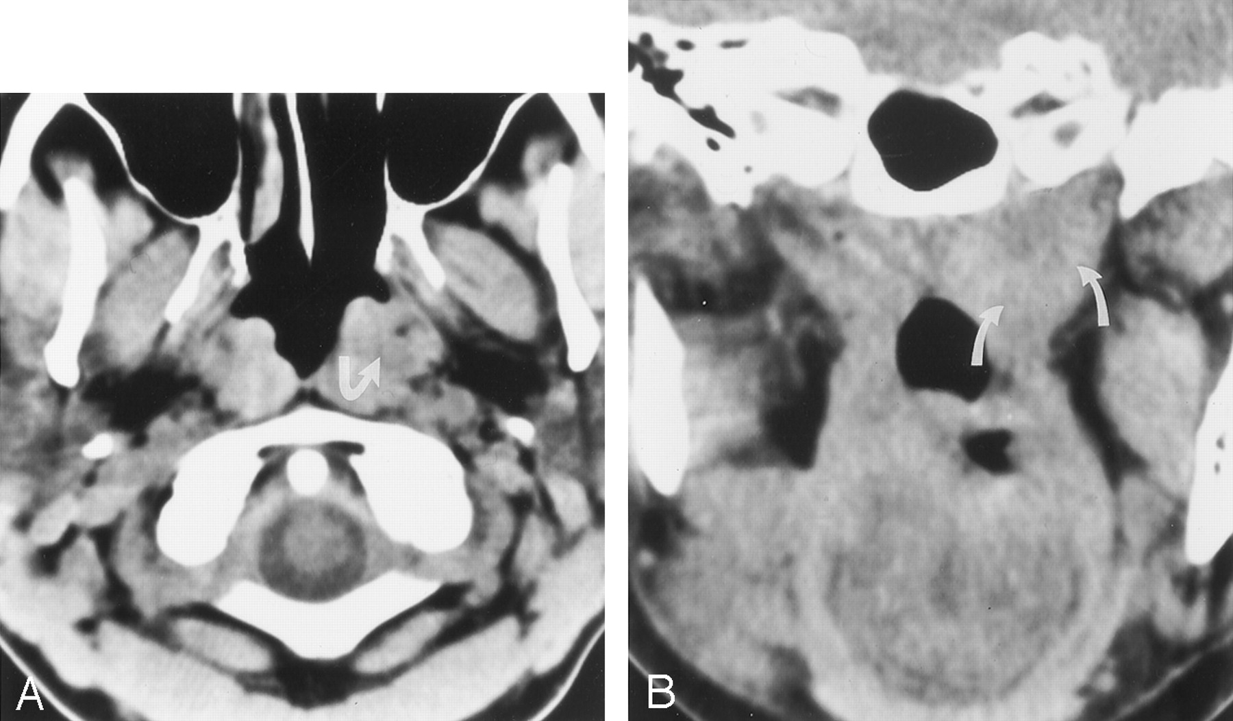
  • Fig 2.
    • Download figure
    • Open in new tab
    • Download powerpoint
    Fig 2.

    49-year-old woman with known cancer of the right side of the tongue and a second primary cancer of the nasopharynx detected at the time of image reinterpretation.

    A, Nonenhanced axial CT image shows asymmetry of the nasopharynx, with increased tissue on the left (arrow) and obliteration of the fat along the deep musculature (levator and tensor veli palatini muscles).

    B, Nonenhanced coronal CT image again shows increased tissue at the left nasopharynx (arrows). Subsequent biopsy revealed carcinoma.
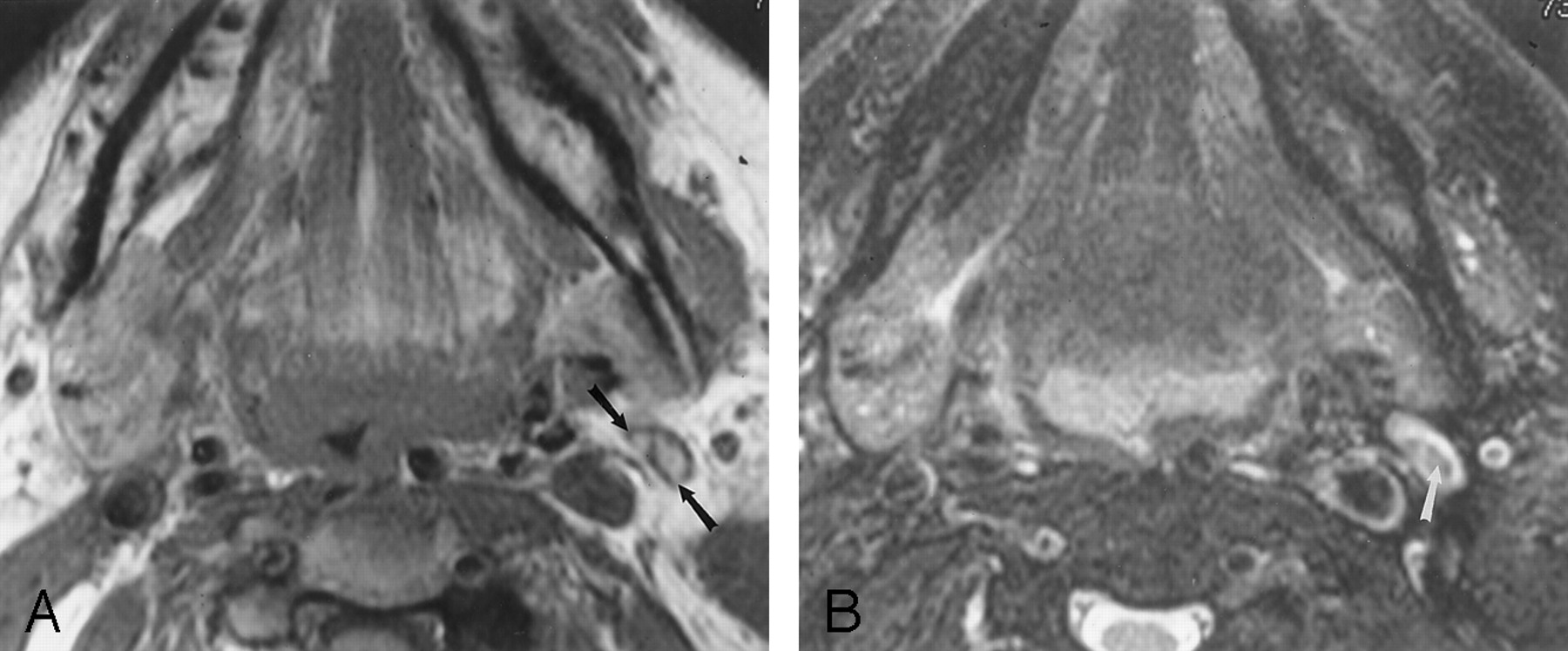
  • Fig 3.
    • Download figure
    • Open in new tab
    • Download powerpoint
    Fig 3.

    80-year-old woman with primary pharyngeal cancer. A normal-sized, fat-replaced left jugulogastric lymph node was interpreted as abnormal because of inhomogeneous signal intensity.

    A, Axial nonenhanced T1-weighted (600/17/1 [repetition time/echo time/excitations]) MR image shows intrinsic high signal intensity in the lymph node (arrows) consistent with fat.

    B, Axial fat-suppressed T2-weighted (4000/80/1) MR image obtained at the same level as that in A shows hypointensity in the hilum of this node (arrow) consistent with suppressed fat.

Tables

  • Figures
    • View popup
    TABLE 1:

    Categorized changes in image interpretation

    Type of ChangeNo. of Cases
    Nodal
     Missed pathologic nodes18
      Cervical14
      Retropharyngeal4
     Nodes misinterpreted as pathologic3
     Submandibular gland mistaken for nodes2
    Changes related to tumor extension
     Missed submucosal extension5
      Parapharyngeal3
      Preepiglottic2
     Presence or absence of cartilage invasion5
     Presence or absence of perineural spread4
     Underestimation of size or extent of tumor4
     Overestimation of size or extent of tumor3
    Changes related to primary neoplasm
     Primary cancer missed on imaging8
      Oral cavity6
      Pharynx2
     Normal anatomy mistaken for primary neoplasm3
    Missed second primary neoplasm6
    Missed metastasis2
    Missed middle cerebral artery aneurysm1
    • View popup
    TABLE 2:

    Change in tumor staging and means of verification of the change in image interpretation

    Patient No.Change in TNMVerification of Change in InterpretationPatient No.Change in TNMVerification of Change in Interpretation
    1T ↑, N ↑Pathologic24T ↑Pathologic
    2T ↑Pathologic25N ↑Pathologic
    3T ↓, N ↓Pathologic26T ↑, N ↑Pathologic
    4T ↑Pathologic27N ↑Pathologic
    5T ↑Pathologic28T ↑Pathologic
    6N ↑Pathologic29T ↑Pathologic
    7N ↑Pathologic30N ↑Pathologic
    8T ↓Pathologic31N ↑Pathologic
    9T ↑, N ↑Pathologic32T ↑Pathologic
    10T ↑Pathologic33N ↑Pathologic
    11T ↑Pathologic34N ↑Radiologic
    12M ↑Pathologic35T ↑Radiologic
    13N ↓Pathologic36T ↑, N ↑Radiologic
    14T ↓Pathologic37N ↓Radiologic
    15T ↑Pathologic38T ↑, N ↑Radiologic
    16N ↑Pathologic39T ↓Radiologic
    17T ↓, N ↓Pathologic40N ↑Radiologic
    18T ↑Pathologic41M ↑Radiologic
    19N ↑Pathologic42N ↑Radiologic
    20N ↑Pathologic43N ↑Radiologic
    21T ↑Pathologic44N ↓Clinical/imaging
    22T ↑Pathologic45T ↑Clinical/imaging
    23T ↑, N ↓Pathologic46T ↑, N ↑Clinical/imaging
    • Note.—↑ indicates upstaged; ↓, downstaged; Radiologic, characteristic radiologic findings; Clinical/imaging, clinical and imaging follow-up.

PreviousNext
Back to top

In this issue

American Journal of Neuroradiology: 23 (10)
American Journal of Neuroradiology
Vol. 23, Issue 10
1 Nov 2002
  • Table of Contents
  • Index by author
Advertisement
Print
Download PDF
Email Article

Thank you for your interest in spreading the word on American Journal of Neuroradiology.

NOTE: We only request your email address so that the person you are recommending the page to knows that you wanted them to see it, and that it is not junk mail. We do not capture any email address.

Enter multiple addresses on separate lines or separate them with commas.
Reinterpretation of Cross-Sectional Images in Patients with Head and Neck Cancer in the Setting of a Multidisciplinary Cancer Center
(Your Name) has sent you a message from American Journal of Neuroradiology
(Your Name) thought you would like to see the American Journal of Neuroradiology web site.
CAPTCHA
This question is for testing whether or not you are a human visitor and to prevent automated spam submissions.
Cite this article
Laurie A. Loevner, Adina I. Sonners, Brian J. Schulman, Kerstin Slawek, Randal S. Weber, David I. Rosenthal, Gul Moonis, Ara A. Chalian
Reinterpretation of Cross-Sectional Images in Patients with Head and Neck Cancer in the Setting of a Multidisciplinary Cancer Center
American Journal of Neuroradiology Nov 2002, 23 (10) 1622-1626;

Citation Manager Formats

  • BibTeX
  • Bookends
  • EasyBib
  • EndNote (tagged)
  • EndNote 8 (xml)
  • Medlars
  • Mendeley
  • Papers
  • RefWorks Tagged
  • Ref Manager
  • RIS
  • Zotero
0 Responses
Respond to this article
Share
Bookmark this article
Reinterpretation of Cross-Sectional Images in Patients with Head and Neck Cancer in the Setting of a Multidisciplinary Cancer Center
Laurie A. Loevner, Adina I. Sonners, Brian J. Schulman, Kerstin Slawek, Randal S. Weber, David I. Rosenthal, Gul Moonis, Ara A. Chalian
American Journal of Neuroradiology Nov 2002, 23 (10) 1622-1626;
del.icio.us logo Twitter logo Facebook logo Mendeley logo
  • Tweet Widget
  • Facebook Like
  • Google Plus One

Jump to section

  • Article
    • Abstract
    • Methods
    • Results
    • Discussion
    • Conclusion
    • Footnotes
    • References
  • Figures & Data
  • Info & Metrics
  • Responses
  • References
  • PDF

Related Articles

  • No related articles found.
  • PubMed
  • Google Scholar

Cited By...

  • Shift Volume Directly Impacts Neuroradiology Error Rate at a Large Academic Medical Center: The Case for Volume Limits
  • Recommendations in Second Opinion Reports of Neurologic Head and Neck Imaging: Frequency, Referring Clinicians Compliance, and Diagnostic Yield
  • Risk Factors for Perceptual-versus-Interpretative Errors in Diagnostic Neuroradiology
  • Impact of Neuroradiology-Based Peer Review on Head and Neck Radiotherapy Target Delineation
  • Impact of Center Size and Experience on Outcomes in Head and Neck Cancer
  • Interobserver Agreement in the Interpretation of Outpatient Head CT Scans in an Academic Neuroradiology Practice
  • Larynx Preservation for Patients With Locally Advanced Laryngeal Cancer
  • Crossref
  • Google Scholar

This article has not yet been cited by articles in journals that are participating in Crossref Cited-by Linking.

More in this TOC Section

  • Correlation of Apparent Diffusion Coefficient at 3T with Prognostic Parameters of Retinoblastoma
  • Parathyroid Lesions: Characterization with Dual-Phase Arterial and Venous Enhanced CT of the Neck
  • Efficacy of Diffusion-Weighted Imaging for the Differentiation between Lymphomas and Carcinomas of the Nasopharynx and Oropharynx: Correlations of Apparent Diffusion Coefficients and Histologic Features
Show more HEAD AND NECK

Similar Articles

Advertisement

Indexed Content

  • Current Issue
  • Accepted Manuscripts
  • Article Preview
  • Past Issues
  • Editorials
  • Editor's Choice
  • Fellows' Journal Club
  • Letters to the Editor
  • Video Articles

Cases

  • Case Collection
  • Archive - Case of the Week
  • Archive - Case of the Month
  • Archive - Classic Case

Special Collections

  • AJNR Awards
  • ASNR Foundation Special Collection
  • Most Impactful AJNR Articles
  • Photon-Counting CT
  • Spinal CSF Leak Articles (Jan 2020-June 2024)

More from AJNR

  • Trainee Corner
  • Imaging Protocols
  • MRI Safety Corner

Multimedia

  • AJNR Podcasts
  • AJNR Scantastics

Resources

  • Turnaround Time
  • Submit a Manuscript
  • Submit a Video Article
  • Submit an eLetter to the Editor/Response
  • Manuscript Submission Guidelines
  • Statistical Tips
  • Fast Publishing of Accepted Manuscripts
  • Graphical Abstract Preparation
  • Imaging Protocol Submission
  • Evidence-Based Medicine Level Guide
  • Publishing Checklists
  • Author Policies
  • Become a Reviewer/Academy of Reviewers
  • News and Updates

About Us

  • About AJNR
  • Editorial Board
  • Editorial Board Alumni
  • Alerts
  • Permissions
  • Not an AJNR Subscriber? Join Now
  • Advertise with Us
  • Librarian Resources
  • Feedback
  • Terms and Conditions
  • AJNR Editorial Board Alumni

American Society of Neuroradiology

  • Not an ASNR Member? Join Now

© 2025 by the American Society of Neuroradiology All rights, including for text and data mining, AI training, and similar technologies, are reserved.
Print ISSN: 0195-6108 Online ISSN: 1936-959X

Powered by HighWire