Advertisement

Main menu

  • Home
  • Content
    • Current Issue
    • Accepted Manuscripts
    • Article Preview
    • Past Issue Archive
    • Video Articles
    • AJNR Case Collection
    • Case of the Week Archive
    • Case of the Month Archive
    • Classic Case Archive
  • Special Collections
    • AJNR Awards
    • ASNR Foundation Special Collection
    • Most Impactful AJNR Articles
    • Photon-Counting CT
    • Spinal CSF Leak Articles (Jan 2020-June 2024)
  • Multimedia
    • AJNR Podcasts
    • AJNR SCANtastic
    • Trainee Corner
    • MRI Safety Corner
    • Imaging Protocols
  • For Authors
    • Submit a Manuscript
    • Submit a Video Article
    • Submit an eLetter to the Editor/Response
    • Manuscript Submission Guidelines
    • Statistical Tips
    • Fast Publishing of Accepted Manuscripts
    • Graphical Abstract Preparation
    • Imaging Protocol Submission
    • Author Policies
  • About Us
    • About AJNR
    • Editorial Board
    • Editorial Board Alumni
  • More
    • Become a Reviewer/Academy of Reviewers
    • Subscribers
    • Permissions
    • Alerts
    • Feedback
    • Advertisers
    • ASNR Home

User menu

  • Alerts
  • Log in

Search

  • Advanced search
American Journal of Neuroradiology

American Journal of Neuroradiology

ASHNR American Society of Functional Neuroradiology ASHNR American Society of Pediatric Neuroradiology ASSR
  • Alerts
  • Log in

Advanced Search

  • Home
  • Content
    • Current Issue
    • Accepted Manuscripts
    • Article Preview
    • Past Issue Archive
    • Video Articles
    • AJNR Case Collection
    • Case of the Week Archive
    • Case of the Month Archive
    • Classic Case Archive
  • Special Collections
    • AJNR Awards
    • ASNR Foundation Special Collection
    • Most Impactful AJNR Articles
    • Photon-Counting CT
    • Spinal CSF Leak Articles (Jan 2020-June 2024)
  • Multimedia
    • AJNR Podcasts
    • AJNR SCANtastic
    • Trainee Corner
    • MRI Safety Corner
    • Imaging Protocols
  • For Authors
    • Submit a Manuscript
    • Submit a Video Article
    • Submit an eLetter to the Editor/Response
    • Manuscript Submission Guidelines
    • Statistical Tips
    • Fast Publishing of Accepted Manuscripts
    • Graphical Abstract Preparation
    • Imaging Protocol Submission
    • Author Policies
  • About Us
    • About AJNR
    • Editorial Board
    • Editorial Board Alumni
  • More
    • Become a Reviewer/Academy of Reviewers
    • Subscribers
    • Permissions
    • Alerts
    • Feedback
    • Advertisers
    • ASNR Home
  • Follow AJNR on Twitter
  • Visit AJNR on Facebook
  • Follow AJNR on Instagram
  • Join AJNR on LinkedIn
  • RSS Feeds

AJNR is seeking candidates for the AJNR Podcast Editor. Read the position description.

Research ArticleBRAIN

Predicting Cerebral Ischemic Infarct Volume with Diffusion and Perfusion MR Imaging

Pamela W. Schaefer, George J. Hunter, Julian He, Leena M. Hamberg, A. Gregory Sorensen, Lee H. Schwamm, Walter J. Koroshetz and R. Gilberto Gonzalez
American Journal of Neuroradiology November 2002, 23 (10) 1785-1794;
Pamela W. Schaefer
  • Find this author on Google Scholar
  • Find this author on PubMed
  • Search for this author on this site
George J. Hunter
  • Find this author on Google Scholar
  • Find this author on PubMed
  • Search for this author on this site
Julian He
  • Find this author on Google Scholar
  • Find this author on PubMed
  • Search for this author on this site
Leena M. Hamberg
  • Find this author on Google Scholar
  • Find this author on PubMed
  • Search for this author on this site
A. Gregory Sorensen
  • Find this author on Google Scholar
  • Find this author on PubMed
  • Search for this author on this site
Lee H. Schwamm
  • Find this author on Google Scholar
  • Find this author on PubMed
  • Search for this author on this site
Walter J. Koroshetz
  • Find this author on Google Scholar
  • Find this author on PubMed
  • Search for this author on this site
R. Gilberto Gonzalez
  • Find this author on Google Scholar
  • Find this author on PubMed
  • Search for this author on this site
  • Article
  • Figures & Data
  • Info & Metrics
  • Responses
  • References
  • PDF
Loading

Article Figures & Data

Figures

  • Tables
  • Fig 1.
    • Download figure
    • Open in new tab
    • Download powerpoint
    Fig 1.

    A–D, Scatterplots demonstrate initial lesion volume with each technique versus final lesion volume at follow-up (F-U) in 81 patients.

    A, Initial DW imaging versus follow-up: r2 = 0.9, slope = 1 .24 ± 0.08 (95% confidence limits).

    B, Initial CBV versus follow-up: r2 = 0.84, slope = 1 .22 ± 0.11.

    C, Initial CBF versus follow-up: r2 = 0.35, slope = 0.44 ± 0.09.

    D, Initial MTT versus follow-up: r2 = 0.22, slope = 0.32 ± 0.08.

    r2 indicates coefficient of determination.

  • Fig 2.
    • Download figure
    • Open in new tab
    • Download powerpoint
    Fig 2.

    A–F, Scatterplots show initial lesion volume versus final infarct volume in patients with a perfusion-diffusion mismatch (perfusion lesion more than 20% larger than the diffusion lesion).

    A and B, DW-CBV mismatch group: For DW imaging versus follow-up, the regression line was significantly different from the line of identity (P < .001). For CBV versus follow-up, the regression line was not significantly different from the line of identity (P = .18).

    C and D, DW-CBF mismatch group: For DW imaging versus follow-up and for CBF versus follow-up, the line of regression was significantly different from the line of identity (P < .001).

    E and F, DW-MTT mismatch group: For DW imaging versus follow-up and for MTT versus follow-up, the line of regression was significantly different from the line of identity (P < .001). r2 indicates coefficient of determination.

  • Fig 3.
    • Download figure
    • Open in new tab
    • Download powerpoint
    Fig 3.

    A, Axial DW image demonstrates an infarct involving the left basal ganglia, insula, and subinsular region.

    B, CBV map shows the lesion is larger than the initial DW imaging abnormality; it also involves the left frontal and parietal opercula.

    C and D, CBF (C) and MTT (D) abnormalities involve most of the left middle cerebral artery distribution.

    E, Follow-up (F/u) T2-weighted image 10 days later demonstrates a lesion similar in size to that of the initial CBV abnormality (B).
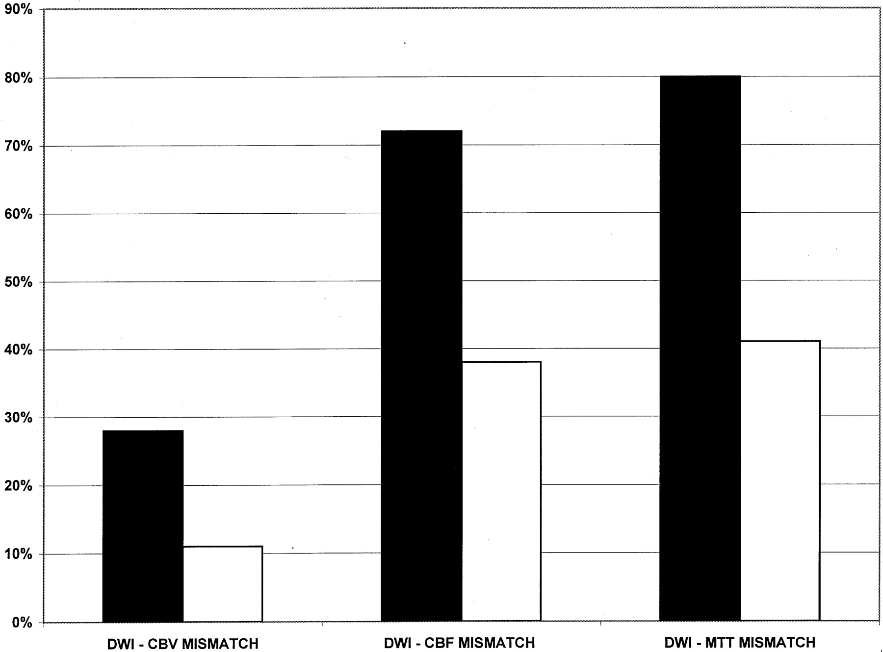
  • Fig 4.
    • Download figure
    • Open in new tab
    • Download powerpoint
    Fig 4.

    Percentage of patients with diffusion-perfusion mismatches for proximal (black bars) and nonproximal (white bars) infarctions.

Tables

  • Figures
    • View popup
    TABLE:

    Sensitivity and specificity of DW imaging and perfusion parameters in 134 patients

    TechniqueFinal DiagnosisSensitivity (%)Specificity (%)PPV (%)NPV (%)FPFN
    PositiveNegative
    DWI
     Positive10119496998116
     Negative626
    CBV
     Positive7807410010050028
     Negative2828
    CBF
     Positive89184969959118
     Negative1826
    MTT
     Positive89184969959118
     Negative1826
    • Note.—FN indicates false-negative findings; FP, false-positive findings; NPV, negative predictive value; PPV, positive predictive value.

PreviousNext
Back to top

In this issue

American Journal of Neuroradiology: 23 (10)
American Journal of Neuroradiology
Vol. 23, Issue 10
1 Nov 2002
  • Table of Contents
  • Index by author
Advertisement
Print
Download PDF
Email Article

Thank you for your interest in spreading the word on American Journal of Neuroradiology.

NOTE: We only request your email address so that the person you are recommending the page to knows that you wanted them to see it, and that it is not junk mail. We do not capture any email address.

Enter multiple addresses on separate lines or separate them with commas.
Predicting Cerebral Ischemic Infarct Volume with Diffusion and Perfusion MR Imaging
(Your Name) has sent you a message from American Journal of Neuroradiology
(Your Name) thought you would like to see the American Journal of Neuroradiology web site.
CAPTCHA
This question is for testing whether or not you are a human visitor and to prevent automated spam submissions.
Cite this article
Pamela W. Schaefer, George J. Hunter, Julian He, Leena M. Hamberg, A. Gregory Sorensen, Lee H. Schwamm, Walter J. Koroshetz, R. Gilberto Gonzalez
Predicting Cerebral Ischemic Infarct Volume with Diffusion and Perfusion MR Imaging
American Journal of Neuroradiology Nov 2002, 23 (10) 1785-1794;

Citation Manager Formats

  • BibTeX
  • Bookends
  • EasyBib
  • EndNote (tagged)
  • EndNote 8 (xml)
  • Medlars
  • Mendeley
  • Papers
  • RefWorks Tagged
  • Ref Manager
  • RIS
  • Zotero
0 Responses
Respond to this article
Share
Bookmark this article
Predicting Cerebral Ischemic Infarct Volume with Diffusion and Perfusion MR Imaging
Pamela W. Schaefer, George J. Hunter, Julian He, Leena M. Hamberg, A. Gregory Sorensen, Lee H. Schwamm, Walter J. Koroshetz, R. Gilberto Gonzalez
American Journal of Neuroradiology Nov 2002, 23 (10) 1785-1794;
del.icio.us logo Twitter logo Facebook logo Mendeley logo
  • Tweet Widget
  • Facebook Like
  • Google Plus One

Jump to section

  • Article
    • Abstract
    • Methods
    • Results
    • Discussion
    • Conclusion
    • Acknowledgments
    • Footnotes
    • References
  • Figures & Data
  • Info & Metrics
  • Responses
  • References
  • PDF

Related Articles

  • No related articles found.
  • PubMed
  • Google Scholar

Cited By...

  • Left angular gyrus disconnection impairs multiplication fact retrieval
  • A Simplified Model for Intravoxel Incoherent Motion Perfusion Imaging of the Brain
  • Validity of Shape as a Predictive Biomarker of Final Infarct Volume in Acute Ischemic Stroke
  • Comparison of 10 TTP and Tmax Estimation Techniques for MR Perfusion-Diffusion Mismatch Quantification in Acute Stroke
  • Microembolism and Catheter Ablation II: Effects of Cerebral Microemboli Injection in a Canine Model
  • Ventral Premotor Cortex May Be Required for Dynamic Changes in the Feeling of Limb Ownership: A Lesion Study
  • Recommendations for Imaging of Acute Ischemic Stroke: A Scientific Statement From the American Heart Association
  • Early Neutrophilia Is Associated With Volume of Ischemic Tissue in Acute Stroke
  • Response to Letters by Lee et al and Lev et al
  • Cerebral Blood Flow Thresholds in Acute Stroke Triage
  • Apparent Diffusion Coefficient Thresholds Do Not Predict the Response to Acute Stroke Thrombolysis
  • Awareness of the Functioning of One's Own Limbs Mediated by the Insular Cortex?
  • Recommendations for Comprehensive Stroke Centers: A Consensus Statement From the Brain Attack Coalition
  • Posterior thalamic hemorrhage induces "pusher syndrome"
  • Crossref
  • Google Scholar

This article has not yet been cited by articles in journals that are participating in Crossref Cited-by Linking.

More in this TOC Section

  • Usefulness of Quantitative Susceptibility Mapping for the Diagnosis of Parkinson Disease
  • White Matter Alterations in the Brains of Patients with Active, Remitted, and Cured Cushing Syndrome: A DTI Study
  • Qualitative and Quantitative Analysis of MR Imaging Findings in Patients with Middle Cerebral Artery Stroke Implanted with Mesenchymal Stem Cells
Show more BRAIN

Similar Articles

Advertisement

Indexed Content

  • Current Issue
  • Accepted Manuscripts
  • Article Preview
  • Past Issues
  • Editorials
  • Editor's Choice
  • Fellows' Journal Club
  • Letters to the Editor
  • Video Articles

Cases

  • Case Collection
  • Archive - Case of the Week
  • Archive - Case of the Month
  • Archive - Classic Case

Special Collections

  • AJNR Awards
  • ASNR Foundation Special Collection
  • Most Impactful AJNR Articles
  • Photon-Counting CT
  • Spinal CSF Leak Articles (Jan 2020-June 2024)

More from AJNR

  • Trainee Corner
  • Imaging Protocols
  • MRI Safety Corner

Multimedia

  • AJNR Podcasts
  • AJNR Scantastics

Resources

  • Turnaround Time
  • Submit a Manuscript
  • Submit a Video Article
  • Submit an eLetter to the Editor/Response
  • Manuscript Submission Guidelines
  • Statistical Tips
  • Fast Publishing of Accepted Manuscripts
  • Graphical Abstract Preparation
  • Imaging Protocol Submission
  • Evidence-Based Medicine Level Guide
  • Publishing Checklists
  • Author Policies
  • Become a Reviewer/Academy of Reviewers
  • News and Updates

About Us

  • About AJNR
  • Editorial Board
  • Editorial Board Alumni
  • Alerts
  • Permissions
  • Not an AJNR Subscriber? Join Now
  • Advertise with Us
  • Librarian Resources
  • Feedback
  • Terms and Conditions
  • AJNR Editorial Board Alumni

American Society of Neuroradiology

  • Not an ASNR Member? Join Now

© 2025 by the American Society of Neuroradiology All rights, including for text and data mining, AI training, and similar technologies, are reserved.
Print ISSN: 0195-6108 Online ISSN: 1936-959X

Powered by HighWire