Advertisement

Main menu

  • Home
  • Content
    • Current Issue
    • Accepted Manuscripts
    • Article Preview
    • Past Issue Archive
    • Video Articles
    • AJNR Case Collection
    • Case of the Week Archive
    • Case of the Month Archive
    • Classic Case Archive
  • Special Collections
    • AJNR Awards
    • ASNR Foundation Special Collection
    • Most Impactful AJNR Articles
    • Photon-Counting CT
    • Spinal CSF Leak Articles (Jan 2020-June 2024)
  • Multimedia
    • AJNR Podcasts
    • AJNR SCANtastic
    • Trainee Corner
    • MRI Safety Corner
    • Imaging Protocols
  • For Authors
    • Submit a Manuscript
    • Submit a Video Article
    • Submit an eLetter to the Editor/Response
    • Manuscript Submission Guidelines
    • Statistical Tips
    • Fast Publishing of Accepted Manuscripts
    • Graphical Abstract Preparation
    • Imaging Protocol Submission
    • Author Policies
  • About Us
    • About AJNR
    • Editorial Board
    • Editorial Board Alumni
  • More
    • Become a Reviewer/Academy of Reviewers
    • Subscribers
    • Permissions
    • Alerts
    • Feedback
    • Advertisers
    • ASNR Home

User menu

  • Alerts
  • Log in

Search

  • Advanced search
American Journal of Neuroradiology

American Journal of Neuroradiology

ASHNR American Society of Functional Neuroradiology ASHNR American Society of Pediatric Neuroradiology ASSR
  • Alerts
  • Log in

Advanced Search

  • Home
  • Content
    • Current Issue
    • Accepted Manuscripts
    • Article Preview
    • Past Issue Archive
    • Video Articles
    • AJNR Case Collection
    • Case of the Week Archive
    • Case of the Month Archive
    • Classic Case Archive
  • Special Collections
    • AJNR Awards
    • ASNR Foundation Special Collection
    • Most Impactful AJNR Articles
    • Photon-Counting CT
    • Spinal CSF Leak Articles (Jan 2020-June 2024)
  • Multimedia
    • AJNR Podcasts
    • AJNR SCANtastic
    • Trainee Corner
    • MRI Safety Corner
    • Imaging Protocols
  • For Authors
    • Submit a Manuscript
    • Submit a Video Article
    • Submit an eLetter to the Editor/Response
    • Manuscript Submission Guidelines
    • Statistical Tips
    • Fast Publishing of Accepted Manuscripts
    • Graphical Abstract Preparation
    • Imaging Protocol Submission
    • Author Policies
  • About Us
    • About AJNR
    • Editorial Board
    • Editorial Board Alumni
  • More
    • Become a Reviewer/Academy of Reviewers
    • Subscribers
    • Permissions
    • Alerts
    • Feedback
    • Advertisers
    • ASNR Home
  • Follow AJNR on Twitter
  • Visit AJNR on Facebook
  • Follow AJNR on Instagram
  • Join AJNR on LinkedIn
  • RSS Feeds

AJNR is seeking candidates for the AJNR Podcast Editor. Read the position description.

Research ArticleBRAIN

Comparison of Two Superparamagnetic Viral-Sized Iron Oxide Particles Ferumoxides and Ferumoxtran-10 with a Gadolinium Chelate in Imaging Intracranial Tumors

Peter Varallyay, Gary Nesbit, Leslie L. Muldoon, Randal R. Nixon, Johnny Delashaw, James I. Cohen, Annie Petrillo, Doris Rink and Edward A. Neuwelt
American Journal of Neuroradiology April 2002, 23 (4) 510-519;
Peter Varallyay
  • Find this author on Google Scholar
  • Find this author on PubMed
  • Search for this author on this site
Gary Nesbit
  • Find this author on Google Scholar
  • Find this author on PubMed
  • Search for this author on this site
Leslie L. Muldoon
  • Find this author on Google Scholar
  • Find this author on PubMed
  • Search for this author on this site
Randal R. Nixon
  • Find this author on Google Scholar
  • Find this author on PubMed
  • Search for this author on this site
Johnny Delashaw
  • Find this author on Google Scholar
  • Find this author on PubMed
  • Search for this author on this site
James I. Cohen
  • Find this author on Google Scholar
  • Find this author on PubMed
  • Search for this author on this site
Annie Petrillo
  • Find this author on Google Scholar
  • Find this author on PubMed
  • Search for this author on this site
Doris Rink
  • Find this author on Google Scholar
  • Find this author on PubMed
  • Search for this author on this site
Edward A. Neuwelt
  • Find this author on Google Scholar
  • Find this author on PubMed
  • Search for this author on this site
  • Article
  • Figures & Data
  • Info & Metrics
  • Responses
  • References
  • PDF
Loading

Article Figures & Data

Figures

  • Tables
  • Fig 1.
    • Download figure
    • Open in new tab
    • Download powerpoint
    Fig 1.

    Patient 2 with anaplastic oligodendroglioma.

    A, Gadolinium-enhanced SE T1-weighted image shows an intensely enhancing tumor nodule in the posterior right temporal lobe.

    B and C, Four hours after ferumoxides infusion, SE T1-weighted image (B) shows only a tiny area of increased signal intensity (arrow), whereas the GRE T2*-weighted image (C) demonstrates no decreased signal intensity (arrow). The rounded hypointense lateral region in C is from a craniotomy plate.

  • Fig 2.
    • Download figure
    • Open in new tab
    • Download powerpoint
    Fig 2.

    Patient 5 with anaplastic oligodendroglioma.

    A and B, SE T1-weighted images obtained 6 hours (A) and 24 hours (B) after ferumoxtran infusion show progressive peripheral and patchy central enhancement in the left temporal tumor.
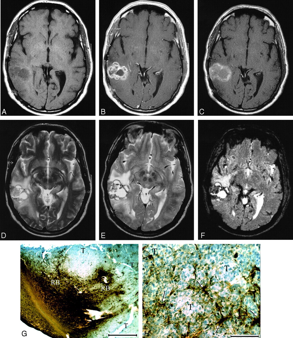
  • Fig 3.
    • Download figure
    • Open in new tab
    • Download powerpoint
    Fig 3.

    Patient 9 with anaplastic oligodendroglioma.

    A and B, Nonenhanced (A) and gadolinium-enhanced (B) SE T1-weighted images of the right temporal tumor. The gadolinium-enhanced image shows evidence of strong, lobulated peripheral enhancement with a central nonenhancing zone.

    C, At 24 hours after ferumoxtran infusion, SE T1-weighted image demonstrates marked high signal intensity in a similar distribution but with less peripheral lobulation compared with the gadolinium-enhanced image. Also note that the non–gadolinium-enhancing central zone became isointense to white matter, suggesting some ferumoxtran accumulation.

    D–F, Fast SE T2-weighted image obtained before ferumoxtran infusion (D) and fast SE T2-weighted (E) and GRE T2*-weighted (F) images obtained 24 hours after ferumoxtran infusion show a heterogeneous tumor mass with peripheral decreased signal intensity that is more prominent on the GRE T2*-weighted image. The distribution of the low-signal-intensity areas is similar to that of the high-signal-intensity areas on the SE T1-weighted image in C.

    G and H, Photomicrographs from histologic staining for iron (DAB-enhanced Perls stain). In G (original magnification ×7.5; bar indicates 1 mm), tumor (T) and reactive brain interface (RB) show the intense staining for iron at the periphery of the tumor. In H (original magnification ×100; bar indicates 0.1 mm), cellular iron staining at the tumor–reactive brain interface shows iron uptake by the parenchymal cells with fibrillar processes (arrows) rather than by the round tumor cells (T).
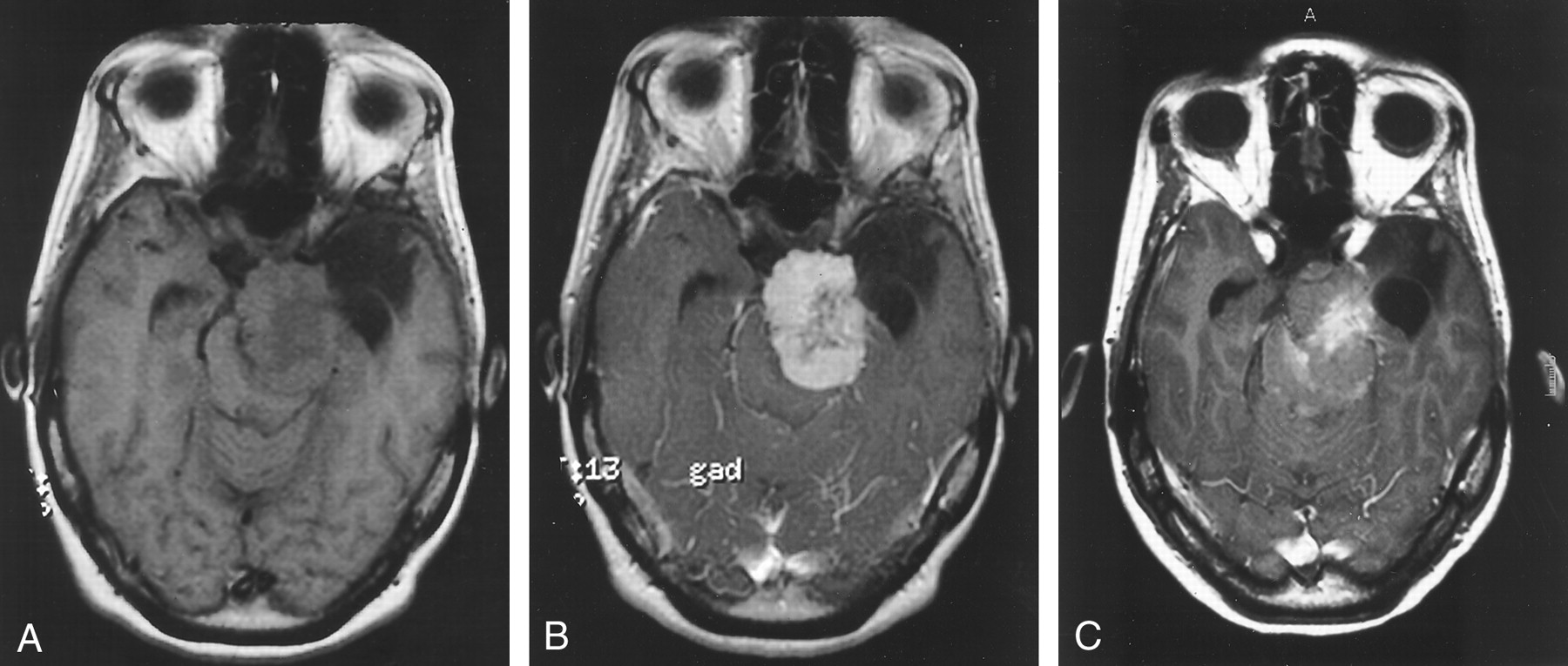
  • Fig 4.
    • Download figure
    • Open in new tab
    • Download powerpoint
    Fig 4.

    Patient 12 with meningioma, after radiation therapy.

    A and B, Nonenhanced (A) and gadolinium-enhanced (B) SE T1-weighted images. The image in B shows evidence of strong enhancement, except in the central region.

    C, At 24 hours after ferumoxtran infusion, SE T1-weighted image shows strong ferumoxtran enhancement in the less gadolinium-enhancing central region and only minimal ferumoxtran enhancement in the surrounding intensely gadolinium-enhancing portion.

  • Fig 5.
    • Download figure
    • Open in new tab
    • Download powerpoint
    Fig 5.

    Patient 6 with oligodendroglioma.

    A, Gadolinium-enhanced SE T1-weighted image shows a large left frontotemporal mass (arrow) with only small areas of enhancement in its anterior aspect.

    B and C, At 24 hours after ferumoxtran infusion, SE T1-weighted (B) and GRE T2*-weighted (C) images demonstrate no signal intensity changes (arrow), indicating no accumulation of iron.

  • Fig 6.
    • Download figure
    • Open in new tab
    • Download powerpoint
    Fig 6.

    Quantitation of SE T1-weighted MR images. Normalized mean SE T1 signal intensity values with standard mean error in areas of absent, minimal, and maximal ferumoxtran enhancement in the tumor as well as in brain around tumor (BAT) (n = 15 patients). a indicates P < .05 for gadolinium (Gd) enhancement compared with precontrast images; b, P < .05 for gadolinium enhancement compared with ferumoxtran (Combidex) enhancement; c, P < .001 compared with precontrast image; d, P < .005 compared with precontrast images.

  • Fig 7.
    • Download figure
    • Open in new tab
    • Download powerpoint
    Fig 7.

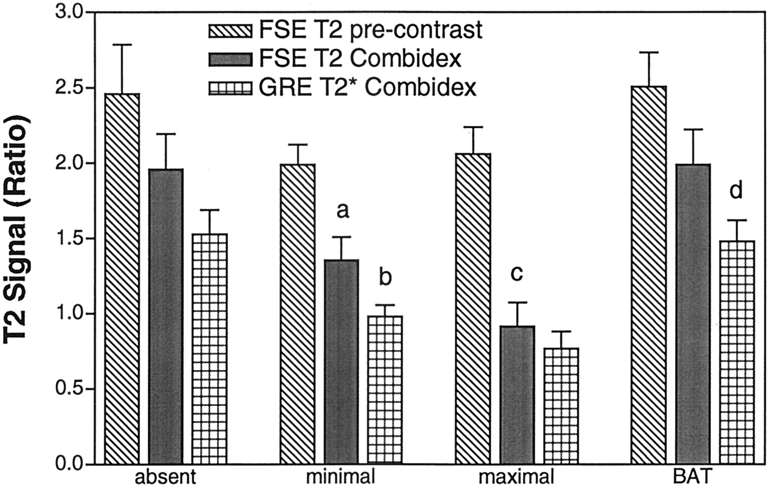
    Quantitation of fast SE T2-weighted and GRE T2*-weighted MR images. Normalized mean T2 signal intensity values with standard mean error in areas of absent, minimal, and maximal ferumoxtran enhancement in the tumor as well as in brain around tumor (BAT) (n = 15 patients). a indicates P < .01 compared with the precontrast fast SE (FSE) T2-weighted images; b, P < .005 comparing ferumoxtran (Combidex)-enhanced GRE T2*-weighted images with ferumoxtran-enhanced fast SE T2-weighted images; c, P < .001 compared with precontrast fast SE T2-weighted images; d, P < .05 comparing ferumoxtran-enhanced GRE T2*-weighted images with ferumoxtran-enhanced fast SE T2-weighted images.

Tables

  • Figures
    • View popup
    TABLE

    Demographics and imaging results

    Patient No./Sex/Age (y)Histologic DiagnosisTumor LocationUSPIOIron UptakeT1 Signal Intensity ChangeT2 Signal Intensity Change
    1/F/48Metastatic large cell carcinomaCerebellumFerumoxides−−−
    2/M/46Anaplastic oligodendragliomaR temporoparietalFerumoxides+*+−
    3/F/53MeningiomaL frontalFerumoxides−−−
    4/M/54Gliblastoma multiformeL thalamus, parietalFerumoxtran-10+++
    5/M/55Anaplastic oligodendragliomaL temporalFerumoxtran-10+†++
    6/M/54OligodendrogliomaL frontotemporalFerumoxtran-10−−−
    7/F/47HamartomaL temporalFerumoxtran-10+*+−
    8/F/30MedulloblastomaCerebellumFerumoxtran-10+†++
    9/M/58Anaplastic oligodendragliomaR temporoparietalFerumoxtran-10+†++
    10/M/55Squamous cell carcinomaNasopharynx with skull base invasionFerumoxtran-10+++
    11/M/50Squamous cell carcinomaNasopharynx with skull base invasionFerumoxtran-10+++
    12/F/42MeningiomaL petroclivalFerumoxtran-10+†++
    13/M/57OligodendrogliomaR parietalFerumoxtran-10−−−
    14/M/55Anaplastic gliomaL temporalFerumoxtran-10+*+−
    15/F/54Glioblastoma multiformeR temporoparietalFerumoxtran-10+++
    16/F/66Glioblastoma multiformeR frontalFerumoxtran-10+†++
    17/F/57Anaplastic oligodendragliomaR frontalFerumoxtran-10+†++
    18/M/54Pituitary adenomaIntra- and suprasellarFerumoxtran-10+++
    19/M/39Glioblastoma multiformeL fronto-opercularFerumoxtran-10+++
    20/M/46Anaplastic oligodendragliomaR posterior temporalFerumoxtran-10+†++
    • Note.—L indicates left; R, right; +, yes; −, no.

    • * Iron accumulation was only minimal compared with gadolinium enhancement.

    • † Ferumoxtran enhancement as measured with ROI (see Methods) was regionally more prominent than gadolinium enhancement.

PreviousNext
Back to top

In this issue

American Journal of Neuroradiology: 23 (4)
American Journal of Neuroradiology
Vol. 23, Issue 4
1 Apr 2002
  • Table of Contents
  • Index by author
Advertisement
Print
Download PDF
Email Article

Thank you for your interest in spreading the word on American Journal of Neuroradiology.

NOTE: We only request your email address so that the person you are recommending the page to knows that you wanted them to see it, and that it is not junk mail. We do not capture any email address.

Enter multiple addresses on separate lines or separate them with commas.
Comparison of Two Superparamagnetic Viral-Sized Iron Oxide Particles Ferumoxides and Ferumoxtran-10 with a Gadolinium Chelate in Imaging Intracranial Tumors
(Your Name) has sent you a message from American Journal of Neuroradiology
(Your Name) thought you would like to see the American Journal of Neuroradiology web site.
CAPTCHA
This question is for testing whether or not you are a human visitor and to prevent automated spam submissions.
Cite this article
Peter Varallyay, Gary Nesbit, Leslie L. Muldoon, Randal R. Nixon, Johnny Delashaw, James I. Cohen, Annie Petrillo, Doris Rink, Edward A. Neuwelt
Comparison of Two Superparamagnetic Viral-Sized Iron Oxide Particles Ferumoxides and Ferumoxtran-10 with a Gadolinium Chelate in Imaging Intracranial Tumors
American Journal of Neuroradiology Apr 2002, 23 (4) 510-519;

Citation Manager Formats

  • BibTeX
  • Bookends
  • EasyBib
  • EndNote (tagged)
  • EndNote 8 (xml)
  • Medlars
  • Mendeley
  • Papers
  • RefWorks Tagged
  • Ref Manager
  • RIS
  • Zotero
0 Responses
Respond to this article
Share
Bookmark this article
Comparison of Two Superparamagnetic Viral-Sized Iron Oxide Particles Ferumoxides and Ferumoxtran-10 with a Gadolinium Chelate in Imaging Intracranial Tumors
Peter Varallyay, Gary Nesbit, Leslie L. Muldoon, Randal R. Nixon, Johnny Delashaw, James I. Cohen, Annie Petrillo, Doris Rink, Edward A. Neuwelt
American Journal of Neuroradiology Apr 2002, 23 (4) 510-519;
del.icio.us logo Twitter logo Facebook logo Mendeley logo
  • Tweet Widget
  • Facebook Like
  • Google Plus One

Jump to section

  • Article
    • Abstract
    • Methods
    • Results
    • Discussion
    • Conclusion
    • Acknowledgments
    • Footnotes
    • References
  • Figures & Data
  • Info & Metrics
  • Responses
  • References
  • PDF

Related Articles

  • No related articles found.
  • PubMed
  • Google Scholar

Cited By...

  • Magnetic Resonance Imaging Investigation of Macrophages in Acute Cardiac Allograft Rejection After Heart Transplantation
  • USPIO-enhanced MRI of highly invasive and highly metastasizing transplanted human squamous cell carcinoma: an experimental study
  • New Frontiers in Translational Research in Neuro-oncology and the Blood-Brain Barrier: Report of the Tenth Annual Blood-Brain Barrier Disruption Consortium Meeting
  • Crossref
  • Google Scholar

This article has not yet been cited by articles in journals that are participating in Crossref Cited-by Linking.

More in this TOC Section

  • Usefulness of Quantitative Susceptibility Mapping for the Diagnosis of Parkinson Disease
  • White Matter Alterations in the Brains of Patients with Active, Remitted, and Cured Cushing Syndrome: A DTI Study
  • Qualitative and Quantitative Analysis of MR Imaging Findings in Patients with Middle Cerebral Artery Stroke Implanted with Mesenchymal Stem Cells
Show more BRAIN

Similar Articles

Advertisement

Indexed Content

  • Current Issue
  • Accepted Manuscripts
  • Article Preview
  • Past Issues
  • Editorials
  • Editor's Choice
  • Fellows' Journal Club
  • Letters to the Editor
  • Video Articles

Cases

  • Case Collection
  • Archive - Case of the Week
  • Archive - Case of the Month
  • Archive - Classic Case

Special Collections

  • AJNR Awards
  • ASNR Foundation Special Collection
  • Most Impactful AJNR Articles
  • Photon-Counting CT
  • Spinal CSF Leak Articles (Jan 2020-June 2024)

More from AJNR

  • Trainee Corner
  • Imaging Protocols
  • MRI Safety Corner

Multimedia

  • AJNR Podcasts
  • AJNR Scantastics

Resources

  • Turnaround Time
  • Submit a Manuscript
  • Submit a Video Article
  • Submit an eLetter to the Editor/Response
  • Manuscript Submission Guidelines
  • Statistical Tips
  • Fast Publishing of Accepted Manuscripts
  • Graphical Abstract Preparation
  • Imaging Protocol Submission
  • Evidence-Based Medicine Level Guide
  • Publishing Checklists
  • Author Policies
  • Become a Reviewer/Academy of Reviewers
  • News and Updates

About Us

  • About AJNR
  • Editorial Board
  • Editorial Board Alumni
  • Alerts
  • Permissions
  • Not an AJNR Subscriber? Join Now
  • Advertise with Us
  • Librarian Resources
  • Feedback
  • Terms and Conditions
  • AJNR Editorial Board Alumni

American Society of Neuroradiology

  • Not an ASNR Member? Join Now

© 2025 by the American Society of Neuroradiology All rights, including for text and data mining, AI training, and similar technologies, are reserved.
Print ISSN: 0195-6108 Online ISSN: 1936-959X

Powered by HighWire