Advertisement

Main menu

  • Home
  • Content
    • Current Issue
    • Accepted Manuscripts
    • Article Preview
    • Past Issue Archive
    • Video Articles
    • AJNR Case Collection
    • Case of the Week Archive
    • Case of the Month Archive
    • Classic Case Archive
  • Special Collections
    • AJNR Awards
    • ASNR Foundation Special Collection
    • Most Impactful AJNR Articles
    • Photon-Counting CT
    • Spinal CSF Leak Articles (Jan 2020-June 2024)
  • Multimedia
    • AJNR Podcasts
    • AJNR SCANtastic
    • Trainee Corner
    • MRI Safety Corner
    • Imaging Protocols
  • For Authors
    • Submit a Manuscript
    • Submit a Video Article
    • Submit an eLetter to the Editor/Response
    • Manuscript Submission Guidelines
    • Statistical Tips
    • Fast Publishing of Accepted Manuscripts
    • Graphical Abstract Preparation
    • Imaging Protocol Submission
    • Author Policies
  • About Us
    • About AJNR
    • Editorial Board
    • Editorial Board Alumni
  • More
    • Become a Reviewer/Academy of Reviewers
    • Subscribers
    • Permissions
    • Alerts
    • Feedback
    • Advertisers
    • ASNR Home

User menu

  • Alerts
  • Log in

Search

  • Advanced search
American Journal of Neuroradiology

American Journal of Neuroradiology

ASHNR American Society of Functional Neuroradiology ASHNR American Society of Pediatric Neuroradiology ASSR
  • Alerts
  • Log in

Advanced Search

  • Home
  • Content
    • Current Issue
    • Accepted Manuscripts
    • Article Preview
    • Past Issue Archive
    • Video Articles
    • AJNR Case Collection
    • Case of the Week Archive
    • Case of the Month Archive
    • Classic Case Archive
  • Special Collections
    • AJNR Awards
    • ASNR Foundation Special Collection
    • Most Impactful AJNR Articles
    • Photon-Counting CT
    • Spinal CSF Leak Articles (Jan 2020-June 2024)
  • Multimedia
    • AJNR Podcasts
    • AJNR SCANtastic
    • Trainee Corner
    • MRI Safety Corner
    • Imaging Protocols
  • For Authors
    • Submit a Manuscript
    • Submit a Video Article
    • Submit an eLetter to the Editor/Response
    • Manuscript Submission Guidelines
    • Statistical Tips
    • Fast Publishing of Accepted Manuscripts
    • Graphical Abstract Preparation
    • Imaging Protocol Submission
    • Author Policies
  • About Us
    • About AJNR
    • Editorial Board
    • Editorial Board Alumni
  • More
    • Become a Reviewer/Academy of Reviewers
    • Subscribers
    • Permissions
    • Alerts
    • Feedback
    • Advertisers
    • ASNR Home
  • Follow AJNR on Twitter
  • Visit AJNR on Facebook
  • Follow AJNR on Instagram
  • Join AJNR on LinkedIn
  • RSS Feeds

AJNR is seeking candidates for the AJNR Podcast Editor. Read the position description.

OtherBRAIN

Emerging Patterns of Diffusion-Weighted MR Imaging in Creutzfeldt-Jakob Disease: Case Report and Review of the Literature

Yang Mao-Draayer, Steven P. Braff, Keith J. Nagle, William Pendlebury, Paul L. Penar and Robert E. Shapiro
American Journal of Neuroradiology April 2002, 23 (4) 550-556;
Yang Mao-Draayer
  • Find this author on Google Scholar
  • Find this author on PubMed
  • Search for this author on this site
Steven P. Braff
  • Find this author on Google Scholar
  • Find this author on PubMed
  • Search for this author on this site
Keith J. Nagle
  • Find this author on Google Scholar
  • Find this author on PubMed
  • Search for this author on this site
William Pendlebury
  • Find this author on Google Scholar
  • Find this author on PubMed
  • Search for this author on this site
Paul L. Penar
  • Find this author on Google Scholar
  • Find this author on PubMed
  • Search for this author on this site
Robert E. Shapiro
  • Find this author on Google Scholar
  • Find this author on PubMed
  • Search for this author on this site

Fig 1.

Fig 1.
  • Download figure
  • Open in new tab
  • Download powerpoint
Fig 1.
  • Download figure
  • Open in new tab
  • Download powerpoint
Fig 1.
  • Download figure
  • Open in new tab
  • Download powerpoint
Fig 1.

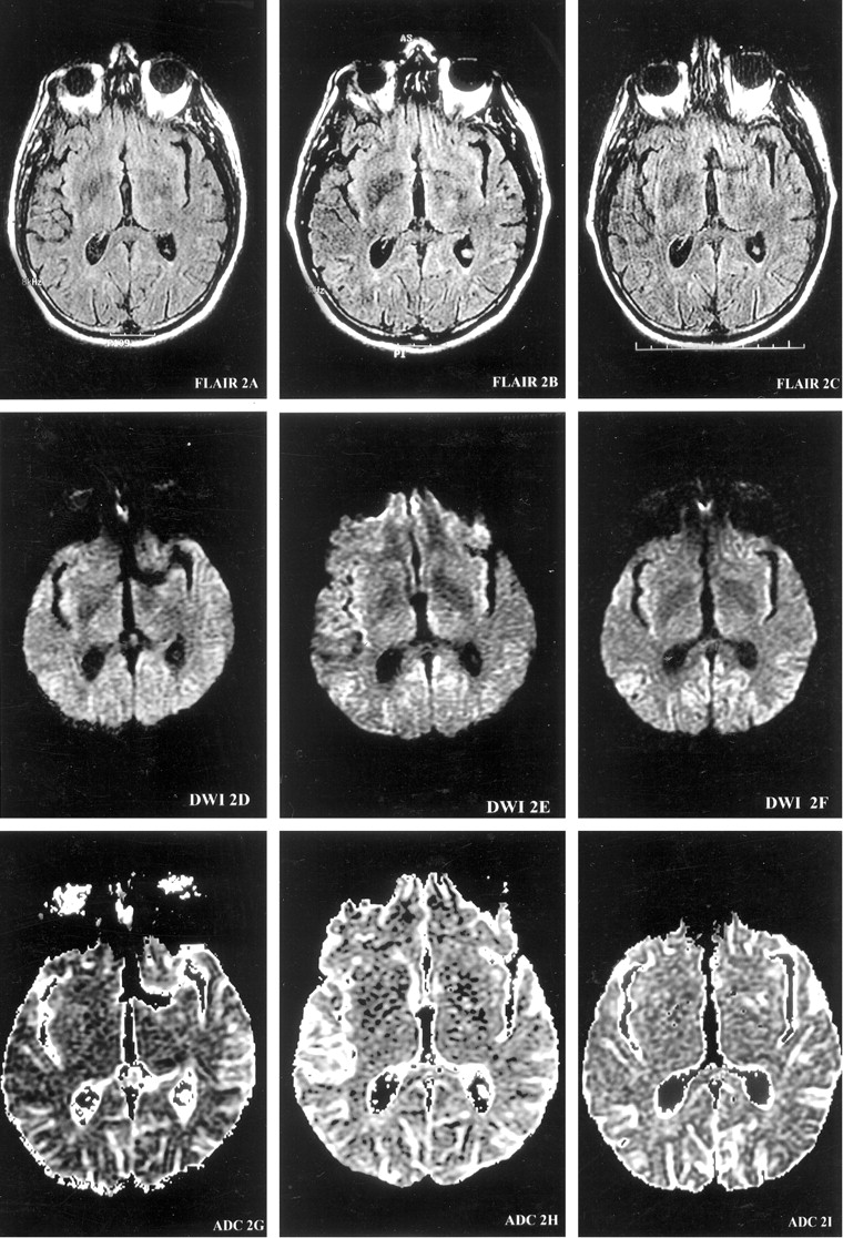
Serial MR images demonstrate the evolution of ribbon-like cortical signal intensity abnormalities. MR images are presented at each of three brain axial levels (1–3), at each of three time points from the onset of symptoms (left columns, 4 months; middle columns, 5.5 months; right columns, 6 months) for FLAIR (top row), DW imaging (middle row), and ADC (bottom row) studies. At 4 months from onset of symptoms, DW images demonstrate gyriform increased signal intensity predominantly in the right temporal cortex (DWI1D) with decreased ADC signal intensity consistent with restricted diffusion (ADC1G). At 5.5 months from onset, the hyperintense signals on DW images involve more cortical gyri, extending into the left temporoparietal cortex (DWI3E). At 6 months from onset, DW images (DWI1F, DWI2F, and DWI3F) show ribbon-like areas of hyperintensity involving the right temporoparietooccipital cortex, as well as the left frontotempoparietal cortex, extending into the parafalcine occipital region.

Images on this page were obtained at level 1.

(continued) Images on this page were obtained at level 2.

(continued) Images on this page were obtained at level 3.

  View article

In this issue

American Journal of Neuroradiology: 46 (5)
American Journal of Neuroradiology
Vol. 46, Issue 5
1 May 2025
  • Table of Contents
  • Index by author
  • Complete Issue (PDF)
Print
Download PDF
Email Article

Thank you for your interest in spreading the word on American Journal of Neuroradiology.

NOTE: We only request your email address so that the person you are recommending the page to knows that you wanted them to see it, and that it is not junk mail. We do not capture any email address.

Enter multiple addresses on separate lines or separate them with commas.
Fig 1.
(Your Name) has sent you a message from American Journal of Neuroradiology
(Your Name) thought you would like to see the American Journal of Neuroradiology web site.
CAPTCHA
This question is for testing whether or not you are a human visitor and to prevent automated spam submissions.
Cite this article
Yang Mao-Draayer, Steven P. Braff, Keith J. Nagle, William Pendlebury, Paul L. Penar, Robert E. Shapiro
Emerging Patterns of Diffusion-Weighted MR Imaging in Creutzfeldt-Jakob Disease: Case Report and Review of the Literature
American Journal of Neuroradiology Apr 2002, 23 (4) 550-556;

Citation Manager Formats

  • BibTeX
  • Bookends
  • EasyBib
  • EndNote (tagged)
  • EndNote 8 (xml)
  • Medlars
  • Mendeley
  • Papers
  • RefWorks Tagged
  • Ref Manager
  • RIS
  • Zotero
0 Responses
Respond to this article
Share
Bookmark this article
Emerging Patterns of Diffusion-Weighted MR Imaging in Creutzfeldt-Jakob Disease: Case Report and Review of the Literature
Yang Mao-Draayer, Steven P. Braff, Keith J. Nagle, William Pendlebury, Paul L. Penar, Robert E. Shapiro
American Journal of Neuroradiology Apr 2002, 23 (4) 550-556;
del.icio.us logo Twitter logo Facebook logo Mendeley logo
  • Tweet Widget
  • Facebook Like
  • Google Plus One

Related Articles

  • No related articles found.
  • PubMed
  • Google Scholar

Cited By...

  • Neuroimaging findings in human prion disease
  • Advances in diagnosing Creutzfeldt-Jakob disease with MRI and CSF 14-3-3 protein analysis
  • Crossref
  • Google Scholar

This article has not yet been cited by articles in journals that are participating in Crossref Cited-by Linking.

More in this TOC Section

  • Optimal MRI Sequence for Identifying Occlusion Location in Acute Stroke: Which Value of Time-Resolved Contrast-Enhanced MRA?
  • Evaluating the Effects of White Matter Multiple Sclerosis Lesions on the Volume Estimation of 6 Brain Tissue Segmentation Methods
  • Quiet PROPELLER MRI Techniques Match the Quality of Conventional PROPELLER Brain Imaging Techniques
Show more BRAIN

Similar Articles

Advertisement

Indexed Content

  • Current Issue
  • Accepted Manuscripts
  • Article Preview
  • Past Issues
  • Editorials
  • Editor's Choice
  • Fellows' Journal Club
  • Letters to the Editor
  • Video Articles

Cases

  • Case Collection
  • Archive - Case of the Week
  • Archive - Case of the Month
  • Archive - Classic Case

Special Collections

  • AJNR Awards
  • ASNR Foundation Special Collection
  • Most Impactful AJNR Articles
  • Photon-Counting CT
  • Spinal CSF Leak Articles (Jan 2020-June 2024)

More from AJNR

  • Trainee Corner
  • Imaging Protocols
  • MRI Safety Corner

Multimedia

  • AJNR Podcasts
  • AJNR Scantastics

Resources

  • Turnaround Time
  • Submit a Manuscript
  • Submit a Video Article
  • Submit an eLetter to the Editor/Response
  • Manuscript Submission Guidelines
  • Statistical Tips
  • Fast Publishing of Accepted Manuscripts
  • Graphical Abstract Preparation
  • Imaging Protocol Submission
  • Evidence-Based Medicine Level Guide
  • Publishing Checklists
  • Author Policies
  • Become a Reviewer/Academy of Reviewers
  • News and Updates

About Us

  • About AJNR
  • Editorial Board
  • Editorial Board Alumni
  • Alerts
  • Permissions
  • Not an AJNR Subscriber? Join Now
  • Advertise with Us
  • Librarian Resources
  • Feedback
  • Terms and Conditions
  • AJNR Editorial Board Alumni

American Society of Neuroradiology

  • Not an ASNR Member? Join Now

© 2025 by the American Society of Neuroradiology All rights, including for text and data mining, AI training, and similar technologies, are reserved.
Print ISSN: 0195-6108 Online ISSN: 1936-959X

Powered by HighWire