
Article Figures & Data
Figures
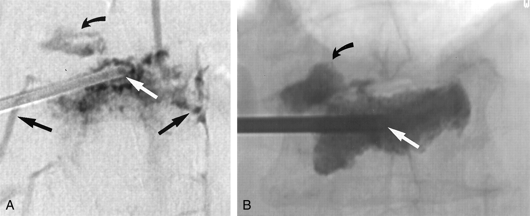
- Fig 1.
Images in a 77-year-old woman with an L1 vertebral body fracture.
A, AP digital subtraction venogram shows the tip of an 11-gauge needle (straight white arrow) at the midline of the vertebral body. Multiple routes of contrast material egress are present, including routes through the superior endplate (curved black arrow) and bilateral paravertebral veins (straight black arrows).
B, AP plain radiograph obtained after vertebroplasty shows that the tip of the needle remains at the midline (white arrow). The needle position has not been altered because direct or rapid venous filling during venography was not observed. Cement fills most of the vertebral body, and it has also extravasated into the superior disk space (black arrow), in the exact same pattern as that predicted by using the venogram in A.
- Fig 2.
Images in a 83-year-old woman with a T6 vertebral body fracture.
A, Lateral digital subtraction venogram shows the tip of the needle (straight white arrow) in the midportion of the vertebral body. Contrast material exits rapidly via a prevertebral vein (straight black arrow) and empties into the hemiazygos vein (curved black arrow). The rapid venous filling warranted an increase in the viscosity of the cement to minimize potential complications.
B, Lateral plain radiograph obtained after vertebroplasty shows that cement fills most of the vertebral body and that is has extravasated into both the superior and inferior endplates (arrows). No evidence for prevertebral cement extravasation is present.


Tables
Criterion Group 1 Group 2 No. of patients 24 24 Men 5 8 Women 19 16 Patient age (y) 74 (52–92) 73 (47–87) Level 42 42 Mid thoracic, T5–T8 7 10 Lower thoracic, T9–T12 8 13 Upper lumbar, L1–L3 17 13 Lower lumbar, L4–L5 10 6 Compression (%) 30.4 (10–70) 33.6 (5–90) Amount of PMMA (mL) 4.65 3.09 Approach Unipediculate 27 37 Bipediculate 15 5 Note.—Data in parentheses are ranges.
Patients Group 1 Group 2 With pain improvement 19 of 20 (95) 21 of 22 (95) Without pain 14 of 20 (70) 14 of 22 (64) With preoperative impaired mobility 11 of 20 (55) 12 of 22 (55) With mobility improvement 11 of 11 (100) 12 of 12 (100) Note.—Follow-up data were available in 20 (83%) of 24 patients in group 1 and in 22 (92%) of 24 patients in group 2. Data in parentheses are percentages.
Postoperative Outcome Mean Points P Value Group 1 Group 2 Pain* 1.3 1.8 .50 Impaired mobility† 0.35 0.27 .43 * Pain was assessed by using an ordinal scale of 0–10, on which 0 represented no pain, and 10 represented the worst pain the patient had ever had.
† Mobility was assessed by using a five-point scale as follows: 0 indicated that the patient was walking without assistance; 1, walking with assistance; 2, wheelchair bound; 3, restricted to sitting in bed; and 4, restricted to lying flat in bed.
Compartment Group 1 Group 2 P Value Epidural 7 10 .33 Paravertebral 7 7 >.99 Prevertebral 3 4 .34 Intervertebral disk space 9 13 .4 Compartment Group 1 Group 2 P Value Epidural (mm) 4.14 2.90 .23 Paravertebral (mm) 4.43 5.86 .58 Prevertebral (mm) 4.67 8.75 .44 Intervertebral disk space (mm3) 617 272 .26
In this issue


Jump to section
Related Articles
Cited By...
- Minimally Invasive Techniques for the Treatment of Osteoporotic Vertebral Fractures
- Percutaneous vertebroplasty and balloon kyphoplasty for the treatment of osteoporotic vertebral compression fractures and osteolytic tumours
- Radiation Dose to the Operator during Vertebroplasty: Prospective Comparison of the Use of 1-cc Syringes versus an Injection Device