
Article Figures & Data
Figures
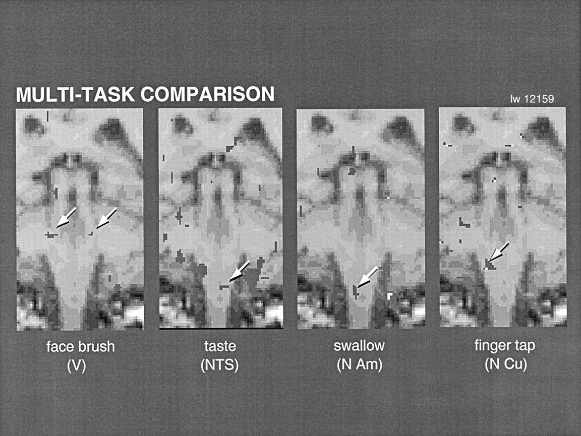
- Fig 1.
Coronal images obtained with motor tasks and sensory stimuli used to activate different brain regions (arrows) in the same individual. These tasks and stimuli correspond to the trigeminal (V), solitary (NTS), ambiguus (N Am), and cuneate (N Cu) nuclei.
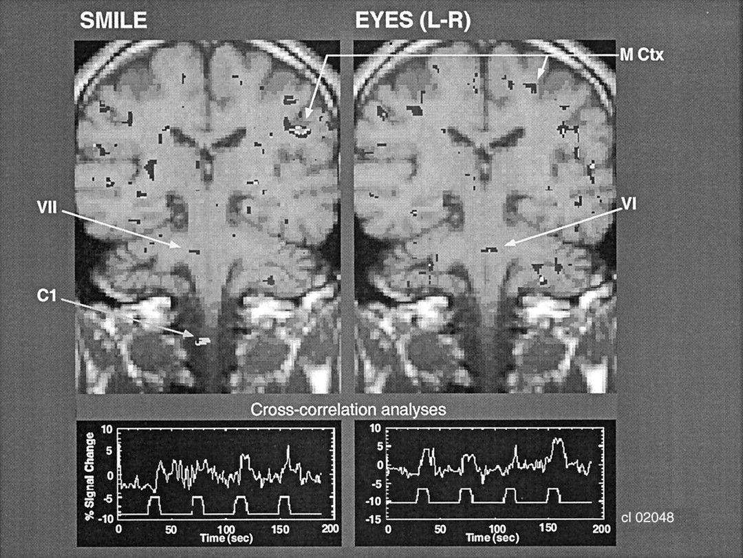
- Fig 2.
Coronal images show that facial and eye movements activate different adjacent brain regions that correspond to the facial and abducens CN nuclei, respectively. Cross-correlation analysis was performed between the onset and termination of the movements and the BOLD signal change in these pontine regions of activation. C1 indicates the C1 level of the spinal cord; M Ctx, motor cortex; VI, CN VI, and VII, CN VII.
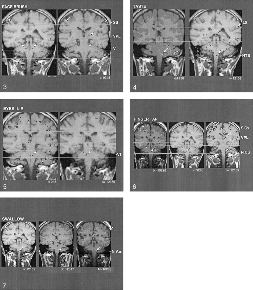
- Fig 3.
Coronal images show the similarity of activation (arrows) produced by brushing the face, which corresponds to the region of the trigeminal main sensory nucleus (V), in two individuals. Note activation of nucleus ventralis posteromedialis (VPL) in one of the individuals and of somatosensory cortex (SS) in both individuals.
- Fig 4.
Coronal images show the similarity of activation (arrows) in the region of the NTS, produced by tasting a sweet-sour-salty-bitter mixture, in two individuals. LS indicates the lateral sulcus.
- Fig 5.
Coronal images shows the similarity of activation (arrows) in the region of the abducens nucleus (VI), produced by voluntary left-right eye movement, in two individuals.
- Fig 6.
Coronal images shows the similarity of activation (arrows) in the region of the nucleus cuneatus (N Cu), produced by tapping the fingers against the thumbs, in three different individuals. Note the activation of the VPL and somatosensory cortex (S Cx) in the finger homuncular region as well.
- Fig 7.
Coronal images shows the similarity of activation (arrows) in the region of the nucleus ambiguus (N Am) in three individuals. These findings were correlated with dry swallowing (ie, Mendelsohn maneuver).
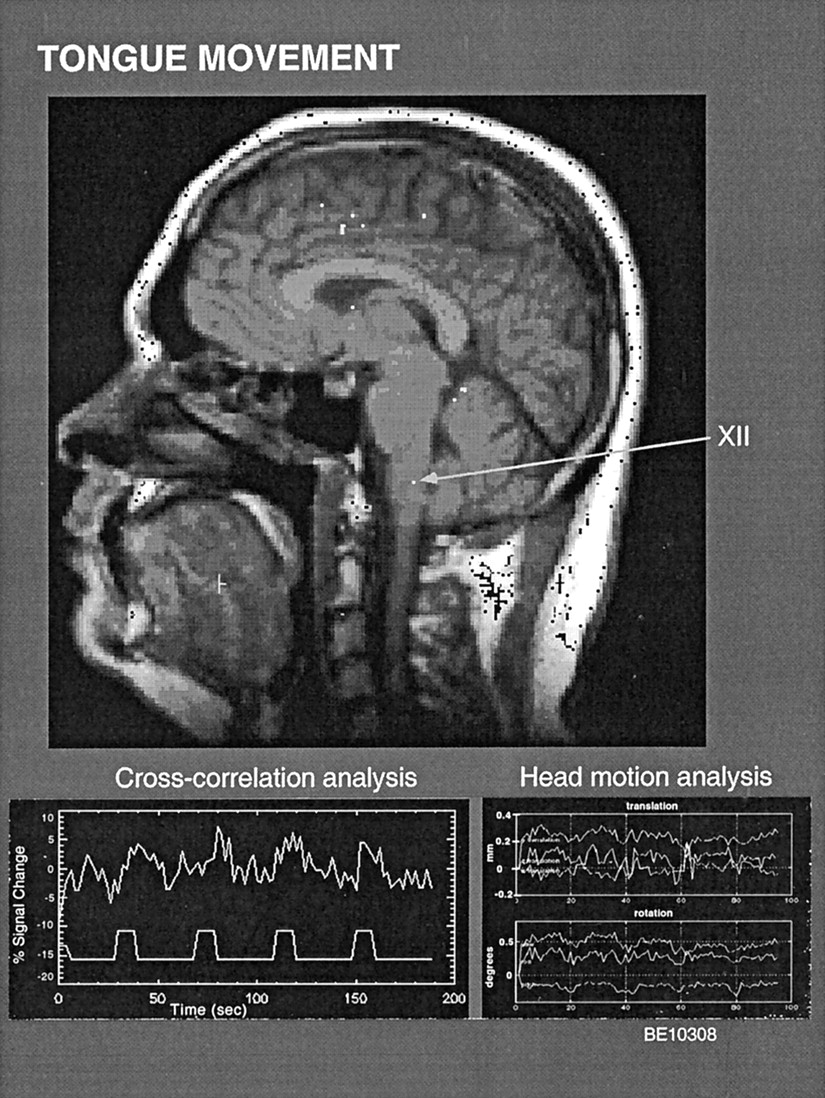
- Fig 8.
Sagittal view in an individual shows activation in the region of the hypoglossal nucleus (XII) and in the muscle of the tongue, in relation to pushing the tongue forcefully and repeatedly against the hard palate. Also shown are the findings from cross-correlation analysis in this region of interest (ROI) and the head movement analysis for this trial. Head movement remained less than 1 mm for the duration of the trial.
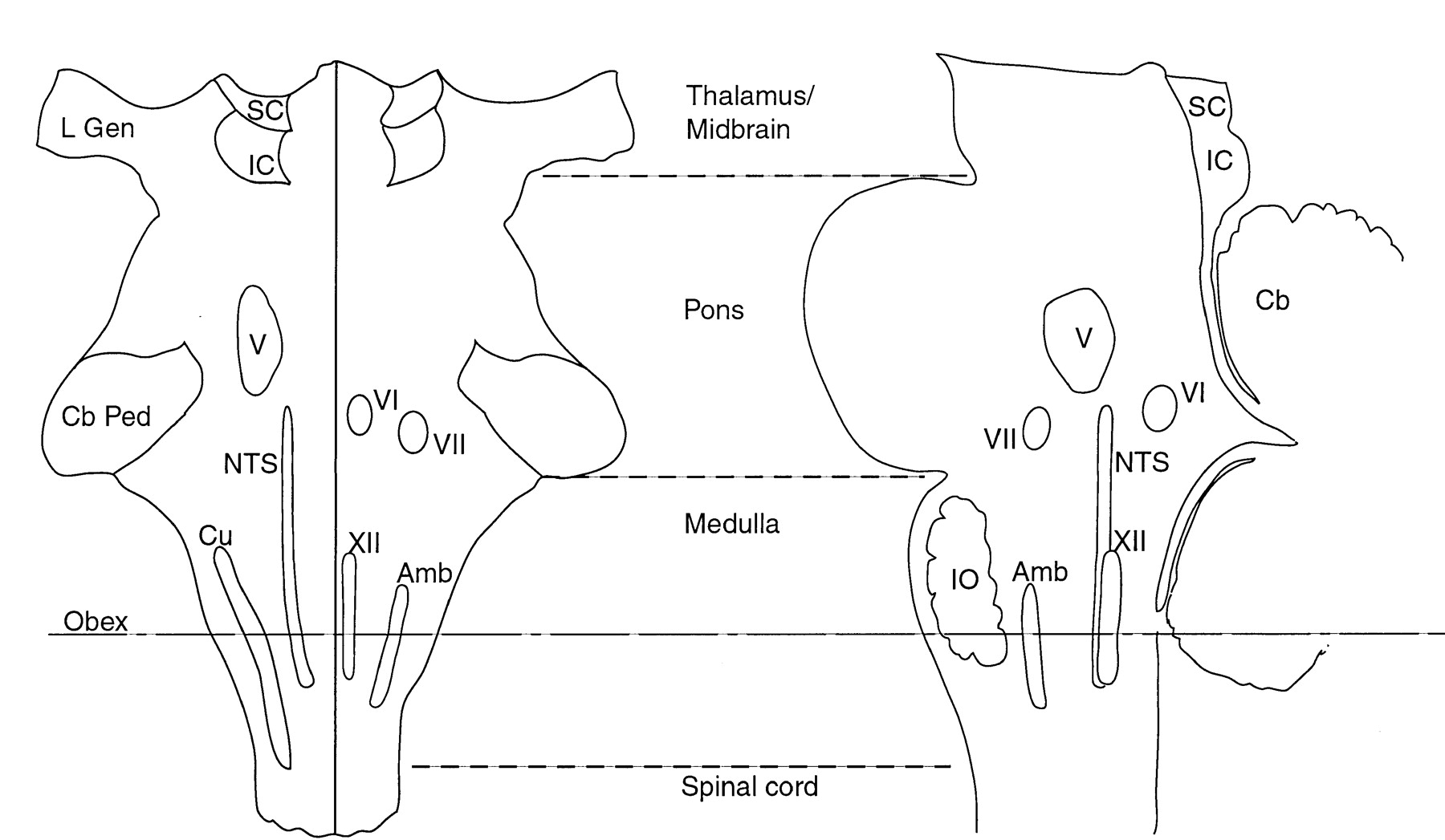
- Fig 9.
Semi-diagrammatic representation of the location of the lower brainstem nuclei analyzed in the present study, for comparison with fMRI findings (adapted from reference 5, with minor adjustments based on data in reference 2). Amb indicates the nucleus ambiguus; Cb, cerebellum; Cb ped, cerebellar peduncle; Cu, nucleus cuneatus; IC, inferior colliculus; IO, inferior olive; L Gen, lateral geniculate nucleus; NTS, NTS; SC, superior colliculus; V, main sensory trigeminal nucleus; VI, abducens nucleus; VII, facial nucleus; and XII, hypoglossal nucleus. For reference, the obex delineates the caudal angle of the rhomboid fossa or fourth ventricle.
- Fig 10.
Comparison of cross-correlation analysis and SPM methods with the same data set. The cross-correlation analysis (top left) revealed that tongue tapping against the hard palate activated cervical spinal cord regions C1, C2. and C3, which provide the motor innervation of the strap muscles that stabilize the tongue. Similarly, activation of C1 and C2 is shown with the SPM method in its nonsmoothed mode (top middle), but not in its smoothed mode (top right). The unsmoothed SPM analysis revealed a more discrete and delimited localization, more similar to the cross-correlation analysis than the results of smoothed SPM analysis. Findings from cross-correlation (bottom left, bottom middle) and head motion analyses (bottom right) are also shown. Note also the cortical activation at the level of the homuncular tongue region (near the lateral sulcus, top middle, top right).


Tables
- TABLE 1:
Summary of responses in specific brain region activation during the performance of the specified paradigms
Paradigm Region Subject Positive Responses Percentage (%) 1* 2* 3 4 5 6 7 Face brushing CN V sensory No/yes No/no No No Yes Yes No 3 33 Jaw clenching CN V motor Yes/yes No/yes No Yes No No Yes 5 56 L-F eye movement CN VI No/yes No/yes No Yes Yes No No 4 44 Smiling, puckering CN VII No/yes Yes/no No No No Yes Yes 4 44 Tasting CN X (NTS) No/no No/no Yes Yes Yes Yes No 4 44 Tongue tapping CN XII No/yes Yes/yes No Yes No Yes Yes 6 67 Swallowing Nucleus ambiguus No/yes Yes/yes Yes Yes Yes No Yes 7 78 Finger tapping Nucleus cuneatus No/yes Yes/yes Yes Yes Yes No No 6 67 Tongue tapping C1, C2, C3 Yes/no No/no No No No Yes Yes 3 33 Note.—Each paradigm was applied in each subject.
* Subjects 1 and 2 underwent testing with all paradigms on 2 days. Data are the results of day 1/results of day 2.
In this issue


Jump to section
Related Articles
Cited By...
- Distinguishing the activity of adjacent somatosensory nuclei within the brainstem using 3T fMRI
- Spatial distribution of hand-grasp motor task activity in spinal cord functional magnetic resonance imaging
- Spatial distribution of hand-grasp motor task activity in spinal cord functional magnetic resonance imaging
- {alpha}-Synuclein Induces Progressive Changes in Brain Microstructure and Sensory-Evoked Brain Function That Precedes Locomotor Decline
- Spinal Cord-Midbrain Functional Connectivity Is Related to Perceived Pain Intensity: A Combined Spino-Cortical fMRI Study
- Tactile Sensory and Pain Networks in the Human Spinal Cord and Brain Stem Mapped by Means of Functional MR Imaging
- Abnormal pontine activation in pathological laughing as shown by functional magnetic resonance imaging.
- A Comparison of Visceral and Somatic Pain Processing in the Human Brainstem Using Functional Magnetic Resonance Imaging