Advertisement

Main menu

  • Home
  • Content
    • Current Issue
    • Accepted Manuscripts
    • Article Preview
    • Past Issue Archive
    • Video Articles
    • AJNR Case Collection
    • Case of the Week Archive
    • Case of the Month Archive
    • Classic Case Archive
  • Special Collections
    • AJNR Awards
    • ASNR Foundation Special Collection
    • Most Impactful AJNR Articles
    • Photon-Counting CT
    • Spinal CSF Leak Articles (Jan 2020-June 2024)
  • Multimedia
    • AJNR Podcasts
    • AJNR SCANtastic
    • Trainee Corner
    • MRI Safety Corner
    • Imaging Protocols
  • For Authors
    • Submit a Manuscript
    • Submit a Video Article
    • Submit an eLetter to the Editor/Response
    • Manuscript Submission Guidelines
    • Statistical Tips
    • Fast Publishing of Accepted Manuscripts
    • Graphical Abstract Preparation
    • Imaging Protocol Submission
    • Author Policies
  • About Us
    • About AJNR
    • Editorial Board
    • Editorial Board Alumni
  • More
    • Become a Reviewer/Academy of Reviewers
    • Subscribers
    • Permissions
    • Alerts
    • Feedback
    • Advertisers
    • ASNR Home

User menu

  • Alerts
  • Log in

Search

  • Advanced search
American Journal of Neuroradiology

American Journal of Neuroradiology

ASHNR American Society of Functional Neuroradiology ASHNR American Society of Pediatric Neuroradiology ASSR
  • Alerts
  • Log in

Advanced Search

  • Home
  • Content
    • Current Issue
    • Accepted Manuscripts
    • Article Preview
    • Past Issue Archive
    • Video Articles
    • AJNR Case Collection
    • Case of the Week Archive
    • Case of the Month Archive
    • Classic Case Archive
  • Special Collections
    • AJNR Awards
    • ASNR Foundation Special Collection
    • Most Impactful AJNR Articles
    • Photon-Counting CT
    • Spinal CSF Leak Articles (Jan 2020-June 2024)
  • Multimedia
    • AJNR Podcasts
    • AJNR SCANtastic
    • Trainee Corner
    • MRI Safety Corner
    • Imaging Protocols
  • For Authors
    • Submit a Manuscript
    • Submit a Video Article
    • Submit an eLetter to the Editor/Response
    • Manuscript Submission Guidelines
    • Statistical Tips
    • Fast Publishing of Accepted Manuscripts
    • Graphical Abstract Preparation
    • Imaging Protocol Submission
    • Author Policies
  • About Us
    • About AJNR
    • Editorial Board
    • Editorial Board Alumni
  • More
    • Become a Reviewer/Academy of Reviewers
    • Subscribers
    • Permissions
    • Alerts
    • Feedback
    • Advertisers
    • ASNR Home
  • Follow AJNR on Twitter
  • Visit AJNR on Facebook
  • Follow AJNR on Instagram
  • Join AJNR on LinkedIn
  • RSS Feeds

AJNR is seeking candidates for the AJNR Podcast Editor. Read the position description.

OtherHEAD AND NECK

The Ascending Pharyngeal Artery: Branches, Anastomoses, and Clinical Significance

Lotfi Hacein-Bey, David L. Daniels, John L. Ulmer, Leighton P. Mark, Michelle M. Smith, James M. Strottmann, Douglas Brown, Glenn A. Meyer and Phillip A. Wackym
American Journal of Neuroradiology August 2002, 23 (7) 1246-1256;
Lotfi Hacein-Bey
  • Find this author on Google Scholar
  • Find this author on PubMed
  • Search for this author on this site
David L. Daniels
  • Find this author on Google Scholar
  • Find this author on PubMed
  • Search for this author on this site
John L. Ulmer
  • Find this author on Google Scholar
  • Find this author on PubMed
  • Search for this author on this site
Leighton P. Mark
  • Find this author on Google Scholar
  • Find this author on PubMed
  • Search for this author on this site
Michelle M. Smith
  • Find this author on Google Scholar
  • Find this author on PubMed
  • Search for this author on this site
James M. Strottmann
  • Find this author on Google Scholar
  • Find this author on PubMed
  • Search for this author on this site
Douglas Brown
  • Find this author on Google Scholar
  • Find this author on PubMed
  • Search for this author on this site
Glenn A. Meyer
  • Find this author on Google Scholar
  • Find this author on PubMed
  • Search for this author on this site
Phillip A. Wackym
  • Find this author on Google Scholar
  • Find this author on PubMed
  • Search for this author on this site
  • Article
  • Figures & Data
  • Info & Metrics
  • Responses
  • References
  • PDF
Loading

Article Figures & Data

Figures

  • Tables
  • Fig 1.
    • Download figure
    • Open in new tab
    • Download powerpoint
    Fig 1.

    A and B, Anteroposterior (A) and lateral (B) angiograms depict rare instances of the ascending pharyngeal artery arising from the internal carotid artery.

  • Fig 2.
    • Download figure
    • Open in new tab
    • Download powerpoint
    Fig 2.

    Diagram of the two major trunks of the ascending pharyngeal artery: anteriorly, the extracranial pharyngeal trunk; posteriorly, the neuromeningeal trunk, which is intracranial and enters the posterior fossa through the foramen magnum.
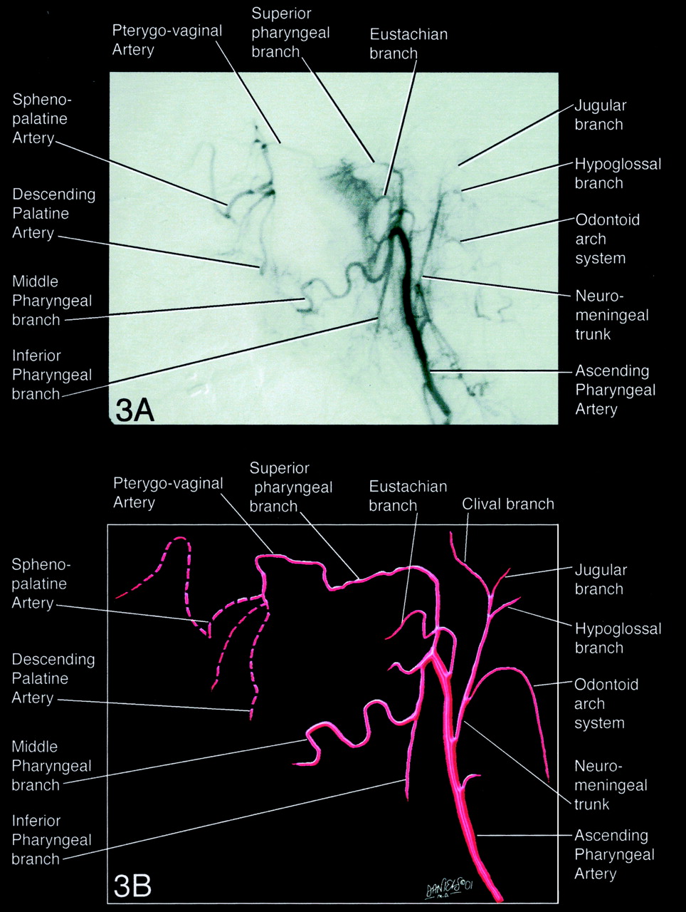
  • Fig 3.
    • Download figure
    • Open in new tab
    • Download powerpoint
    Fig 3.

    Angiogram (A) and diagram (B) of the superior, middle, and inferior pharyngeal branches supplying the pharyngeal submucosal spaces.

  • Fig 4.
    • Download figure
    • Open in new tab
    • Download powerpoint
    Fig 4.

    Diagram of the lateral view of the hypoglossal and jugular branches, the two main divisions of the neuromeningeal trunk. Top, superior view; bottom, inferior view; left, anterior view; right, posterior view.

  • Fig 5.
    • Download figure
    • Open in new tab
    • Download powerpoint
    Fig 5.

    View from above the hypoglossal and jugular branches. Top, superior view; bottom, inferior view; left, anterior view; right, posterior view.

  • Fig 6.
    • Download figure
    • Open in new tab
    • Download powerpoint
    Fig 6.

    Angiogram (A) and diagram (B) of the odontoid arch system that gives rise to several branches that supply the first, second and third cervical roots and the periosteum of the spinous processes and pedicles. Top, superior view; bottom, inferior view; left, anterior view; right, posterior view.

  • Fig 7.
    • Download figure
    • Open in new tab
    • Download powerpoint
    Fig 7.

    Angiogram (A) and diagram (B) of the inferior tympanic artery arising as a separate branch between the pharyngeal and neuropharyngeal trunks. Top, superior view; bottom, inferior view; left, anterior view; right, posterior view.

  • Fig 8.
    • Download figure
    • Open in new tab
    • Download powerpoint
    Fig 8.

    Diagram depicting ascending pharyngeal artery anastomoses.
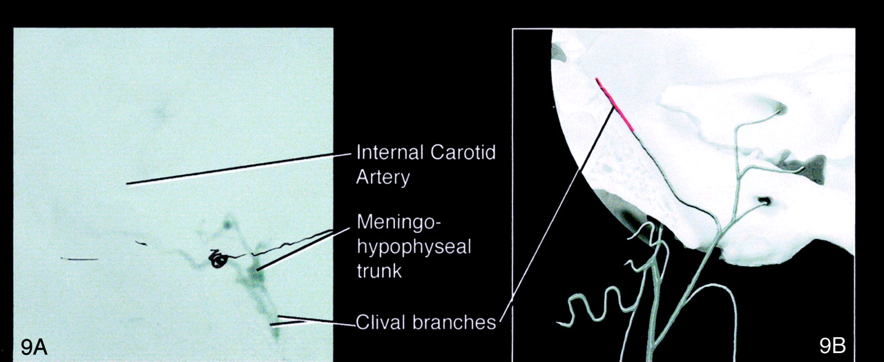
  • Fig 9.
    • Download figure
    • Open in new tab
    • Download powerpoint
    Fig 9.

    Angiogram (A) and diagram (B) of the ascending pharyngeal artery communication with the internal carotid artery to the lateral clival branch of the jugular artery.

  • Fig 10.
    • Download figure
    • Open in new tab
    • Download powerpoint
    Fig 10.

    Angiogram (A) and diagram (B) of the ascending pharyngeal artery anastomoses to the vertebral artery via the hypoglossal branch and musculospinal branch. Top, superior view; bottom, inferior view; left, anterior view; right, posterior view.

  • Fig 11.
    • Download figure
    • Open in new tab
    • Download powerpoint
    Fig 11.

    Angiogram (A) and diagram (B) of anastomoses to the occipital artery via the odontoid arch. Top, superior view; bottom, inferior view; left, anterior view; right, posterior view.
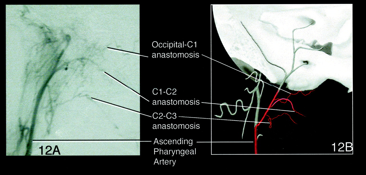
  • Fig 12.
    • Download figure
    • Open in new tab
    • Download powerpoint
    Fig 12.

    Angiogram (A) and diagram (B) of C1-C2 anastomoses to the occipital artery via the common trunk. Top, superior view; bottom, inferior view; left, anterior view; right, posterior view.

  • Fig 13.
    • Download figure
    • Open in new tab
    • Download powerpoint
    Fig 13.

    Angiogram (A) and diagram (B) of anastomoses via the pterygovaginal artery to the accessory meningeal artery. Top, superior view; bottom, inferior view; left, anterior view; right, posterior view.

  • Fig 14.
    • Download figure
    • Open in new tab
    • Download powerpoint
    Fig 14.

    Angiogram (A) and diagram (B) of the ascending pharyngeal artery supplying a meningioma. Top, superior view; bottom, inferior view; left, anterior view; right, posterior view.

  • Fig 15.
    • Download figure
    • Open in new tab
    • Download powerpoint
    Fig 15.

    Angiogram (A) and diagram (B) of the ascending pharyngeal artery supplying a glomus jugulare tumor. Top, superior view; bottom, inferior view; left, anterior view; right, posterior view.

Tables

  • Figures
    • View popup
    TABLE 1:

    Ascending pharyngeal artery anastomoses and foramina

    BranchForamenAnastomoses
    Middle pharyngeal arteryDescending palatine artery (IMA)
    Accessory meningeal artery (IMA)
    Superior pharyngeal arteryForamen lacerumInferolateral trunk to ICA
    Clival branches to meningohypophyseal trunk to ICA
    Pterygovaginal canalPterygovaginal artery to accessory meningeal artery (IMA)
    Foramen lacerumRecurrent artery of the foramen lacerum to ICA
    Inferior tympanic arteryPetrosquamosal branch of middle meningeal artery
    Caroticotympanic artery to ICA
    Stylomastoid artery
    Hypoglossal branchHypoglossal canalVertebral artery
    Jugular branchJugular foramenLateral clival artery to ICA
    Musculospinal branchForamen magnumVertebral artery
    Odontoid arch systemForamen magnumOccipital artery
    • Note.—IMA signifies internal maxillary artery; ICA, internal carotid artery.

PreviousNext
Back to top

In this issue

American Journal of Neuroradiology: 23 (7)
American Journal of Neuroradiology
Vol. 23, Issue 7
1 Aug 2002
  • Table of Contents
  • Index by author
Advertisement
Print
Download PDF
Email Article

Thank you for your interest in spreading the word on American Journal of Neuroradiology.

NOTE: We only request your email address so that the person you are recommending the page to knows that you wanted them to see it, and that it is not junk mail. We do not capture any email address.

Enter multiple addresses on separate lines or separate them with commas.
The Ascending Pharyngeal Artery: Branches, Anastomoses, and Clinical Significance
(Your Name) has sent you a message from American Journal of Neuroradiology
(Your Name) thought you would like to see the American Journal of Neuroradiology web site.
CAPTCHA
This question is for testing whether or not you are a human visitor and to prevent automated spam submissions.
Cite this article
Lotfi Hacein-Bey, David L. Daniels, John L. Ulmer, Leighton P. Mark, Michelle M. Smith, James M. Strottmann, Douglas Brown, Glenn A. Meyer, Phillip A. Wackym
The Ascending Pharyngeal Artery: Branches, Anastomoses, and Clinical Significance
American Journal of Neuroradiology Aug 2002, 23 (7) 1246-1256;

Citation Manager Formats

  • BibTeX
  • Bookends
  • EasyBib
  • EndNote (tagged)
  • EndNote 8 (xml)
  • Medlars
  • Mendeley
  • Papers
  • RefWorks Tagged
  • Ref Manager
  • RIS
  • Zotero
0 Responses
Respond to this article
Share
Bookmark this article
The Ascending Pharyngeal Artery: Branches, Anastomoses, and Clinical Significance
Lotfi Hacein-Bey, David L. Daniels, John L. Ulmer, Leighton P. Mark, Michelle M. Smith, James M. Strottmann, Douglas Brown, Glenn A. Meyer, Phillip A. Wackym
American Journal of Neuroradiology Aug 2002, 23 (7) 1246-1256;
del.icio.us logo Twitter logo Facebook logo Mendeley logo
  • Tweet Widget
  • Facebook Like
  • Google Plus One

Jump to section

  • Article
    • Abstract
    • Anatomy
    • Clinical Relevance of the Ascending Pharyngeal Artery
    • Neurovascular Research Models
    • Conclusion
    • Acknowledgments
    • References
  • Figures & Data
  • Info & Metrics
  • Responses
  • References
  • PDF

Related Articles

  • No related articles found.
  • PubMed
  • Google Scholar

Cited By...

  • Endovascular treatment strategy, technique, and outcomes for dural arteriovenous fistulas of the marginal sinus region
  • Anatomical Structures, Cell Types, and Biomarkers Tables Plus 3D Reference Organs in Support of a Human Reference Atlas
  • Variant ascending pharyngeal artery maintaining flow in a subocclusive internal carotid artery
  • Transvenous coil embolization with intra-operative cone beam CT assistance in the treatment of hypoglossal canal dural arteriovenous fistulae
  • The road less traveled: transarterial embolization of dural arteriovenous fistulas via the ascending pharyngeal artery
  • Onyx embolization of dural arteriovenous fistulas of the cavernous sinus through the superior pharyngeal branch of the ascending pharyngeal artery
  • Onyx embolization of dural arteriovenous fistulas of the cavernous sinus through the superior pharyngeal branch of the ascending pharyngeal artery
  • Severe epistaxis after nasogastric tube insertion requiring arterial embolisation
  • Balloon-augmented Onyx embolization of a dural arteriovenous fistula arising from the neuromeningeal trunk of the ascending pharyngeal artery: technical report
  • Dangerous Extracranial-Intracranial Anastomoses and Supply to the Cranial Nerves: Vessels the Neurointerventionalist Needs to Know
  • Crossref
  • Google Scholar

This article has not yet been cited by articles in journals that are participating in Crossref Cited-by Linking.

More in this TOC Section

  • Correlation of Apparent Diffusion Coefficient at 3T with Prognostic Parameters of Retinoblastoma
  • Parathyroid Lesions: Characterization with Dual-Phase Arterial and Venous Enhanced CT of the Neck
  • Kimura Disease: CT and MR Imaging Findings
Show more HEAD AND NECK

Similar Articles

Advertisement

Indexed Content

  • Current Issue
  • Accepted Manuscripts
  • Article Preview
  • Past Issues
  • Editorials
  • Editor's Choice
  • Fellows' Journal Club
  • Letters to the Editor
  • Video Articles

Cases

  • Case Collection
  • Archive - Case of the Week
  • Archive - Case of the Month
  • Archive - Classic Case

Special Collections

  • AJNR Awards
  • ASNR Foundation Special Collection
  • Most Impactful AJNR Articles
  • Photon-Counting CT
  • Spinal CSF Leak Articles (Jan 2020-June 2024)

More from AJNR

  • Trainee Corner
  • Imaging Protocols
  • MRI Safety Corner

Multimedia

  • AJNR Podcasts
  • AJNR Scantastics

Resources

  • Turnaround Time
  • Submit a Manuscript
  • Submit a Video Article
  • Submit an eLetter to the Editor/Response
  • Manuscript Submission Guidelines
  • Statistical Tips
  • Fast Publishing of Accepted Manuscripts
  • Graphical Abstract Preparation
  • Imaging Protocol Submission
  • Evidence-Based Medicine Level Guide
  • Publishing Checklists
  • Author Policies
  • Become a Reviewer/Academy of Reviewers
  • News and Updates

About Us

  • About AJNR
  • Editorial Board
  • Editorial Board Alumni
  • Alerts
  • Permissions
  • Not an AJNR Subscriber? Join Now
  • Advertise with Us
  • Librarian Resources
  • Feedback
  • Terms and Conditions
  • AJNR Editorial Board Alumni

American Society of Neuroradiology

  • Not an ASNR Member? Join Now

© 2025 by the American Society of Neuroradiology All rights, including for text and data mining, AI training, and similar technologies, are reserved.
Print ISSN: 0195-6108 Online ISSN: 1936-959X

Powered by HighWire