Advertisement

Main menu

  • Home
  • Content
    • Current Issue
    • Accepted Manuscripts
    • Article Preview
    • Past Issue Archive
    • Video Articles
    • AJNR Case Collection
    • Case of the Week Archive
    • Case of the Month Archive
    • Classic Case Archive
  • Special Collections
    • AJNR Awards
    • ASNR Foundation Special Collection
    • Most Impactful AJNR Articles
    • Photon-Counting CT
    • Spinal CSF Leak Articles (Jan 2020-June 2024)
  • Multimedia
    • AJNR Podcasts
    • AJNR SCANtastic
    • Trainee Corner
    • MRI Safety Corner
    • Imaging Protocols
  • For Authors
    • Submit a Manuscript
    • Submit a Video Article
    • Submit an eLetter to the Editor/Response
    • Manuscript Submission Guidelines
    • Statistical Tips
    • Fast Publishing of Accepted Manuscripts
    • Graphical Abstract Preparation
    • Imaging Protocol Submission
    • Author Policies
  • About Us
    • About AJNR
    • Editorial Board
    • Editorial Board Alumni
  • More
    • Become a Reviewer/Academy of Reviewers
    • Subscribers
    • Permissions
    • Alerts
    • Feedback
    • Advertisers
    • ASNR Home

User menu

  • Alerts
  • Log in

Search

  • Advanced search
American Journal of Neuroradiology

American Journal of Neuroradiology

ASHNR American Society of Functional Neuroradiology ASHNR American Society of Pediatric Neuroradiology ASSR
  • Alerts
  • Log in

Advanced Search

  • Home
  • Content
    • Current Issue
    • Accepted Manuscripts
    • Article Preview
    • Past Issue Archive
    • Video Articles
    • AJNR Case Collection
    • Case of the Week Archive
    • Case of the Month Archive
    • Classic Case Archive
  • Special Collections
    • AJNR Awards
    • ASNR Foundation Special Collection
    • Most Impactful AJNR Articles
    • Photon-Counting CT
    • Spinal CSF Leak Articles (Jan 2020-June 2024)
  • Multimedia
    • AJNR Podcasts
    • AJNR SCANtastic
    • Trainee Corner
    • MRI Safety Corner
    • Imaging Protocols
  • For Authors
    • Submit a Manuscript
    • Submit a Video Article
    • Submit an eLetter to the Editor/Response
    • Manuscript Submission Guidelines
    • Statistical Tips
    • Fast Publishing of Accepted Manuscripts
    • Graphical Abstract Preparation
    • Imaging Protocol Submission
    • Author Policies
  • About Us
    • About AJNR
    • Editorial Board
    • Editorial Board Alumni
  • More
    • Become a Reviewer/Academy of Reviewers
    • Subscribers
    • Permissions
    • Alerts
    • Feedback
    • Advertisers
    • ASNR Home
  • Follow AJNR on Twitter
  • Visit AJNR on Facebook
  • Follow AJNR on Instagram
  • Join AJNR on LinkedIn
  • RSS Feeds

AJNR is seeking candidates for the AJNR Podcast Editor. Read the position description.

OtherBRAIN

Serial Proton MR Spectroscopy and Diffusion Imaging Findings in HIV-Related Herpes Simplex Encephalitis

Philipp G. Sämann, Jürgen Schlegel, Georg Müller, Franz Prantl, Christoph Emminger and Dorothee P. Auer
American Journal of Neuroradiology November 2003, 24 (10) 2015-2019;
Philipp G. Sämann
  • Find this author on Google Scholar
  • Find this author on PubMed
  • Search for this author on this site
Jürgen Schlegel
  • Find this author on Google Scholar
  • Find this author on PubMed
  • Search for this author on this site
Georg Müller
  • Find this author on Google Scholar
  • Find this author on PubMed
  • Search for this author on this site
Franz Prantl
  • Find this author on Google Scholar
  • Find this author on PubMed
  • Search for this author on this site
Christoph Emminger
  • Find this author on Google Scholar
  • Find this author on PubMed
  • Search for this author on this site
Dorothee P. Auer
  • Find this author on Google Scholar
  • Find this author on PubMed
  • Search for this author on this site
  • Article
  • Figures & Data
  • Info & Metrics
  • Responses
  • References
  • PDF
Loading

Article Figures & Data

Figures

  • Tables
  • Fig 1.
    • Download figure
    • Open in new tab
    • Download powerpoint
    Fig 1.

    A and B, Axial and coronal T2-weighted images (1.5 T, TR/TE = 3800/84 and 3500/75, respectively) show extensive areas of abnormally high signal intensity in the splenium and the perispenial, cingulate, and the bilateral perisplenial white matter. Note sparing of the hippocampi and the temporal and insular cortices. C–F, Diffusion-weighted images (upper row [C and E]) and corresponding apparent diffusion coefficient false-color maps (lower row) of week 8 (D) and week 26 (F) after disease onset. Note that the CSF signal intensity is nulled. C and D, Lesions on diffusion-weighted images 4 weeks after the first MR study (C) show hypointense central area with a hyperintense rim (arrows) and high regional mean diffusivity. E and F, Note resolution of diffusion-weighted hyperintensities and peripheral normalization of regional mean diffusivity, indicating irreversible necrosis and gliosis formation in areas with regional mean diffusivity exceeding about 1.4 ×10⁻³ mm2/s, whereas initially hyperintense areas on diffusion-weighted images, mainly representing T2 shine-through, without corresponding reduction of regional mean diffusivity reflect potentially reversible inflammatory changes. (Image acquisition: TR/TE = 2200/120, tetrahedral gradient acquisition scheme [15] with b values of 330, 798, and 1320 s/mm2)

  • Fig 2.
    • Download figure
    • Open in new tab
    • Download powerpoint
    Fig 2.

    A–C, 1H-MR spectra (automated stimulated-echo acquisition mode; TR/TM/TE = 3000/13.7/30, LCModel fit) of the right perisplenial lesion (voxel volume 7.2–8.0 mL; cf voxel position in 1A) in the 4th (A), 8th (B), and 26th (C) week after symptom onset. D, Note the time course indicating partial resolution of the acute inflammatory changes (decrease of Cho, Lac, and Lip), only incomplete recovery of NAA but evolving gliotic reaction (increase of mI). Mean σ ± SD of unaffected parietal white matter from 15 male HIV-positive subjects (mean age, 40.9; years; range, 28–63 years) are given on the left for comparison. Metabolite concentrations were estimated by using LCModel (Provencher S., http://s-provencher.com).
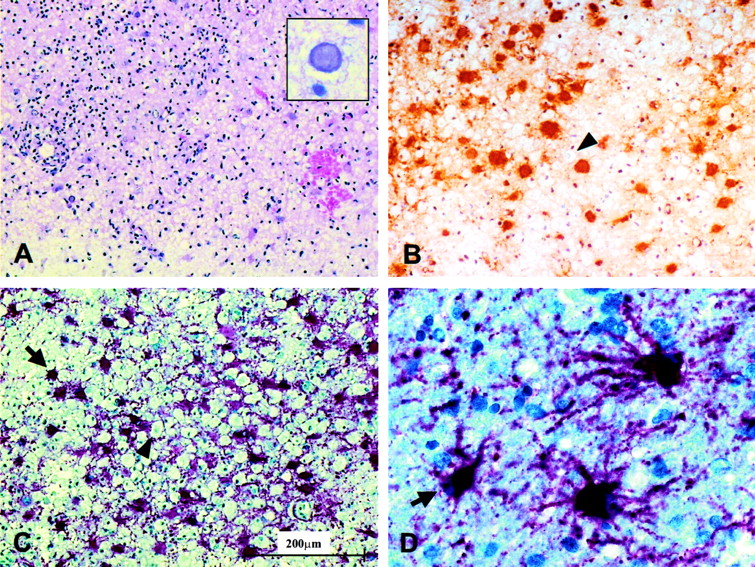
  • Fig 3.
    • Download figure
    • Open in new tab
    • Download powerpoint
    Fig 3.

    Histopathologic examination of the biopsies of the left cingulate gyrus revealed the morphology of a hemorrhagic-necrotizing inflammation (A, [H&E staining]) with multiple cells containing Cowdry type A intranuclear inclusions (insert). Immunohistochemical detection of HSV antigen is depicted in B. By using an antibody directed against the glial fibrillary acid protein, a prominent gliosis with multiple reactive astrocytes (C and D) intermixed with macrophages (B and C) could be detected. Astrocytes and macrophages are marked by arrows and arrowheads, respectively.

Tables

  • Figures
  • Regional Mean Diffusivity (D) in the 8th and 26th Week

    RegionMean diffusivity D (× 10−3 mm2/s; mean ± SD [% relative to control])
    8th week26th week
    Right frontal white matter (control)0.672 ± .094 (100)0.722 ± .089 (100)
    Left perisplenial1.258 ± .214 (187)1.177 ± .108 (163)
    Right perisplenial1.084 ± 0.283 (161)1.232 ± .215 (171)
    Left cingulate1.038 ± .217 (154)0.995 ± .141 (138)
    Left parahippocampal0.920 ± .158 (137)0.771 ± .110 (107)
    Right parahippocampal0.843 ± .121 (125)0.788 ± .101 (109)
    • Note—Numbers in parentheses are percentages.

PreviousNext
Back to top

In this issue

American Journal of Neuroradiology: 24 (10)
American Journal of Neuroradiology
Vol. 24, Issue 10
1 Nov 2003
  • Table of Contents
  • Index by author
Advertisement
Print
Download PDF
Email Article

Thank you for your interest in spreading the word on American Journal of Neuroradiology.

NOTE: We only request your email address so that the person you are recommending the page to knows that you wanted them to see it, and that it is not junk mail. We do not capture any email address.

Enter multiple addresses on separate lines or separate them with commas.
Serial Proton MR Spectroscopy and Diffusion Imaging Findings in HIV-Related Herpes Simplex Encephalitis
(Your Name) has sent you a message from American Journal of Neuroradiology
(Your Name) thought you would like to see the American Journal of Neuroradiology web site.
CAPTCHA
This question is for testing whether or not you are a human visitor and to prevent automated spam submissions.
Cite this article
Philipp G. Sämann, Jürgen Schlegel, Georg Müller, Franz Prantl, Christoph Emminger, Dorothee P. Auer
Serial Proton MR Spectroscopy and Diffusion Imaging Findings in HIV-Related Herpes Simplex Encephalitis
American Journal of Neuroradiology Nov 2003, 24 (10) 2015-2019;

Citation Manager Formats

  • BibTeX
  • Bookends
  • EasyBib
  • EndNote (tagged)
  • EndNote 8 (xml)
  • Medlars
  • Mendeley
  • Papers
  • RefWorks Tagged
  • Ref Manager
  • RIS
  • Zotero
0 Responses
Respond to this article
Share
Bookmark this article
Serial Proton MR Spectroscopy and Diffusion Imaging Findings in HIV-Related Herpes Simplex Encephalitis
Philipp G. Sämann, Jürgen Schlegel, Georg Müller, Franz Prantl, Christoph Emminger, Dorothee P. Auer
American Journal of Neuroradiology Nov 2003, 24 (10) 2015-2019;
del.icio.us logo Twitter logo Facebook logo Mendeley logo
  • Tweet Widget
  • Facebook Like
  • Google Plus One

Jump to section

  • Article
    • Abstract
    • Case Report
    • Discussion
    • Conclusion
    • References
  • Figures & Data
  • Info & Metrics
  • Responses
  • References
  • PDF

Related Articles

  • No related articles found.
  • PubMed
  • Google Scholar

Cited By...

  • No citing articles found.
  • Crossref
  • Google Scholar

This article has not yet been cited by articles in journals that are participating in Crossref Cited-by Linking.

More in this TOC Section

  • Optimal MRI Sequence for Identifying Occlusion Location in Acute Stroke: Which Value of Time-Resolved Contrast-Enhanced MRA?
  • Evaluating the Effects of White Matter Multiple Sclerosis Lesions on the Volume Estimation of 6 Brain Tissue Segmentation Methods
  • Quiet PROPELLER MRI Techniques Match the Quality of Conventional PROPELLER Brain Imaging Techniques
Show more BRAIN

Similar Articles

Advertisement

Indexed Content

  • Current Issue
  • Accepted Manuscripts
  • Article Preview
  • Past Issues
  • Editorials
  • Editor's Choice
  • Fellows' Journal Club
  • Letters to the Editor
  • Video Articles

Cases

  • Case Collection
  • Archive - Case of the Week
  • Archive - Case of the Month
  • Archive - Classic Case

Special Collections

  • AJNR Awards
  • ASNR Foundation Special Collection
  • Most Impactful AJNR Articles
  • Photon-Counting CT
  • Spinal CSF Leak Articles (Jan 2020-June 2024)

More from AJNR

  • Trainee Corner
  • Imaging Protocols
  • MRI Safety Corner

Multimedia

  • AJNR Podcasts
  • AJNR Scantastics

Resources

  • Turnaround Time
  • Submit a Manuscript
  • Submit a Video Article
  • Submit an eLetter to the Editor/Response
  • Manuscript Submission Guidelines
  • Statistical Tips
  • Fast Publishing of Accepted Manuscripts
  • Graphical Abstract Preparation
  • Imaging Protocol Submission
  • Evidence-Based Medicine Level Guide
  • Publishing Checklists
  • Author Policies
  • Become a Reviewer/Academy of Reviewers
  • News and Updates

About Us

  • About AJNR
  • Editorial Board
  • Editorial Board Alumni
  • Alerts
  • Permissions
  • Not an AJNR Subscriber? Join Now
  • Advertise with Us
  • Librarian Resources
  • Feedback
  • Terms and Conditions
  • AJNR Editorial Board Alumni

American Society of Neuroradiology

  • Not an ASNR Member? Join Now

© 2025 by the American Society of Neuroradiology All rights, including for text and data mining, AI training, and similar technologies, are reserved.
Print ISSN: 0195-6108 Online ISSN: 1936-959X

Powered by HighWire