Advertisement

Main menu

  • Home
  • Content
    • Current Issue
    • Accepted Manuscripts
    • Article Preview
    • Past Issue Archive
    • Video Articles
    • AJNR Case Collection
    • Case of the Week Archive
    • Case of the Month Archive
    • Classic Case Archive
  • Special Collections
    • AJNR Awards
    • ASNR Foundation Special Collection
    • Most Impactful AJNR Articles
    • Photon-Counting CT
    • Spinal CSF Leak Articles (Jan 2020-June 2024)
  • Multimedia
    • AJNR Podcasts
    • AJNR SCANtastic
    • Trainee Corner
    • MRI Safety Corner
    • Imaging Protocols
  • For Authors
    • Submit a Manuscript
    • Submit a Video Article
    • Submit an eLetter to the Editor/Response
    • Manuscript Submission Guidelines
    • Statistical Tips
    • Fast Publishing of Accepted Manuscripts
    • Graphical Abstract Preparation
    • Imaging Protocol Submission
    • Author Policies
  • About Us
    • About AJNR
    • Editorial Board
    • Editorial Board Alumni
  • More
    • Become a Reviewer/Academy of Reviewers
    • Subscribers
    • Permissions
    • Alerts
    • Feedback
    • Advertisers
    • ASNR Home

User menu

  • Alerts
  • Log in

Search

  • Advanced search
American Journal of Neuroradiology

American Journal of Neuroradiology

ASHNR American Society of Functional Neuroradiology ASHNR American Society of Pediatric Neuroradiology ASSR
  • Alerts
  • Log in

Advanced Search

  • Home
  • Content
    • Current Issue
    • Accepted Manuscripts
    • Article Preview
    • Past Issue Archive
    • Video Articles
    • AJNR Case Collection
    • Case of the Week Archive
    • Case of the Month Archive
    • Classic Case Archive
  • Special Collections
    • AJNR Awards
    • ASNR Foundation Special Collection
    • Most Impactful AJNR Articles
    • Photon-Counting CT
    • Spinal CSF Leak Articles (Jan 2020-June 2024)
  • Multimedia
    • AJNR Podcasts
    • AJNR SCANtastic
    • Trainee Corner
    • MRI Safety Corner
    • Imaging Protocols
  • For Authors
    • Submit a Manuscript
    • Submit a Video Article
    • Submit an eLetter to the Editor/Response
    • Manuscript Submission Guidelines
    • Statistical Tips
    • Fast Publishing of Accepted Manuscripts
    • Graphical Abstract Preparation
    • Imaging Protocol Submission
    • Author Policies
  • About Us
    • About AJNR
    • Editorial Board
    • Editorial Board Alumni
  • More
    • Become a Reviewer/Academy of Reviewers
    • Subscribers
    • Permissions
    • Alerts
    • Feedback
    • Advertisers
    • ASNR Home
  • Follow AJNR on Twitter
  • Visit AJNR on Facebook
  • Follow AJNR on Instagram
  • Join AJNR on LinkedIn
  • RSS Feeds

AJNR is seeking candidates for the AJNR Podcast Editor. Read the position description.

OtherHEAD AND NECK

MR Imaging Features of Nasopharyngeal Tuberculosis: Report of Three Cases and Literature Review

Ann D. King, Anil T. Ahuja, Gary M.K. Tse, Andrew C.A. van Hasselt and Amy B.W. Chan
American Journal of Neuroradiology February 2003, 24 (2) 279-282;
Ann D. King
  • Find this author on Google Scholar
  • Find this author on PubMed
  • Search for this author on this site
Anil T. Ahuja
  • Find this author on Google Scholar
  • Find this author on PubMed
  • Search for this author on this site
Gary M.K. Tse
  • Find this author on Google Scholar
  • Find this author on PubMed
  • Search for this author on this site
Andrew C.A. van Hasselt
  • Find this author on Google Scholar
  • Find this author on PubMed
  • Search for this author on this site
Amy B.W. Chan
  • Find this author on Google Scholar
  • Find this author on PubMed
  • Search for this author on this site
  • Article
  • Figures & Data
  • Info & Metrics
  • Responses
  • References
  • PDF
Loading

Article Figures & Data

Figures

  • Tables
  • Fig 1.
    • Download figure
    • Open in new tab
    • Download powerpoint
    Fig 1.

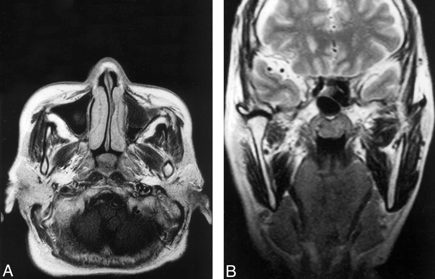
    Images from the case of a 63-year-old woman who presented with right-sided cervical lymphadenopathy (case 1).

    A, Axial contrast-enhanced T1-weighted image (505/12/2 [TR/TE/NEX]) of the nasopharynx shows a discrete moderately enhancing polypoid mass in the central roof and upper posterior wall at the site of the adenoids.

    B, Coronal T2-weighted image (2500/100/3) shows a discrete polypoid mass of intermediate T2 signal intensity in the central roof/upper posterior wall at the site of the adenoids.

  • Fig 2.
    • Download figure
    • Open in new tab
    • Download powerpoint
    Fig 2.

    Images from the case of a 29-year-old woman with bilateral cervical lymphadenopathy (case 2).

    A, Axial contrast-enhanced T1-weighted image (499/12/2) of the nasopharynx shows a discrete moderately enhancing polypoid mass in the central roof and upper posterior wall, with a small focus of necrosis at the site of the adenoids.

    B, Coronal contrast-enhanced T1-weighted image (425/13/2) shows a moderately enhancing polypoid mass in the central roof/upper posterior wall with a small focus of necrosis at the site of the adenoids.

  • Fig 3.
    • Download figure
    • Open in new tab
    • Download powerpoint
    Fig 3.

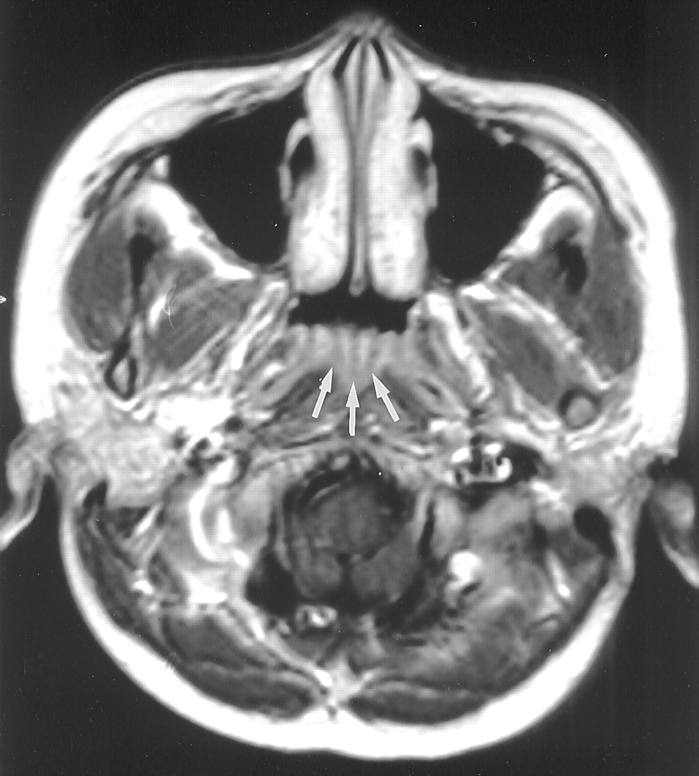
    Image from the case of a 61-year-old man with postnasal drip (case 3). Axial contrast-enhanced T1-weighted image (506/12/2) of the nasopharynx shows mild diffuse mucosal thickening in the left side of the roof of the nasopharynx (arrows).

  • Fig 4.
    • Download figure
    • Open in new tab
    • Download powerpoint
    Fig 4.

    Axial contrast-enhanced T1-weighted image (500/15/2) of a nasopharynx shows the septations in normal lymphoid tissue (arrows).

Tables

  • Figures
  • Literature review of the imaging features of nasopharyngeal tuberculosis

    ReferenceSymptomsRadiologic Features on MR ImagesDiagnosis
    Köktenera (4)Incidental finding of a nasopharyngeal mass on cranial MR image for headacheMucosal thickening in the left fossa of Rosenmüller; no extension outside the nasopharynxPrimary tuberculosis; nasopharyngeal biopsy revealed granulomatous necrosis
    Percodani et al (1)Left lateral pharyngeal pain, hearing loss, nasal obstruction, postnasal dripSoft tissue in the left lateral wall with extension into the longus capitisTuberculosis; nasopharyngeal biopsy revealed giant epithelioid granulomata; mycobacterium tuberculosis isolated on culture
    Chopra et al (2)Nasal obstruction and fullness in both ears.2- to 3-cm polypoid mass in the midline of the superior nasopharynx extending to the left fossa of Rosenmüller; no bone or CNS invasionPrimary tuberculosis; nasopharyngeal biopsy revealed necrotising granulomas; mycobacterium tuberculosis isolated on culture
PreviousNext
Back to top

In this issue

American Journal of Neuroradiology: 24 (2)
American Journal of Neuroradiology
Vol. 24, Issue 2
1 Feb 2003
  • Table of Contents
  • Index by author
Advertisement
Print
Download PDF
Email Article

Thank you for your interest in spreading the word on American Journal of Neuroradiology.

NOTE: We only request your email address so that the person you are recommending the page to knows that you wanted them to see it, and that it is not junk mail. We do not capture any email address.

Enter multiple addresses on separate lines or separate them with commas.
MR Imaging Features of Nasopharyngeal Tuberculosis: Report of Three Cases and Literature Review
(Your Name) has sent you a message from American Journal of Neuroradiology
(Your Name) thought you would like to see the American Journal of Neuroradiology web site.
CAPTCHA
This question is for testing whether or not you are a human visitor and to prevent automated spam submissions.
Cite this article
Ann D. King, Anil T. Ahuja, Gary M.K. Tse, Andrew C.A. van Hasselt, Amy B.W. Chan
MR Imaging Features of Nasopharyngeal Tuberculosis: Report of Three Cases and Literature Review
American Journal of Neuroradiology Feb 2003, 24 (2) 279-282;

Citation Manager Formats

  • BibTeX
  • Bookends
  • EasyBib
  • EndNote (tagged)
  • EndNote 8 (xml)
  • Medlars
  • Mendeley
  • Papers
  • RefWorks Tagged
  • Ref Manager
  • RIS
  • Zotero
0 Responses
Respond to this article
Share
Bookmark this article
MR Imaging Features of Nasopharyngeal Tuberculosis: Report of Three Cases and Literature Review
Ann D. King, Anil T. Ahuja, Gary M.K. Tse, Andrew C.A. van Hasselt, Amy B.W. Chan
American Journal of Neuroradiology Feb 2003, 24 (2) 279-282;
del.icio.us logo Twitter logo Facebook logo Mendeley logo
  • Tweet Widget
  • Facebook Like
  • Google Plus One

Jump to section

  • Article
    • Abstract
    • Case Reports
    • Discussion
    • Conclusion
    • References
  • Figures & Data
  • Info & Metrics
  • Responses
  • References
  • PDF

Related Articles

  • No related articles found.
  • PubMed
  • Google Scholar

Cited By...

  • Tuberculosis of the cavum revealed by acute facial pain
  • Crossref
  • Google Scholar

This article has not yet been cited by articles in journals that are participating in Crossref Cited-by Linking.

More in this TOC Section

  • Correlation of Apparent Diffusion Coefficient at 3T with Prognostic Parameters of Retinoblastoma
  • Parathyroid Lesions: Characterization with Dual-Phase Arterial and Venous Enhanced CT of the Neck
  • Kimura Disease: CT and MR Imaging Findings
Show more HEAD AND NECK

Similar Articles

Advertisement

Indexed Content

  • Current Issue
  • Accepted Manuscripts
  • Article Preview
  • Past Issues
  • Editorials
  • Editor's Choice
  • Fellows' Journal Club
  • Letters to the Editor
  • Video Articles

Cases

  • Case Collection
  • Archive - Case of the Week
  • Archive - Case of the Month
  • Archive - Classic Case

Special Collections

  • AJNR Awards
  • ASNR Foundation Special Collection
  • Most Impactful AJNR Articles
  • Photon-Counting CT
  • Spinal CSF Leak Articles (Jan 2020-June 2024)

More from AJNR

  • Trainee Corner
  • Imaging Protocols
  • MRI Safety Corner

Multimedia

  • AJNR Podcasts
  • AJNR Scantastics

Resources

  • Turnaround Time
  • Submit a Manuscript
  • Submit a Video Article
  • Submit an eLetter to the Editor/Response
  • Manuscript Submission Guidelines
  • Statistical Tips
  • Fast Publishing of Accepted Manuscripts
  • Graphical Abstract Preparation
  • Imaging Protocol Submission
  • Evidence-Based Medicine Level Guide
  • Publishing Checklists
  • Author Policies
  • Become a Reviewer/Academy of Reviewers
  • News and Updates

About Us

  • About AJNR
  • Editorial Board
  • Editorial Board Alumni
  • Alerts
  • Permissions
  • Not an AJNR Subscriber? Join Now
  • Advertise with Us
  • Librarian Resources
  • Feedback
  • Terms and Conditions
  • AJNR Editorial Board Alumni

American Society of Neuroradiology

  • Not an ASNR Member? Join Now

© 2025 by the American Society of Neuroradiology All rights, including for text and data mining, AI training, and similar technologies, are reserved.
Print ISSN: 0195-6108 Online ISSN: 1936-959X

Powered by HighWire