Advertisement

Main menu

  • Home
  • Content
    • Current Issue
    • Accepted Manuscripts
    • Article Preview
    • Past Issue Archive
    • Video Articles
    • AJNR Case Collection
    • Case of the Week Archive
    • Case of the Month Archive
    • Classic Case Archive
  • Special Collections
    • AJNR Awards
    • ASNR Foundation Special Collection
    • Most Impactful AJNR Articles
    • Photon-Counting CT
    • Spinal CSF Leak Articles (Jan 2020-June 2024)
  • Multimedia
    • AJNR Podcasts
    • AJNR SCANtastic
    • Trainee Corner
    • MRI Safety Corner
    • Imaging Protocols
  • For Authors
    • Submit a Manuscript
    • Submit a Video Article
    • Submit an eLetter to the Editor/Response
    • Manuscript Submission Guidelines
    • Statistical Tips
    • Fast Publishing of Accepted Manuscripts
    • Graphical Abstract Preparation
    • Imaging Protocol Submission
    • Author Policies
  • About Us
    • About AJNR
    • Editorial Board
    • Editorial Board Alumni
  • More
    • Become a Reviewer/Academy of Reviewers
    • Subscribers
    • Permissions
    • Alerts
    • Feedback
    • Advertisers
    • ASNR Home

User menu

  • Alerts
  • Log in

Search

  • Advanced search
American Journal of Neuroradiology

American Journal of Neuroradiology

ASHNR American Society of Functional Neuroradiology ASHNR American Society of Pediatric Neuroradiology ASSR
  • Alerts
  • Log in

Advanced Search

  • Home
  • Content
    • Current Issue
    • Accepted Manuscripts
    • Article Preview
    • Past Issue Archive
    • Video Articles
    • AJNR Case Collection
    • Case of the Week Archive
    • Case of the Month Archive
    • Classic Case Archive
  • Special Collections
    • AJNR Awards
    • ASNR Foundation Special Collection
    • Most Impactful AJNR Articles
    • Photon-Counting CT
    • Spinal CSF Leak Articles (Jan 2020-June 2024)
  • Multimedia
    • AJNR Podcasts
    • AJNR SCANtastic
    • Trainee Corner
    • MRI Safety Corner
    • Imaging Protocols
  • For Authors
    • Submit a Manuscript
    • Submit a Video Article
    • Submit an eLetter to the Editor/Response
    • Manuscript Submission Guidelines
    • Statistical Tips
    • Fast Publishing of Accepted Manuscripts
    • Graphical Abstract Preparation
    • Imaging Protocol Submission
    • Author Policies
  • About Us
    • About AJNR
    • Editorial Board
    • Editorial Board Alumni
  • More
    • Become a Reviewer/Academy of Reviewers
    • Subscribers
    • Permissions
    • Alerts
    • Feedback
    • Advertisers
    • ASNR Home
  • Follow AJNR on Twitter
  • Visit AJNR on Facebook
  • Follow AJNR on Instagram
  • Join AJNR on LinkedIn
  • RSS Feeds

AJNR is seeking candidates for the AJNR Podcast Editor. Read the position description.

Research ArticleHEAD AND NECK

Assessment of Metastatic Cervical Adenopathy Using Dynamic Contrast-Enhanced MR Imaging

Nancy J. Fischbein, Susan M. Noworolski, Roland G. Henry, Michael J. Kaplan, William P. Dillon and Sarah J. Nelson
American Journal of Neuroradiology March 2003, 24 (3) 301-311;
Nancy J. Fischbein
  • Find this author on Google Scholar
  • Find this author on PubMed
  • Search for this author on this site
Susan M. Noworolski
  • Find this author on Google Scholar
  • Find this author on PubMed
  • Search for this author on this site
Roland G. Henry
  • Find this author on Google Scholar
  • Find this author on PubMed
  • Search for this author on this site
Michael J. Kaplan
  • Find this author on Google Scholar
  • Find this author on PubMed
  • Search for this author on this site
William P. Dillon
  • Find this author on Google Scholar
  • Find this author on PubMed
  • Search for this author on this site
Sarah J. Nelson
  • Find this author on Google Scholar
  • Find this author on PubMed
  • Search for this author on this site
  • Article
  • Figures & Data
  • Info & Metrics
  • Responses
  • References
  • PDF
Loading

Article Figures & Data

Figures

  • Tables
  • Fig 1.
    • Download figure
    • Open in new tab
    • Download powerpoint
    Fig 1.

    Patient 21, a 62-year-old man who was previously treated for squamous cell carcinoma of the floor of the mouth with primary and level I lymph node resection at an outside institution.

    A, Coil-corrected axial view fast spin-echo T2-weighted image with fat saturation (4000/90/4) shows pathologically enlarged left neck nodes in level IIA and superficial to the sternocleidomastoid muscle (white arrows). The level IIA node shows internal areas of irregular low signal intensity (arrowheads), an appearance we would describe as nodal heterogeneity but not nodal necrosis. Note that it would be impossible to exclude these small, irregular areas from region of interest analysis. Also note the soft tissue deformity and absence of the ipsilateral submandibular gland due to previous resection.

    B, Coil-corrected axial view contrast-enhanced T1-weighted image with fat saturation (600/20/2), obtained at the same level as that shown in A, shows slightly irregular enhancement of both nodes, with the irregularity clearly more pronounced in the level IIA node. Both nodes were pathologically confirmed to be tumor-involved.
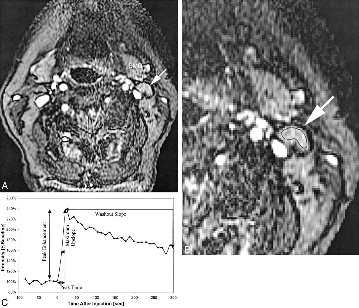
  • Fig 2.
    • Download figure
    • Open in new tab
    • Download powerpoint
    Fig 2.

    Patient 16, an 83-year-old woman with newly diagnosed squamous cell carcinoma of the lateral tongue.

    A, Axial view fast spoiled gradient recalled image (10.4/2.3; flip angle, 30 degrees), obtained during bolus administration of contrast agent, shows a normal appearing left level IIA lymph node (arrow). SMG, submandibular gland.

    B, Same image as that shown in A, magnified to emphasize the area of interest and with a region of interest indicator placed over the level IIA node. Pathologic analysis showed that this lymph node was not a tumor-involved node.

    C, Plot of signal intensity versus time generated from the region of interest placed around the cervical lymph node shown in B. Derivation of peak time, peak enhancement, maximum up-slope, and washout slope are illustrated.
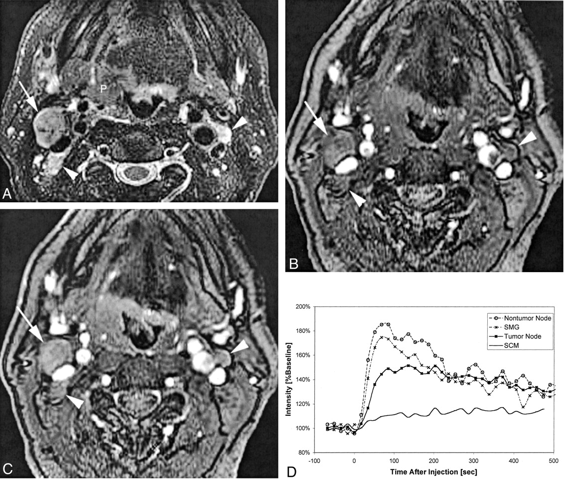
  • Fig 3.
    • Download figure
    • Open in new tab
    • Download powerpoint
    Fig 3.

    Patient 6, a 57-year-old man with right oropharyngeal cancer and multiple palpable nodes who underwent pharyngectomy, right modified radical neck dissection, and left supraomohyoid neck dissection.

    A, Coil-corrected fast spin-echo T2-weighted image with fat saturation (4000/90/4) shows the primary tumor (P), a dominant tumor-involved right level IIA node (white arrow), and two smaller tumor-involved level IIA lymph nodes (white arrowheads) on the right and left sides of the neck.

    B, Baseline image (10.4/2.3; flip angle, 30 degrees) from the dynamic sequence shows the same tumor-involved nodes 117 seconds after contrast medium injection.

    C, Enhanced dynamic image (10.4/2.3; flip angle, 30 degrees) shows the same tumor-involved nodes.

    D, Plot of signal intensity versus time after injection of contrast agent, generated from regions of interest placed on the right level IIA tumor-involved node, a left level III non-tumor-involved node (not included on these images), the submandibular gland (SMG), and the sternocleidomastoid muscle (SCM). The different curve morphologies generated from these different tissues can be appreciated as shown by this comparison plot.
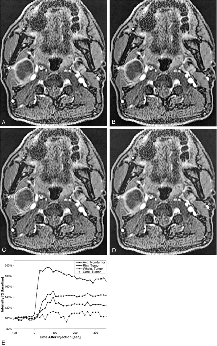
  • Fig 4.
    • Download figure
    • Open in new tab
    • Download powerpoint
    Fig 4.

    Patient 7, a 52-year-old man who had previously undergone partial glossectomy for squamous cell carcinoma of the tongue, received no additional therapy, and then presented with a palpable right neck mass. Fine needle aspiration confirmed recurrent squamous cell carcinoma in the neck, and MR imaging revealed right cervical necrotic lymphadenopathy. The patient then underwent re-excision of the primary site and a right radical neck dissection.

    A, Coil-corrected axial view enhanced dynamic image (10.4/2.3; flip angle, 30 degrees), acquired 195 s into the acquisition, shows a large, centrally necrotic, right level IIA lymph node.

    B, Same image as that shown in A, with the region of interest encompassing the whole node shown.

    C, Same image as that shown in A, with the region of interest encompassing only the rim shown.

    D, Same image as that shown in A, with the region of interest encompassing only the necrotic core shown.

    E, Plot of signal intensity versus time from this node, with curves generated from regions of interest placed on the whole node, the rim only, and the necrotic core only. Note that the “rim” curve is very similar to the “whole” curve but appears reduced in magnitude by a scaling factor related to the necrotic core. All these curves appear very different from the average non-tumor curve that is shown for comparison, but a statistically significant comparison was not possible because of the small number of grossly necrotic nodes.

Tables

  • Figures
    • View popup
    TABLE 1:

    Patient demographic information

    Patient No.Age (y)/SexDiagnosisPreoperative Staging*Nodal Status on Conventional Images (CT Scan or MR Image)Postoperative Staging
    144/FSCC tongueT3N1CT: negative (N1 based on palpation)T3N2c
    259/MSCC floor of mouthT2N0MR: negativeT2N0
    356/MSCC buccal mucosaT2N0MR: negativeT1N0
    464/FSCC oropharynxT3N0CT:negativeT2N0
    553/MSCC oropharynxT3N2aMR: multiple abnormal nodesT3N2a
    657/MSCC oropharynxT4N2cMR: multiple abnormal nodesT4N2c
    752/MSCC tongueTxN2b†MR: multiple abnormal nodesTxN2b
    872/MSCC retromolar trigoneT2N2bMR: multiple abnormal nodesT1N2b
    972/FSCC nasal cavityTxN1†CT: single necrotic nodeTxN1
    1063/MSCC tongueT3N0MR: negativeT2N0
    1171/MSCC tongueT3N0MR: negative (but N1 based on PET)T3N1‡
    1279/FSCC alveolar ridgeT3N2bMR: multiple abnormal nodesT4N2b
    1374/MSCC retromolar trigoneT2N0MR: negativeT2N1‡
    1465/FSCC alveolar ridgeT4N0MR: negativeT4N0
    1547/MSCC tongueTxN2b†CT: multiple abnormal nodesTxN2b
    1683/FSCC tongueT1N0MR: negativeT1N0
    1752/MSCC tongueT1N0MR: negativeT1N0
    1844/FSCC tongueT1N0MR: negativeT1N0
    1966/MSCC alveolar ridgeT4N0MR: negativeT4N0
    2085/FSCC buccal mucosaT4N0CT: negativeT4N1‡
    2162/MSCC floor of mouthTxN2b†CT: multiple abnormal nodesTxN2b
    • Note.—F indicates female; M, male; SCC, squamous cell carcinoma; PET, positron emission tomography.

    • * All patients were staged as M0. Preoperative staging was based on clinical palpation and conventional anatomic imaging with CT and/or MR imaging, not high resolution MR imaging.

    • † Primary site previously excised (hence, the “Tx” designation), but no radiation or other therapy to the neck previously received. Patient evaluated and treated for neck disease.

    • ‡ Three patients preoperatively staged as N0 were upstaged to N1 based on pathologic findings.

    • View popup
    TABLE 2:

    Dynamic contrast-enhanced MR imaging features of tumor involved versus non-tumor-involved nodes

    No.Peak Time (s)Peak Enhancement (%baseline)Maximum Slope (%baseline/min)Washout Slope (%baseline/min)
    Tumor-involved25111 (68)176 (30)193 (123)−7.6 (5.3)
    Non-tumor-involved4346 (29)198 (46)324 (224)−12.2 (8.1)
    P value<.001<.05<.01<.05
    • Note.—Number in parentheses indicates SD. Also note that the washout slope for non-tumor-involved nodes is based on N = 40 rather than 43 because late data points were not acquired for three nodes.

    • View popup
    TABLE 3:

    Dynamic contrast-enhanced MR imaging characterization of submandibular gland and sternocleidomastoid muscle

    TissuePeak Time (s)Peak Enhancement (%baseline)Maximum Slope (%baseline/min)Washout Slope (%baseline/min)
    SMG50 (41)206 (67)333 (186)−11.4 (6.6)
    SCM264 (102)125 (10)49 (36)−3.0 (10.9)
    • Note.—Number in parentheses indicates SD. SMG, submandibular gland; SCM, sternocleidomastoid muscle.

    • View popup
    TABLE 4:

    Subanalysis of whole necrotic node versus peripheral rim only

    TissuePeak Time (s)Peak Enhancement (%baseline)Maximum Slope (%baseline/min)Washout Slope (%baseline/min)
    Whole necrotic node114 (22)131 (14)57 (12)−0.1 (0.001)
    Peripheral rim only136 (48)180 (49)171 (132)−0.2 (0.05)
    Normal node46 (29)198 (46)324 (224)−12.2 (8.1)
    • Note.—P values cannot be calculated because of the small number of necrotic nodes.

PreviousNext
Back to top

In this issue

American Journal of Neuroradiology: 24 (3)
American Journal of Neuroradiology
Vol. 24, Issue 3
1 Mar 2003
  • Table of Contents
  • Index by author
Advertisement
Print
Download PDF
Email Article

Thank you for your interest in spreading the word on American Journal of Neuroradiology.

NOTE: We only request your email address so that the person you are recommending the page to knows that you wanted them to see it, and that it is not junk mail. We do not capture any email address.

Enter multiple addresses on separate lines or separate them with commas.
Assessment of Metastatic Cervical Adenopathy Using Dynamic Contrast-Enhanced MR Imaging
(Your Name) has sent you a message from American Journal of Neuroradiology
(Your Name) thought you would like to see the American Journal of Neuroradiology web site.
CAPTCHA
This question is for testing whether or not you are a human visitor and to prevent automated spam submissions.
Cite this article
Nancy J. Fischbein, Susan M. Noworolski, Roland G. Henry, Michael J. Kaplan, William P. Dillon, Sarah J. Nelson
Assessment of Metastatic Cervical Adenopathy Using Dynamic Contrast-Enhanced MR Imaging
American Journal of Neuroradiology Mar 2003, 24 (3) 301-311;

Citation Manager Formats

  • BibTeX
  • Bookends
  • EasyBib
  • EndNote (tagged)
  • EndNote 8 (xml)
  • Medlars
  • Mendeley
  • Papers
  • RefWorks Tagged
  • Ref Manager
  • RIS
  • Zotero
0 Responses
Respond to this article
Share
Bookmark this article
Assessment of Metastatic Cervical Adenopathy Using Dynamic Contrast-Enhanced MR Imaging
Nancy J. Fischbein, Susan M. Noworolski, Roland G. Henry, Michael J. Kaplan, William P. Dillon, Sarah J. Nelson
American Journal of Neuroradiology Mar 2003, 24 (3) 301-311;
del.icio.us logo Twitter logo Facebook logo Mendeley logo
  • Tweet Widget
  • Facebook Like
  • Google Plus One

Jump to section

  • Article
    • Abstract
    • Methods
    • Results
    • Discussion
    • Conclusion
    • Acknowledgments
    • Footnotes
    • References
  • Figures & Data
  • Info & Metrics
  • Responses
  • References
  • PDF

Related Articles

  • No related articles found.
  • PubMed
  • Google Scholar

Cited By...

  • Dynamic Contrast-Enhanced MR Imaging in Head and Neck Cancer: Techniques and Clinical Applications
  • Optimization of Ultrasmall Superparamagnetic Iron Oxide (P904)-enhanced Magnetic Resonance Imaging of Lymph Nodes: Initial Experience in a Mouse Model
  • Multiparametric MR Imaging of Sinonasal Diseases: Time-Signal Intensity Curve- and Apparent Diffusion Coefficient-Based Differentiation between Benign and Malignant Lesions
  • Current Concepts in Lymph Node Imaging
  • Crossref
  • Google Scholar

This article has not yet been cited by articles in journals that are participating in Crossref Cited-by Linking.

More in this TOC Section

  • Correlation of Apparent Diffusion Coefficient at 3T with Prognostic Parameters of Retinoblastoma
  • Parathyroid Lesions: Characterization with Dual-Phase Arterial and Venous Enhanced CT of the Neck
  • Efficacy of Diffusion-Weighted Imaging for the Differentiation between Lymphomas and Carcinomas of the Nasopharynx and Oropharynx: Correlations of Apparent Diffusion Coefficients and Histologic Features
Show more HEAD AND NECK

Similar Articles

Advertisement

Indexed Content

  • Current Issue
  • Accepted Manuscripts
  • Article Preview
  • Past Issues
  • Editorials
  • Editor's Choice
  • Fellows' Journal Club
  • Letters to the Editor
  • Video Articles

Cases

  • Case Collection
  • Archive - Case of the Week
  • Archive - Case of the Month
  • Archive - Classic Case

Special Collections

  • AJNR Awards
  • ASNR Foundation Special Collection
  • Most Impactful AJNR Articles
  • Photon-Counting CT
  • Spinal CSF Leak Articles (Jan 2020-June 2024)

More from AJNR

  • Trainee Corner
  • Imaging Protocols
  • MRI Safety Corner

Multimedia

  • AJNR Podcasts
  • AJNR Scantastics

Resources

  • Turnaround Time
  • Submit a Manuscript
  • Submit a Video Article
  • Submit an eLetter to the Editor/Response
  • Manuscript Submission Guidelines
  • Statistical Tips
  • Fast Publishing of Accepted Manuscripts
  • Graphical Abstract Preparation
  • Imaging Protocol Submission
  • Evidence-Based Medicine Level Guide
  • Publishing Checklists
  • Author Policies
  • Become a Reviewer/Academy of Reviewers
  • News and Updates

About Us

  • About AJNR
  • Editorial Board
  • Editorial Board Alumni
  • Alerts
  • Permissions
  • Not an AJNR Subscriber? Join Now
  • Advertise with Us
  • Librarian Resources
  • Feedback
  • Terms and Conditions
  • AJNR Editorial Board Alumni

American Society of Neuroradiology

  • Not an ASNR Member? Join Now

© 2025 by the American Society of Neuroradiology All rights, including for text and data mining, AI training, and similar technologies, are reserved.
Print ISSN: 0195-6108 Online ISSN: 1936-959X

Powered by HighWire