Advertisement

Main menu

  • Home
  • Content
    • Current Issue
    • Accepted Manuscripts
    • Article Preview
    • Past Issue Archive
    • Video Articles
    • AJNR Case Collection
    • Case of the Week Archive
    • Case of the Month Archive
    • Classic Case Archive
  • Special Collections
    • AJNR Awards
    • ASNR Foundation Special Collection
    • Most Impactful AJNR Articles
    • Photon-Counting CT
    • Spinal CSF Leak Articles (Jan 2020-June 2024)
  • Multimedia
    • AJNR Podcasts
    • AJNR SCANtastic
    • Trainee Corner
    • MRI Safety Corner
    • Imaging Protocols
  • For Authors
    • Submit a Manuscript
    • Submit a Video Article
    • Submit an eLetter to the Editor/Response
    • Manuscript Submission Guidelines
    • Statistical Tips
    • Fast Publishing of Accepted Manuscripts
    • Graphical Abstract Preparation
    • Imaging Protocol Submission
    • Author Policies
  • About Us
    • About AJNR
    • Editorial Board
    • Editorial Board Alumni
  • More
    • Become a Reviewer/Academy of Reviewers
    • Subscribers
    • Permissions
    • Alerts
    • Feedback
    • Advertisers
    • ASNR Home

User menu

  • Alerts
  • Log in

Search

  • Advanced search
American Journal of Neuroradiology

American Journal of Neuroradiology

ASHNR American Society of Functional Neuroradiology ASHNR American Society of Pediatric Neuroradiology ASSR
  • Alerts
  • Log in

Advanced Search

  • Home
  • Content
    • Current Issue
    • Accepted Manuscripts
    • Article Preview
    • Past Issue Archive
    • Video Articles
    • AJNR Case Collection
    • Case of the Week Archive
    • Case of the Month Archive
    • Classic Case Archive
  • Special Collections
    • AJNR Awards
    • ASNR Foundation Special Collection
    • Most Impactful AJNR Articles
    • Photon-Counting CT
    • Spinal CSF Leak Articles (Jan 2020-June 2024)
  • Multimedia
    • AJNR Podcasts
    • AJNR SCANtastic
    • Trainee Corner
    • MRI Safety Corner
    • Imaging Protocols
  • For Authors
    • Submit a Manuscript
    • Submit a Video Article
    • Submit an eLetter to the Editor/Response
    • Manuscript Submission Guidelines
    • Statistical Tips
    • Fast Publishing of Accepted Manuscripts
    • Graphical Abstract Preparation
    • Imaging Protocol Submission
    • Author Policies
  • About Us
    • About AJNR
    • Editorial Board
    • Editorial Board Alumni
  • More
    • Become a Reviewer/Academy of Reviewers
    • Subscribers
    • Permissions
    • Alerts
    • Feedback
    • Advertisers
    • ASNR Home
  • Follow AJNR on Twitter
  • Visit AJNR on Facebook
  • Follow AJNR on Instagram
  • Join AJNR on LinkedIn
  • RSS Feeds

AJNR is seeking candidates for the AJNR Podcast Editor. Read the position description.

Research ArticlePEDIATRICS

MR Ventriculography for the Study of CSF Flow

Vivek B. Joseph, Lakshminarayan Raghuram, Ipeson P. Korah and Ari G. Chacko
American Journal of Neuroradiology March 2003, 24 (3) 373-381;
Vivek B. Joseph
  • Find this author on Google Scholar
  • Find this author on PubMed
  • Search for this author on this site
Lakshminarayan Raghuram
  • Find this author on Google Scholar
  • Find this author on PubMed
  • Search for this author on this site
Ipeson P. Korah
  • Find this author on Google Scholar
  • Find this author on PubMed
  • Search for this author on this site
Ari G. Chacko
  • Find this author on Google Scholar
  • Find this author on PubMed
  • Search for this author on this site
  • Article
  • Figures & Data
  • Info & Metrics
  • Responses
  • References
  • PDF
Loading

Article Figures & Data

Figures

  • Fig 1.
    • Download figure
    • Open in new tab
    • Download powerpoint
    Fig 1.

    65-year old woman with multiple cerebral metastases and hydrocephalus probably secondary to carcinomatous meningitis.

    A, Contrast-enhanced CT scan of the brain shows a peripherally enhancing mass (black arrow) in the right frontal region, with surrounding edema. There is dilatation of the lateral and third ventricles that cannot be explained by the location of the mass. Air from the external ventricular drain is noted in the frontal horns (white arrow). Histopathologic examination revealed this tumor to be a metastatic adenocarcinoma.

    B, Contrast-enhanced CT scan through the posterior fossa shows another enhancing mass (arrow) in the left cerebellar hemisphere. The recesses of the fourth ventricle are well seen.

    C, Contrast-enhanced CT scan shows the dilated upper fourth ventricle with no evidence of basal arachnoiditis.

    D, Axial MR ventriculogram shows contrast material in the lateral and third ventricles.

    E, Coronal MR ventriculogram shows contrast material in both lateral ventricles, open foramina of Monro, and third ventricle. There is no contrast material in the basal subarachnoid cisterns.

    F, Sagittal MR ventriculogram shows contrast material in the lateral, third, and fourth ventricles. Note contrast material exiting the fourth ventricle through the foramen of Magendie into the cisterna magna (arrow).

    G, Axial MR ventriculogram shows contrast material in the lower fourth ventricle exiting the foramen of Magendie. Note the absence of contrast material in the right foramen of Luschka.

    H, Axial MR ventriculogram shows contrast material in the lower fourth ventricle entering the left foramen of Luschka (white arrow). Note the absence of contrast material in the right foramen of Luschka. The absence of contrast material in the basal subarachnoid spaces and the block in the right foramen of Luschka led us to a diagnosis of basal arachnoiditis due to leptomeningeal seeding in this patient with metastatic adenocarcinoma.

  • Fig 2.
    • Download figure
    • Open in new tab
    • Download powerpoint
    Fig 2.

    9-year-old girl with hydrocephalus and hydromyelia.

    A and B, Axial contrast-enhanced CT scans of the brain show dilatation of the lateral, third, and fourth ventricles. There is suspicion of a cystic lesion (arrow in B) in the fourth ventricle. MR ventriculography was performed to study the intraventricular CSF flow and to delineate the fourth ventricle.

    C, Axial MR ventriculogram after injection of gadodiamide into the right lateral ventricle shows contrast material layered in the dependent portion of the right lateral ventricle (arrow) and no contrast material in the left lateral ventricle.

    D, Coronal MR ventriculogram after injection of gadodiamide into the right lateral ventricle shows contrast material limited to the body and temporal horn of the right lateral ventricle (arrow). There is minimal contrast material in the third ventricle, suggesting a partial block in the right foramen of Monro.

    E, Sagittal MR ventriculogram after injection of contrast material into the right lateral ventricle shows minimal contrast material entering the third and fourth ventricles. Note the presence of a syringomyelia in the upper cervical cord (arrow).

    F, Axial MR ventriculogram through the bodies of the lateral ventricles. Contrast material was injected into the left lateral ventricle 100 minutes after the previous injection into the right lateral ventricle. This image shows a more uniform distribution of the contrast material in the right lateral ventricle, with no evidence of layering.

    G, Coronal MR ventriculogram after injection of contrast material into the left lateral ventricle shows good flow of the contrast material through the left foramen of Monro into the third ventricle.

    H, Sagittal MR ventriculogram shows flow of contrast material from the third ventricle into the fourth ventricle with no intraventricular lesion. The syrinx in the upper cervical cord (arrow) is again noted.

  • Fig 3.
    • Download figure
    • Open in new tab
    • Download powerpoint
    Fig 3.

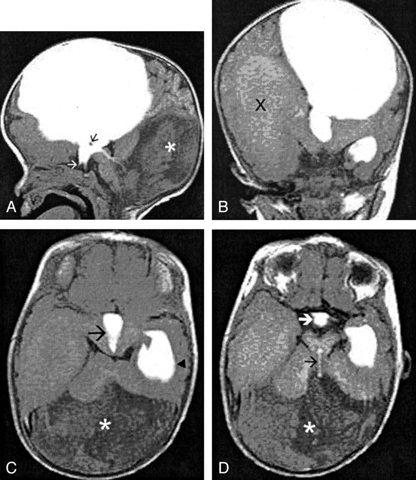
    6-month-old girl with a Dandy-Walker malformation and aqueductal stenosis.

    A, Sagittal MR ventriculogram shows intense contrast in the left lateral and third ventricles. The anterior recesses of the third ventricle (white arrow) and the massa intermedia (black arrow) are clearly visible. There is a thin trickle of contrast material in the cerebral aqueduct, indicating a relative stenosis. The asterisk (*) indicates the posterior fossa cyst.

    B, Coronal MR ventriculogram shows intense contrast in the body of the left lateral ventricle, third ventricle, and the left temporal horn. Contrast in the right lateral ventricle is of less intensity (X).

    C, Axial MR ventriculogram shows contrast in the third ventricle (arrow) and the left temporal horn (arrowhead). Contrast of less intensity fills the right temporal horn. The posterior fossa cyst (*) has no contrast.

    D, Axial MR ventriculogram shows intense contrast in the anteroinferior part of the third ventricle (white arrow) and temporal horn of the left lateral ventricle, and a thin trickle of contrast material in the cerebral aqueduct (black arrow) entering the posterior fossa cyst (*)

  • Fig 4.
    • Download figure
    • Open in new tab
    • Download powerpoint
    Fig 4.

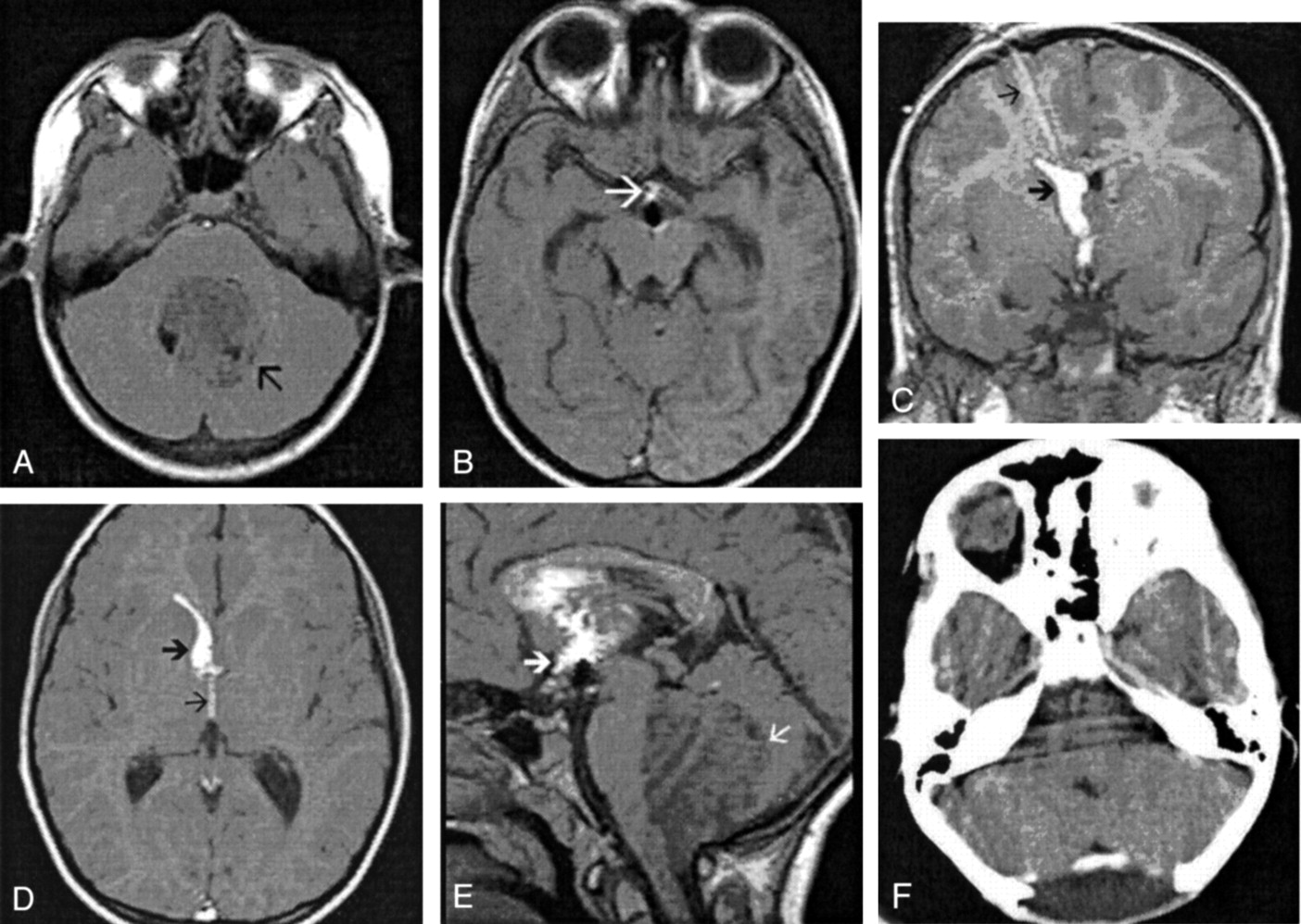
    8-year-old boy with a medulloblastoma and hydrocephalus who underwent an emergency third ventriculostomy before excision of the tumor.

    A, Nonenhanced axial T1-weighted MR image shows a large hypointense tumor (arrow) in the posterior fossa.

    B, Axial MR ventriculogram after third ventriculostomy shows contrast material in the anteroinferior third ventricle (arrow). Note that there is no contrast material in the adjacent suprasellar and sylvian cisterns.

    C, Coronal ventriculogram after third ventriculostomy shows contrast material along the external ventricular drain (thin arrow), the frontal horn of the right lateral ventricle (thick arrow), and the third ventricle. Note that no contrast material is seen in the suprasellar cisterns.

    D, Axial MR ventriculogram after third ventriculostomy shows contrast material in the right frontal horn (thick arrow) and third ventricle (thin arrow).

    E, Sagittal MR ventriculogram after third ventriculostomy shows contrast material in the dependent portion of the lateral ventricle and the third ventricle (thick arrow). There is no contrast material seen in suprasellar cisterns. Note the large hypointense tumor (thin arrow) in the posterior fossa.

    F, Postoperative contrast-enhanced CT scan through the posterior fossa shows complete tumor excision.

  • Fig 5.
    • Download figure
    • Open in new tab
    • Download powerpoint
    Fig 5.

    49-year-old man with a cystic cervicomedullary schwannoma and hydrocephalus who underwent a third ventriculostomy before excision of the tumor.

    A, Axial T1-weighted image through the posterior fossa shows the cystic tumor (arrow) in the left cerebellomedullary angle, compressing the fourth ventricle.

    B, Sagittal T1-weighted image shows the cervicomedullary cystic tumor (arrow) with the solid portion inferiorly.

    C, Axial MR ventriculogram after third ventriculostomy. The contrast material was injected into the right lateral ventricle. Note contrast material in the right temporal horn, in the suprasellar cisterns (thick arrow), and left sylvian cistern (thin arrow).

    D, Axial nonenhanced T1-weighted image for comparison with C.

    E, Coronal MR ventriculogram after third ventriculostomy shows contrast material in the bodies of both lateral ventricles. Note contrast material in the left sylvian cistern (arrow).

    F, Sagittal MR ventriculogram after third ventriculostomy shows contrast material in the lateral, third, and fourth ventricles. Contrast of lesser intensity is seen in the suprasellar cistern (upper arrow). Note obstruction to the flow of contrast material in the fourth ventricle by the cystic tumor (lower arrow).

    G, Sagittal nonenhanced T1-weighted MR image for comparison with F.

PreviousNext
Back to top

In this issue

American Journal of Neuroradiology: 24 (3)
American Journal of Neuroradiology
Vol. 24, Issue 3
1 Mar 2003
  • Table of Contents
  • Index by author
Advertisement
Print
Download PDF
Email Article

Thank you for your interest in spreading the word on American Journal of Neuroradiology.

NOTE: We only request your email address so that the person you are recommending the page to knows that you wanted them to see it, and that it is not junk mail. We do not capture any email address.

Enter multiple addresses on separate lines or separate them with commas.
MR Ventriculography for the Study of CSF Flow
(Your Name) has sent you a message from American Journal of Neuroradiology
(Your Name) thought you would like to see the American Journal of Neuroradiology web site.
CAPTCHA
This question is for testing whether or not you are a human visitor and to prevent automated spam submissions.
Cite this article
Vivek B. Joseph, Lakshminarayan Raghuram, Ipeson P. Korah, Ari G. Chacko
MR Ventriculography for the Study of CSF Flow
American Journal of Neuroradiology Mar 2003, 24 (3) 373-381;

Citation Manager Formats

  • BibTeX
  • Bookends
  • EasyBib
  • EndNote (tagged)
  • EndNote 8 (xml)
  • Medlars
  • Mendeley
  • Papers
  • RefWorks Tagged
  • Ref Manager
  • RIS
  • Zotero
0 Responses
Respond to this article
Share
Bookmark this article
MR Ventriculography for the Study of CSF Flow
Vivek B. Joseph, Lakshminarayan Raghuram, Ipeson P. Korah, Ari G. Chacko
American Journal of Neuroradiology Mar 2003, 24 (3) 373-381;
del.icio.us logo Twitter logo Facebook logo Mendeley logo
  • Tweet Widget
  • Facebook Like
  • Google Plus One

Jump to section

  • Article
    • Abstract
    • Methods
    • Results
    • Discussion
    • Conclusion
    • References
  • Figures & Data
  • Info & Metrics
  • Responses
  • References
  • PDF

Related Articles

  • No related articles found.
  • PubMed
  • Google Scholar

Cited By...

  • Is All "Communicating" Hydrocephalus Really Communicating? Prospective Study on the Value of 3D-Constructive Interference in Steady State Sequence at 3T
  • Prediction of the Jugular Venous Waveform Using a Model of CSF Dynamics
  • Crossref
  • Google Scholar

This article has not yet been cited by articles in journals that are participating in Crossref Cited-by Linking.

More in this TOC Section

  • SyMRI & MR Fingerprinting in Brainstem Myelination
  • Comparison of Image Quality and Radiation Dose in Pediatric Temporal Bone CT Using Photon-Counting Detector CT and Energy-Integrating Detector CT
  • Neonatal Hypocalcemia: Cerebral MRI Abnormalities
Show more Pediatrics

Similar Articles

Advertisement

Indexed Content

  • Current Issue
  • Accepted Manuscripts
  • Article Preview
  • Past Issues
  • Editorials
  • Editor's Choice
  • Fellows' Journal Club
  • Letters to the Editor
  • Video Articles

Cases

  • Case Collection
  • Archive - Case of the Week
  • Archive - Case of the Month
  • Archive - Classic Case

Special Collections

  • AJNR Awards
  • ASNR Foundation Special Collection
  • Most Impactful AJNR Articles
  • Photon-Counting CT
  • Spinal CSF Leak Articles (Jan 2020-June 2024)

More from AJNR

  • Trainee Corner
  • Imaging Protocols
  • MRI Safety Corner

Multimedia

  • AJNR Podcasts
  • AJNR Scantastics

Resources

  • Turnaround Time
  • Submit a Manuscript
  • Submit a Video Article
  • Submit an eLetter to the Editor/Response
  • Manuscript Submission Guidelines
  • Statistical Tips
  • Fast Publishing of Accepted Manuscripts
  • Graphical Abstract Preparation
  • Imaging Protocol Submission
  • Evidence-Based Medicine Level Guide
  • Publishing Checklists
  • Author Policies
  • Become a Reviewer/Academy of Reviewers
  • News and Updates

About Us

  • About AJNR
  • Editorial Board
  • Editorial Board Alumni
  • Alerts
  • Permissions
  • Not an AJNR Subscriber? Join Now
  • Advertise with Us
  • Librarian Resources
  • Feedback
  • Terms and Conditions
  • AJNR Editorial Board Alumni

American Society of Neuroradiology

  • Not an ASNR Member? Join Now

© 2025 by the American Society of Neuroradiology All rights, including for text and data mining, AI training, and similar technologies, are reserved.
Print ISSN: 0195-6108 Online ISSN: 1936-959X

Powered by HighWire