
Article Figures & Data
Figures
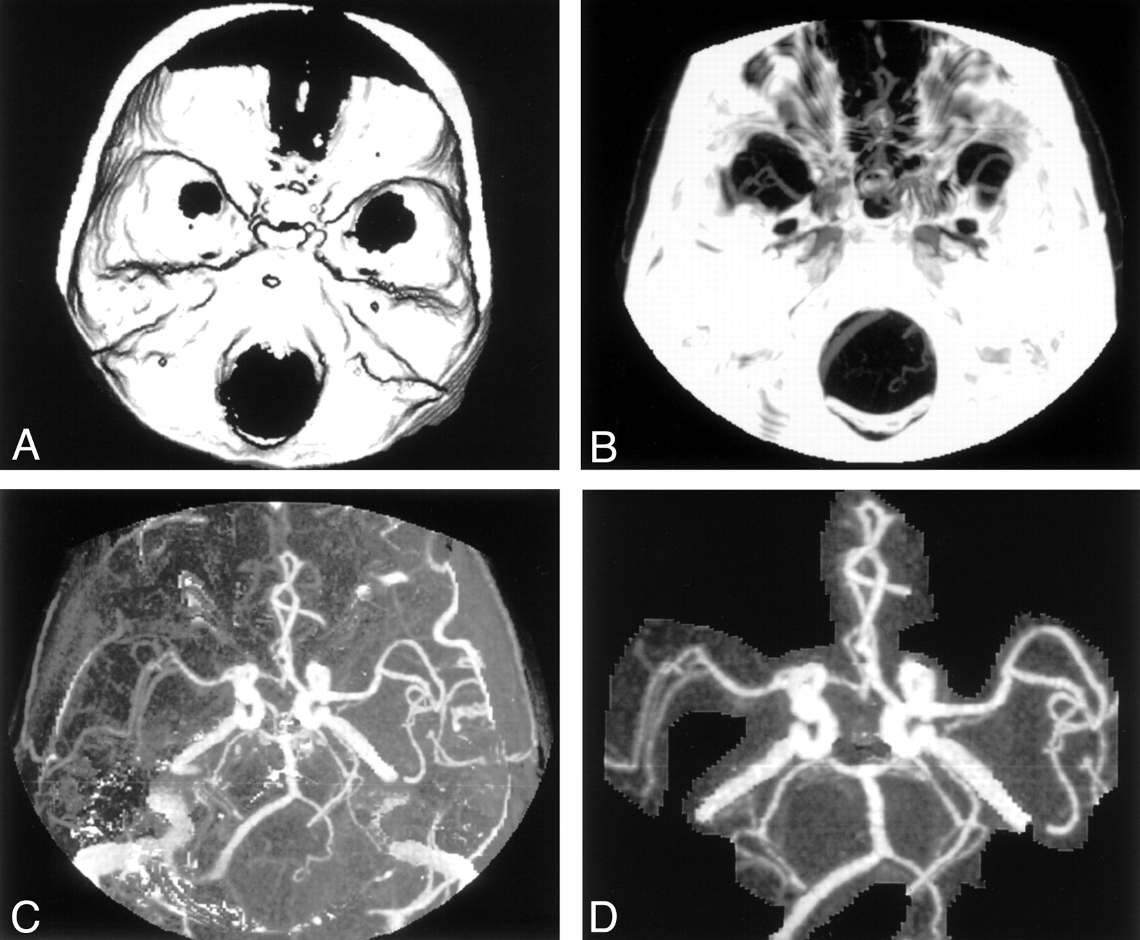
- Fig 1.
Image processing for subtraction CTA.
A, Three-dimensional model of the skull is created from precontrast dataset.
B, MIP created from the contrast-enhanced dataset.
C, Subtracted image after removing A from B, before any editing.
D, Subtracted image with minimal editing.
- Fig 2.
The restraining device is molded around the back of the patient’s head and the angles of the jaw. Inset shows the valve for the suction connection
- Fig 3.
MIP images.
A, MIP CTA image after editing shows residual bone at the base of the skull.
B, MIP DSCTA image better shows the basilar artery, its branches. and the aneurysm.
- Fig 4.
MIP images.
A, MIP CTA image shows that bone obscures the detail of the distal vertebral arteries.
B, MIP DSCTA image clearly shows clear the vertebral arteries and the posterior inferior cerebellar arteries.


Tables
Indications for CTA and DSCTA No. of Patients (n = 30) Suspected or known intracranial aneurysm 14 Carotid occlusive disease 10 Skull-base tumors 3 Cerebral venous thrombosis 3 Grade and Definition Intracranial Findings Neck Findings Circle of Willis Skull-Base Vessels Carotids Vertebral Arteries 1, nondiagnostic Obscured Obscured Obscured Obscured 2, poor quality Poorly visualized Obscured Poorly visualized Obscured 3, acceptable Clearly seen Obscured Clearly seen Obscured 4, good quality Clearly seen Partly obscured Clearly seen Partly obscured 5, excellent Clearly seen Clearly seen Clearly seen Clearly seen Test Operator 1 (n = 30) Operator 2 (n = 30) Mean Median SD Mean Median SD CTA 2.50 3 0.86 2.67 3 0.80 Hard-copy check by operator 3 3.07 3 1.17 2.83 3 1.09 DSCTA 3.37 3 0.81 3.30 3 0.95 Hard copy check by operator 3 3.97 4 0.85 3.83 4 0.95 DSCTA minus CTA* 0.87 1 0.73 0.63 1 0.56 Hard-copy check by Observer 3† 0.90 1 1.03 1.00 1 1.11 * 95% CIs were 0.59, 1.14 for operator 1 and 0.43, 0.84 for operator 2. P values with the t test were <.001 for both operators.
† 95% CIs were 0.52, 1.25 for operator 1 and 0.58, 1.42 for operator 2. P values with the t test were <.001 for both operators.
Observer (n = 30) CTA DSCTA CTA vs DSCTA Mean SD Mean SD Mean 95% CI P Value 1 8.44 2.47 8.88 2.89 −0.44 −1.19, 0.32 .249 2 10.07 4.55 7.53 2.85 2.53 1.08, 3.99 .001 1 and 2 (mean) 9.26 3.10 8.21 2.25 1.05 0.34, 1.75 .005
In this issue


Jump to section
Related Articles
Cited By...
- Bone-Subtracted Spinal CT Angiography Using Nonrigid Registration for Better Visualization of Arterial Feeders in Spinal Arteriovenous Fistulas
- Comparison of Image Quality and Radiation Dose between Fixed Tube Current and Combined Automatic Tube Current Modulation in Craniocervical CT Angiography
- Internal Carotid Artery Stenosis Measurement: Comparison of 3D Computed Rotational Angiography and Conventional Digital Subtraction Angiography