Advertisement

Main menu

  • Home
  • Content
    • Current Issue
    • Accepted Manuscripts
    • Article Preview
    • Past Issue Archive
    • Video Articles
    • AJNR Case Collection
    • Case of the Week Archive
    • Case of the Month Archive
    • Classic Case Archive
  • Special Collections
    • AJNR Awards
    • ASNR Foundation Special Collection
    • Most Impactful AJNR Articles
    • Photon-Counting CT
    • Spinal CSF Leak Articles (Jan 2020-June 2024)
  • Multimedia
    • AJNR Podcasts
    • AJNR SCANtastic
    • Trainee Corner
    • MRI Safety Corner
    • Imaging Protocols
  • For Authors
    • Submit a Manuscript
    • Submit a Video Article
    • Submit an eLetter to the Editor/Response
    • Manuscript Submission Guidelines
    • Statistical Tips
    • Fast Publishing of Accepted Manuscripts
    • Graphical Abstract Preparation
    • Imaging Protocol Submission
    • Author Policies
  • About Us
    • About AJNR
    • Editorial Board
    • Editorial Board Alumni
  • More
    • Become a Reviewer/Academy of Reviewers
    • Subscribers
    • Permissions
    • Alerts
    • Feedback
    • Advertisers
    • ASNR Home

User menu

  • Alerts
  • Log in

Search

  • Advanced search
American Journal of Neuroradiology

American Journal of Neuroradiology

ASHNR American Society of Functional Neuroradiology ASHNR American Society of Pediatric Neuroradiology ASSR
  • Alerts
  • Log in

Advanced Search

  • Home
  • Content
    • Current Issue
    • Accepted Manuscripts
    • Article Preview
    • Past Issue Archive
    • Video Articles
    • AJNR Case Collection
    • Case of the Week Archive
    • Case of the Month Archive
    • Classic Case Archive
  • Special Collections
    • AJNR Awards
    • ASNR Foundation Special Collection
    • Most Impactful AJNR Articles
    • Photon-Counting CT
    • Spinal CSF Leak Articles (Jan 2020-June 2024)
  • Multimedia
    • AJNR Podcasts
    • AJNR SCANtastic
    • Trainee Corner
    • MRI Safety Corner
    • Imaging Protocols
  • For Authors
    • Submit a Manuscript
    • Submit a Video Article
    • Submit an eLetter to the Editor/Response
    • Manuscript Submission Guidelines
    • Statistical Tips
    • Fast Publishing of Accepted Manuscripts
    • Graphical Abstract Preparation
    • Imaging Protocol Submission
    • Author Policies
  • About Us
    • About AJNR
    • Editorial Board
    • Editorial Board Alumni
  • More
    • Become a Reviewer/Academy of Reviewers
    • Subscribers
    • Permissions
    • Alerts
    • Feedback
    • Advertisers
    • ASNR Home
  • Follow AJNR on Twitter
  • Visit AJNR on Facebook
  • Follow AJNR on Instagram
  • Join AJNR on LinkedIn
  • RSS Feeds

AJNR is seeking candidates for the AJNR Podcast Editor. Read the position description.

Research ArticleBRAIN

Assessment of the Collateral Function of the Circle of Willis

Three-Dimensional Time-of-Flight MR Angiography Compared with Transcranial Color-Coded Duplex Sonography

Arjan W.J. Hoksbergen, Charles B.L. Majoie, Frans-Jan H. Hulsmans and Dink A. Legemate
American Journal of Neuroradiology March 2003, 24 (3) 456-462;
Arjan W.J. Hoksbergen
  • Find this author on Google Scholar
  • Find this author on PubMed
  • Search for this author on this site
Charles B.L. Majoie
  • Find this author on Google Scholar
  • Find this author on PubMed
  • Search for this author on this site
Frans-Jan H. Hulsmans
  • Find this author on Google Scholar
  • Find this author on PubMed
  • Search for this author on this site
Dink A. Legemate
  • Find this author on Google Scholar
  • Find this author on PubMed
  • Search for this author on this site
  • Article
  • Figures & Data
  • Info & Metrics
  • Responses
  • References
  • PDF
Loading

Article Figures & Data

Figures

  • Tables
  • Fig 1.
    • Download figure
    • Open in new tab
    • Download powerpoint
    Fig 1.

    Examples and prevalence of circle of Willis anomalies that hamper collateral function. Prevalence is derived from studies by using an external diameter of 1 mm as a threshold for hypoplasia of collateral arteries. A1 indicates the precommunicating part of the anterior cerebral artery; M1, main trunk of the middle cerebral artery; P1, precommunicating part of the posterior cerebral artery; BA, basilar artery; and VA, vertebral artery.
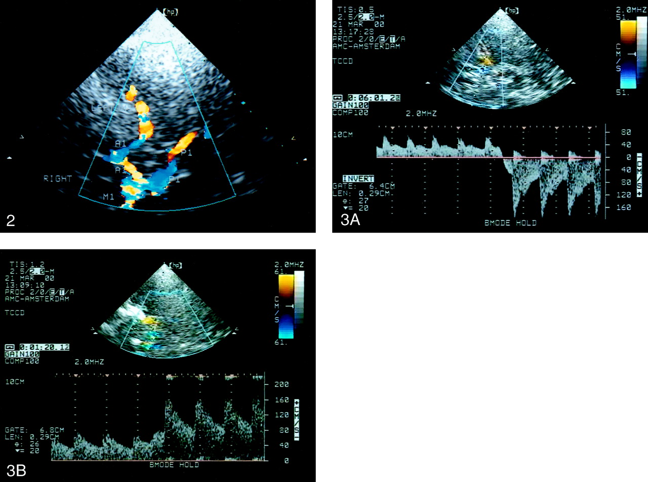
  • Fig 2.
    • Download figure
    • Open in new tab
    • Download powerpoint
    Fig 2.

    TCCD sonogram of the circle of Willis in the left temporal window and axial scanning plane at the level of the mesencephalon. The ipsilateral (left) M1, P1 (red), and A1 (blue) are shown. The contralateral (right) M1 and P1 (blue) and A1 (red) are also shown.

  • Fig 3.
    • Download figure
    • Open in new tab
    • Download powerpoint
    Fig 3.

    Doppler spectra.

    A, Spectrum shows blood flow reversal in the A1 during ipsilateral carotid compression indicating functional patency of the AcoA.

    B, Spectrum shows blood flow velocity increase of more than 20% in the P1 during ipsilateral carotid compression indicating functional patency of the PcoA.
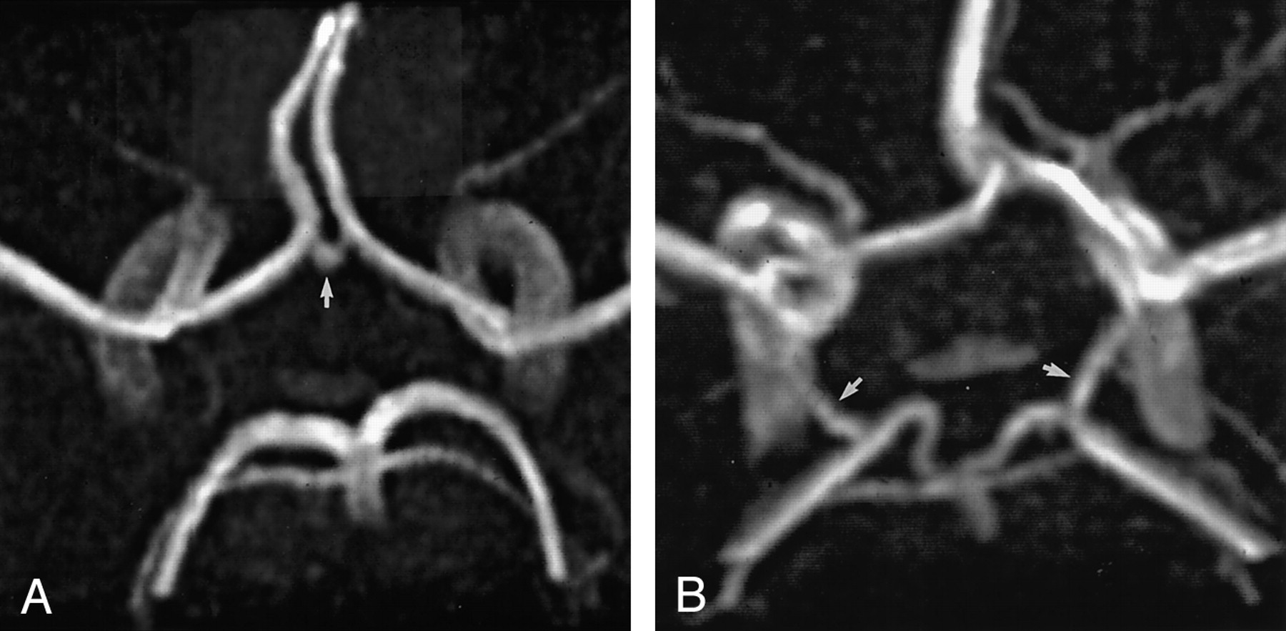
  • Fig 4.
    • Download figure
    • Open in new tab
    • Download powerpoint
    Fig 4.

    3D TOF MR angiograms.

    A, Angiogram of the circle of Willis with the complete anterior configuration shows the AcoA (arrow).

    B, Angiogram of the circle of Willis with the complete posterior configuration shows the right and left PcoAs (arrows)

Tables

  • Figures
    • View popup
    TABLE 1:

    MR angiography compared with conventional angiography for the detection of intracranial collaterals

    Author*No. of SubjectsAcoAPcoA
    Sensitivity, %Specificity, %Sensitivity, %Specificity, %
    Patrux et al (4)548910081100
    Stock et al (5)6267737593
    • * Numbers in parentheses are reference citations.

    • View popup
    TABLE 2:

    Detection of collateral pathways (n = 150) in 50 healthy subjects

    Number of findings
    Detection with MR Angiography*Detection with TCCD*Total
    YesNo
    Observer 1
     Yes1055110
     No182240
     Total12327150
    Observer 2
     Yes1079116
     No161834
     Total12327150
    Diagnostic discrimination
    MeasureObserver 1†Observer 2†
    Sensitivity, %85 (79, 92)87 (81, 93)
    Specificity, %81 (62, 94)67 (46, 84)
    Positive predictive value, %95 (90, 99)92 (86, 96)
    Negative predictive value, %55 (39, 71)53 (35, 70)
    • * Yes indicates functional. No indicates nonfunctional.

    • † Numbers in parentheses are the 95% confidence intervals.

    • View popup
    TABLE 3:

    Interobserver agreement with MR angiography for the detection of collateral pathways (n = 150)*

    Findings, Observer 1†Findings, Observer 2†Total
    YesNo
    Yes1019110
    No152540
    Total11634150
    • * κ = 0.57 (95% confidence interval: 0.42, 0.72).

    • † Yes indicates functional. No indicates nonfunctional.

    • View popup
    TABLE 4:

    Percentage of nonvisualized collaterals in 3D time-of-flight MR angiography studies

    Study*No. of SubjectsAge, yAcoA, %A1, %PcoA, %P1, %
    Healthy volunteers
     Krabbe-Hartkamp et al (3)15055193282
     Current study†5051101227
    Cerebrovascular patients
     Patrux et al (4)5444545568
     Stock et al (5)6252404394
     Hartkamp et al (23)756273212
     Brunereau et al (24)10921–824524412
    • * Numbers in parentheses are reference citations.

    • † Data were derived from the consensus review.

PreviousNext
Back to top

In this issue

American Journal of Neuroradiology: 24 (3)
American Journal of Neuroradiology
Vol. 24, Issue 3
1 Mar 2003
  • Table of Contents
  • Index by author
Advertisement
Print
Download PDF
Email Article

Thank you for your interest in spreading the word on American Journal of Neuroradiology.

NOTE: We only request your email address so that the person you are recommending the page to knows that you wanted them to see it, and that it is not junk mail. We do not capture any email address.

Enter multiple addresses on separate lines or separate them with commas.
Assessment of the Collateral Function of the Circle of Willis
(Your Name) has sent you a message from American Journal of Neuroradiology
(Your Name) thought you would like to see the American Journal of Neuroradiology web site.
CAPTCHA
This question is for testing whether or not you are a human visitor and to prevent automated spam submissions.
Cite this article
Arjan W.J. Hoksbergen, Charles B.L. Majoie, Frans-Jan H. Hulsmans, Dink A. Legemate
Assessment of the Collateral Function of the Circle of Willis
American Journal of Neuroradiology Mar 2003, 24 (3) 456-462;

Citation Manager Formats

  • BibTeX
  • Bookends
  • EasyBib
  • EndNote (tagged)
  • EndNote 8 (xml)
  • Medlars
  • Mendeley
  • Papers
  • RefWorks Tagged
  • Ref Manager
  • RIS
  • Zotero
0 Responses
Respond to this article
Share
Bookmark this article
Assessment of the Collateral Function of the Circle of Willis
Arjan W.J. Hoksbergen, Charles B.L. Majoie, Frans-Jan H. Hulsmans, Dink A. Legemate
American Journal of Neuroradiology Mar 2003, 24 (3) 456-462;
del.icio.us logo Twitter logo Facebook logo Mendeley logo
  • Tweet Widget
  • Facebook Like
  • Google Plus One

Jump to section

  • Article
    • Abstract
    • Methods
    • Results
    • Discussion
    • Conclusion
    • Acknowledgments
    • References
  • Figures & Data
  • Info & Metrics
  • Responses
  • References
  • PDF

Related Articles

  • No related articles found.
  • PubMed
  • Google Scholar

Cited By...

  • Assessment of Collateral Flow in Patients with Carotid Stenosis Using Random Vessel-Encoded Arterial Spin-Labeling: Comparison with Digital Subtraction Angiography
  • Middle Cerebral Artery Dissection: Diagnostic and Prognostic Value of Transcranial Color-Coded Sonography
  • Assessment of Intracranial Collateral Flow by Using Dynamic Arterial Spin Labeling MRA and Transcranial Color-Coded Duplex Ultrasound
  • Crossref
  • Google Scholar

This article has not yet been cited by articles in journals that are participating in Crossref Cited-by Linking.

More in this TOC Section

  • Usefulness of Quantitative Susceptibility Mapping for the Diagnosis of Parkinson Disease
  • White Matter Alterations in the Brains of Patients with Active, Remitted, and Cured Cushing Syndrome: A DTI Study
  • Qualitative and Quantitative Analysis of MR Imaging Findings in Patients with Middle Cerebral Artery Stroke Implanted with Mesenchymal Stem Cells
Show more BRAIN

Similar Articles

Advertisement

Indexed Content

  • Current Issue
  • Accepted Manuscripts
  • Article Preview
  • Past Issues
  • Editorials
  • Editor's Choice
  • Fellows' Journal Club
  • Letters to the Editor
  • Video Articles

Cases

  • Case Collection
  • Archive - Case of the Week
  • Archive - Case of the Month
  • Archive - Classic Case

Special Collections

  • AJNR Awards
  • ASNR Foundation Special Collection
  • Most Impactful AJNR Articles
  • Photon-Counting CT
  • Spinal CSF Leak Articles (Jan 2020-June 2024)

More from AJNR

  • Trainee Corner
  • Imaging Protocols
  • MRI Safety Corner

Multimedia

  • AJNR Podcasts
  • AJNR Scantastics

Resources

  • Turnaround Time
  • Submit a Manuscript
  • Submit a Video Article
  • Submit an eLetter to the Editor/Response
  • Manuscript Submission Guidelines
  • Statistical Tips
  • Fast Publishing of Accepted Manuscripts
  • Graphical Abstract Preparation
  • Imaging Protocol Submission
  • Evidence-Based Medicine Level Guide
  • Publishing Checklists
  • Author Policies
  • Become a Reviewer/Academy of Reviewers
  • News and Updates

About Us

  • About AJNR
  • Editorial Board
  • Editorial Board Alumni
  • Alerts
  • Permissions
  • Not an AJNR Subscriber? Join Now
  • Advertise with Us
  • Librarian Resources
  • Feedback
  • Terms and Conditions
  • AJNR Editorial Board Alumni

American Society of Neuroradiology

  • Not an ASNR Member? Join Now

© 2025 by the American Society of Neuroradiology All rights, including for text and data mining, AI training, and similar technologies, are reserved.
Print ISSN: 0195-6108 Online ISSN: 1936-959X

Powered by HighWire