Advertisement

Main menu

  • Home
  • Content
    • Current Issue
    • Accepted Manuscripts
    • Article Preview
    • Past Issue Archive
    • Video Articles
    • AJNR Case Collection
    • Case of the Week Archive
    • Case of the Month Archive
    • Classic Case Archive
  • Special Collections
    • AJNR Awards
    • ASNR Foundation Special Collection
    • Most Impactful AJNR Articles
    • Photon-Counting CT
    • Spinal CSF Leak Articles (Jan 2020-June 2024)
  • Multimedia
    • AJNR Podcasts
    • AJNR SCANtastic
    • Trainee Corner
    • MRI Safety Corner
    • Imaging Protocols
  • For Authors
    • Submit a Manuscript
    • Submit a Video Article
    • Submit an eLetter to the Editor/Response
    • Manuscript Submission Guidelines
    • Statistical Tips
    • Fast Publishing of Accepted Manuscripts
    • Graphical Abstract Preparation
    • Imaging Protocol Submission
    • Author Policies
  • About Us
    • About AJNR
    • Editorial Board
    • Editorial Board Alumni
  • More
    • Become a Reviewer/Academy of Reviewers
    • Subscribers
    • Permissions
    • Alerts
    • Feedback
    • Advertisers
    • ASNR Home

User menu

  • Alerts
  • Log in

Search

  • Advanced search
American Journal of Neuroradiology

American Journal of Neuroradiology

ASHNR American Society of Functional Neuroradiology ASHNR American Society of Pediatric Neuroradiology ASSR
  • Alerts
  • Log in

Advanced Search

  • Home
  • Content
    • Current Issue
    • Accepted Manuscripts
    • Article Preview
    • Past Issue Archive
    • Video Articles
    • AJNR Case Collection
    • Case of the Week Archive
    • Case of the Month Archive
    • Classic Case Archive
  • Special Collections
    • AJNR Awards
    • ASNR Foundation Special Collection
    • Most Impactful AJNR Articles
    • Photon-Counting CT
    • Spinal CSF Leak Articles (Jan 2020-June 2024)
  • Multimedia
    • AJNR Podcasts
    • AJNR SCANtastic
    • Trainee Corner
    • MRI Safety Corner
    • Imaging Protocols
  • For Authors
    • Submit a Manuscript
    • Submit a Video Article
    • Submit an eLetter to the Editor/Response
    • Manuscript Submission Guidelines
    • Statistical Tips
    • Fast Publishing of Accepted Manuscripts
    • Graphical Abstract Preparation
    • Imaging Protocol Submission
    • Author Policies
  • About Us
    • About AJNR
    • Editorial Board
    • Editorial Board Alumni
  • More
    • Become a Reviewer/Academy of Reviewers
    • Subscribers
    • Permissions
    • Alerts
    • Feedback
    • Advertisers
    • ASNR Home
  • Follow AJNR on Twitter
  • Visit AJNR on Facebook
  • Follow AJNR on Instagram
  • Join AJNR on LinkedIn
  • RSS Feeds

AJNR is seeking candidates for the AJNR Podcast Editor. Read the position description.

Research ArticleBRAIN

Wegener Granuloma in the Fourth Ventricle

Ansgar Berlis, Franziska Petschner, Ingolf Carl Bötefür and Joachim Spreer
American Journal of Neuroradiology March 2003, 24 (3) 523-525;
Ansgar Berlis
  • Find this author on Google Scholar
  • Find this author on PubMed
  • Search for this author on this site
Franziska Petschner
  • Find this author on Google Scholar
  • Find this author on PubMed
  • Search for this author on this site
Ingolf Carl Bötefür
  • Find this author on Google Scholar
  • Find this author on PubMed
  • Search for this author on this site
Joachim Spreer
  • Find this author on Google Scholar
  • Find this author on PubMed
  • Search for this author on this site
  • Article
  • Figures & Data
  • Info & Metrics
  • Responses
  • References
  • PDF
Loading

Abstract

Summary: We report the case of a 57-year-old man with a known history of antineutrophil cytoplasmic antibody-positive Wegener granulomatosis with initial involvement of the upper and lower respiratory tract. MR imaging of the brain was performed because of new onset CNS symptoms (nausea, altered mental status). The images revealed a mass in the fourth ventricle. The mass diminished in size after systemic steroid treatment, as shown by follow-up MR imaging. The patient died in acute respiratory distress secondary to pneumonia. Histopathologic findings confirmed a Wegener granuloma of the fourth ventricle.

Wegener granulomatosis is a rare autoimmune vasculitis of unknown cause characterized by necrotizing granuloma of the respiratory tract, focal necrotizing glomerulonephritis, and systemic vasculitis (1–4). Wegener granulomatosis belongs to the small vessel vasculitides with association to antineutrophil cytoplasmic antibodies, which are usually specific for proteinase 3(5). Although the paranasal sinuses, lungs, skin, and kidneys are most commonly affected, many other organs can be involved. Neurologic involvement in Wegener granulomatosis was described in 25.7% to 54% of patients before the era of effective therapeutic agents, including prednisone and cyclophosphamide (6, 7) and in 22% to 33.6% of patients after the advent of more effective immunosuppressive regimens (2, 4).

Case Report

A 57-year-old man developed dyspnea, bloody rhinitis, and hoarseness 2 years before admission. Radiologic examination of the chest revealed suspicious right pulmonary infiltrates. Pathohistologic workup after right superior lobe resection confirmed the diagnosis of Wegener granulomatosis. An antineutrophil cytoplasmic antibody with specificity for proteinase 3 was detected in the patient’s sera, and immunosuppressive therapy with systemic steroids and oral cyclophosphamide was initiated. Secondary to ineffectiveness or side effects (lymphocytopenia leading to recurrent infections) of the immunosuppressive regimen, several changes of the therapy were necessary during the ensuing months. In addition to steroids that were orally and IV administered at different doses, the immunosuppressive regimens consisted of desoxyspergualin (subcutaneously administered), leflunomid (orally administered), and cyclophosphamide (IV and orally administered) for different intervals. Despite ongoing immunosuppressive therapy, the patient complained of nausea, vomiting, and headache and developed progressive neurologic deficits with skew deviation, slight upbeat nystagmus, and diplopia. MR imaging (Fig 1) showed a well-delineated mass in the inferior fourth ventricle, compressing and infiltrating the surrounding structures with extrusion through the left foramen of Luschka. Our differential diagnoses of the fourth ventricle mass included ependymoma and Wegener granuloma. Biopsy of the tumor was canceled because of the patient’s respiratory insufficiency and poor clinical condition. High doses of prednisone and cyclophospamide had to be administered in addition to antibiotics. Under this treatment, the neurologic symptoms improved. Follow-up MR imaging performed after 4 and 6 weeks showed tumor regression, whereas the nonocclusive hydrocephalus remained the same (Figs 2 and 3). Despite tumor regression, the pulmonary complications progressed. The patient died 4 months later.

Fig 1.
  • Download figure
  • Open in new tab
  • Download powerpoint
Fig 1.

Initial contrast-enhanced T1-weighted MR images obtained in three planes and coronal view diffusion-weighted image (lower right) show inhomogeneous contrast enhancement of a mass at the floor of the fourth ventricle. Compression and infiltration of the left dorsolateral medulla oblongata and the vermis can be seen, with extrusion through the left foramen of Luschka. Concomitant hydrocephalus is revealed, and high signal intensity on the diffusion-weighted image implies dense cellularity of the tumor. Note the thickening of the mucosa with contrast enhancement at the paranasal sinuses, whereas CT scans revealed intact bony structures.

Fig 2.
  • Download figure
  • Open in new tab
  • Download powerpoint
Fig 2.

Follow-up MR images obtained 4 weeks after initial imaging, during immunosuppressive therapy. Approximately 50% tumor regression is revealed. Remaining high signal intensity in the lesion can be seen on the diffusion-weighted image.

Fig 3.
  • Download figure
  • Open in new tab
  • Download powerpoint
Fig 3.

Follow-up MR images obtained 6 weeks after initial imaging show slight additional regression of the tumor but evident reduction of the signal intensity on the diffusion-weighted image and remaining nonocclusive hydrocephalus.

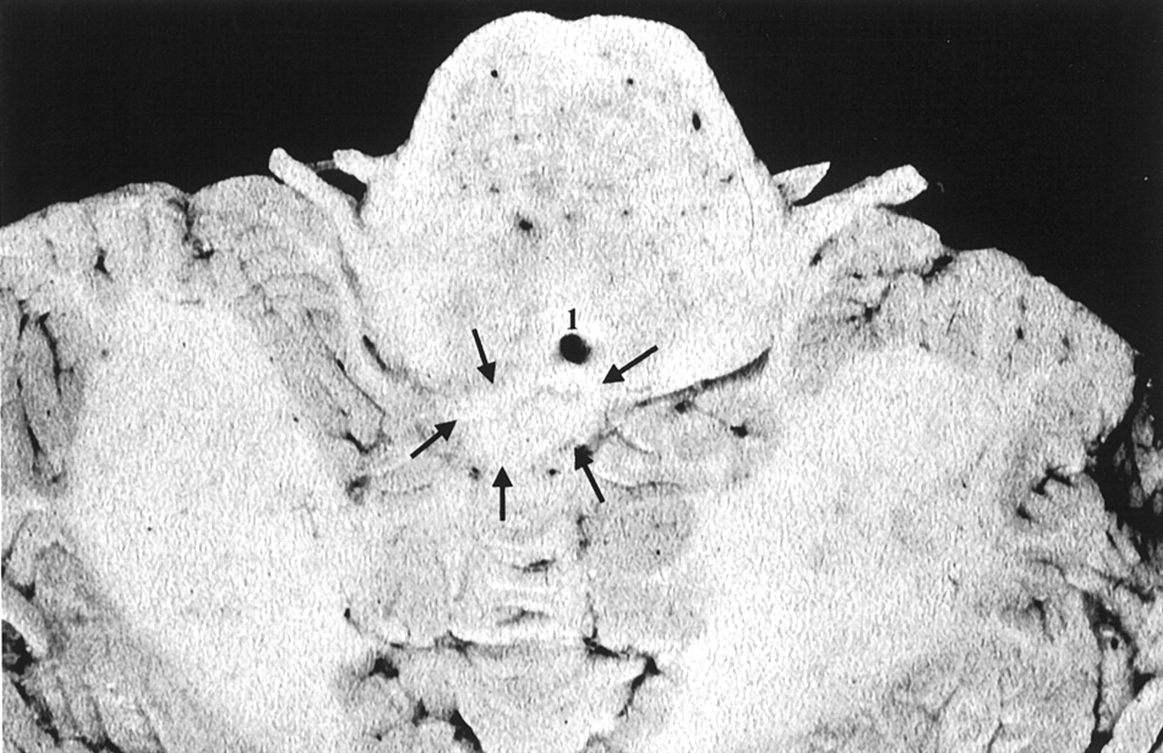
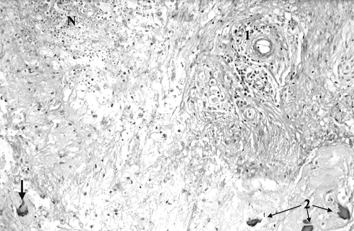
Histopathologic examination (Figs 4–6) showed that the immediate cause of death was pneumonia and cachexia. Slight obstructive hydrocephalus was seen, and a necrotic mass was found in the inferior fourth ventricle, containing infiltrates of lymphocytes, eosinophilic granulocytes, plasma cells, accumulations of macrophages, and typical multinucleated giant cells. Calcifications with surrounding scarred connective tissue were observed, as was necrotizing vasculitis leading to vessel occlusion. The mass originated in the vermis, and diffuse infiltration of the adjacent cerebellum and the medulla oblongata was observed. No signs of acute inflammatory processes, CNS neoplasia, or metastasis were seen. Additionally, pulmonary fibrosis secondary to Wegener granulomatosis was confirmed histologically.

Fig 4.
  • Download figure
  • Open in new tab
  • Download powerpoint
Fig 4.

Image of the cerebellum and medulla oblongata obtained at nearly the same level as the transverse T1-weighted images shown in Figures 1 through 3. Note the poor demarcation of the granuloma (arrows) from surrounding brain tissue (1, foramen of Magendie).

Fig 5.
  • Download figure
  • Open in new tab
  • Download powerpoint
Fig 5.

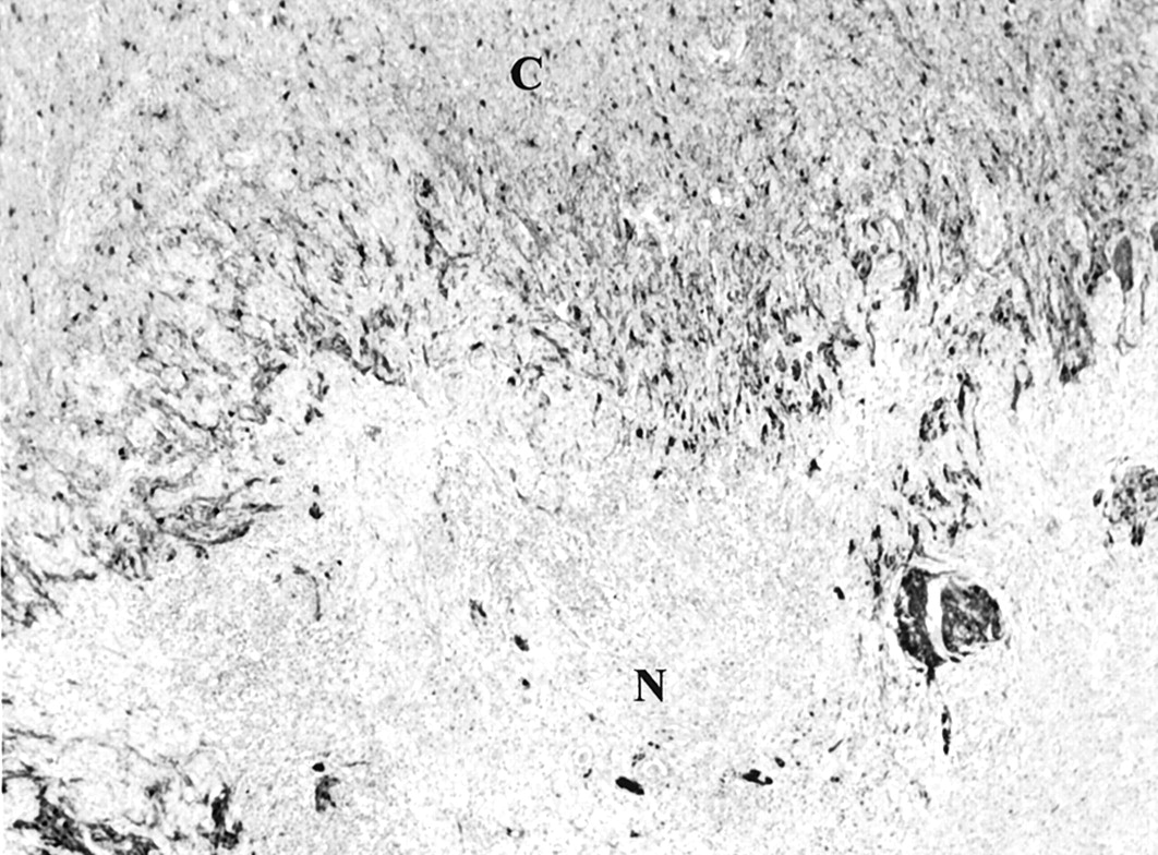
Histologic specimen (glial fibrillary acidic protein stained) with necrotizing mass (N) shows diffuse infiltration of the cerebellum (C).

Fig 6.
  • Download figure
  • Open in new tab
  • Download powerpoint
Fig 6.

Histologic specimen (periodic acid-Schiff stain) shows typical Wegener granulomatosis patterns with necrotizing vasculitis (1), multinucleated giant cell (arrow), and necrotizing lesions (N). Calcifications (2) were detectable with surrounding scarred connective tissue.

Discussion

The estimated incidence of Wegener granulomatosis is 1:100,000 per year (8). In former times, the outcome almost invariably was fatal. The prognosis dramatically improved once sufficient immunosuppressive regimens became available (5). Mahr et al (9) currently report a mortality rate of 37% during a mean follow-up period of 1.9 years.

Neurologic involvement in Wegener granulomatosis has been reported in ≤54% of the cases (2, 6, 7). After the advent of therapeutic agents, including prednisone and cyclophosphamide, neurologic involvement decreased to 22% (4). According to Drachmann (6), there are three different types of CNS involvement: direct granulomatous infiltration from contiguous lesions in the nose and paranasal sinuses, remote lesions from the nose and paranasal sinuses, and vasculitis of the nervous system itself. Peripheral nervous system involvement is present in approximately 10% to 16% of the cases (2, 4), and cerebral and meningeal involvement is rare (reported in 2% to 8% of cases of Wegener granulomatosis) (2, 4, 6, 10). On CT scans and MR images, white matter lesions with or without contrast enhancement, infarcts due to primary manifestations of the disease (eg, vasculitis or arterial occlusion caused by granulomatous vasculitis), and thickening of the dura with contrast enhancement may be found (2, 4, 11–18). Diffuse, symmetric thickening of the entire dura was seen, as was focal, nodular, and plaquelike thickening with mass effect (4, 12–15). Two patients were described as having thickening and contrast enhancement of the dura overlying the thoracic spinal cord (4, 13). The pituitary gland and infundibulum may be involved by means of distant granulomas or direct spread (6, 13, 19, 20). Only a few reports have been published showing subarachnoid hemorrhage (6, 21), intracerebral hemorrhage (6), and arterial occlusion (6, 22). Histologically or radiologically confirmed vasculitis of the CNS in Wegener granulomatosis is very rare (4, 23). Typically, small vessels (50–300 μm) are affected in cases of Wegener granulomatosis (4). In our case, necrotizing vasculitis with vessel occlusions could be detected only within the tumorlike mass whereas generalized or distant focal vasculitis was absent.

The external ophthalmoplegia associated with Wegener granulomatosis is probably related to granulomatous infiltration of the orbit or the cavernous sinus (4, 24). Drachmann (6) found cranial nerve involvement due to remote granulomatous lesions in 2% of his patients. Anderson et al (7) found 65 cases presenting with affection of the cranial nerves. It was assumed that cranial neuropathy occurred secondary to either direct or remote extension of the granulomatous lesion. Cranial nerve involvement has been described in 6.5% (21 of 324 cases) (4) to 9.4% (eight of 85 cases) (2) of the cases.

Remote granulomatous lesions in association with brain parenchyma are the least common form of CNS involvement in cases of Wegener granulomatosis. With respect to the classification of Drachmann (4), our patient presented with a remote granuloma of the fourth ventricle and additional affection of the upper respiratory tract (nose and paranasal sinuses). Because there is no need to declare the nose or paranasal sinuses as a source for a remote lesion, even if the involvement of another classically related organ is known and the contiguity is excluded, the above-mentioned classification is questionable today. In addition to exclusion of a contiguous granuloma by CT findings that showed intact bony structures of the paranasal sinuses, the surrounding leptomeningeal structures were not affected.

Acknowledgments

The authors thank Paul S. Jackson, MD, PhD, and Bill Perry for reviewing the manuscript. We thank Albrecht Hochmuth, MD, Ulrich Pechstein, MD, Hans-Hartmut Peter, MD, Joachim Klisch, MD, and Martin Schumacher, MD, for thoughtful contributions to the manuscript.

References

  1. ↵
    DeRemee RA, McDonald TJ, Harrison EG Jr, Coles DT. Wegener’s granulomatosis: anatomic correlates, a proposed classification. Mayo Clin Proc 1976;51:777–781
    PubMed
  2. ↵
    Fauci AS, Haynes BF, Katz P, Wolff SM. Wegener’s granulomatosis: prospective clinical and therapeutic experience with 85 patients for 21 years. Ann Intern Med 1983;98:76–85
  3. Leavitt RY, Fauci AS, Bloch DA, et al. The American college of Rheumatology 1990 criteria for the classification of Wegener’s granulomatosis. Arthritis Rheumatol 1990;33:1101–1107
  4. ↵
    Nishino H, Rubino FA, DeRemee RA, Swanson JW, Parisi JE. Neurological involvement in Wegener’s granulomatosis: an analysis of 324 consecutive patients at the Mayo Clinic. Ann Neurol 1993;33:4–9
    CrossRefPubMed
  5. ↵
    Yi ES, Colby TV. Wegener’s granulomatosis. Semin Diagn Pathol 2001;18:24–46
    PubMed
  6. ↵
    Drachmann DD. Neurological complications of Wegener’s granulomatosis. Arch Neurol 1963;8:145–155
    CrossRef
  7. ↵
    Anderson JM, Jamieson DG, Jefferson JM. Non-healing granuloma and the nervous system. Q J Med 1975;44:309–323
    Abstract/FREE Full Text
  8. ↵
    Andrassy K, Koderisch J, Rasmussen N, Ritz E. Therapy of Wegener’s granulomatosis and related vasculitides [in German]. Dtsch Med Wochenschr 1989;114:27–29
    PubMed
  9. ↵
    Mahr A, Girard T, Agher R, Guillevin L. Analysis of factors predictive of survival based on 49 patients with systemic Wegener’s granulomatosis and prospective follow-up. Rheumatology 2001;40:492–498
    Abstract/FREE Full Text
  10. ↵
    Hoffman GS, Kerr GS, Leavitt RY, et al. Wegener’s granulomatosis: an analysis of 158 patients. Ann Intern Med 1992;116:488–498
  11. ↵
    Asmus R, Koltze H, Muhle C, et al. MRI of the head in Wegener’s granulomatosis. Adv Exp Med Biol 1993;336:319–321
    PubMed
  12. ↵
    Provenzale JM, Allen NB. Wegener granulomatosis: CT and MR findings. AJNR Am J Neuroradiol 1996;17:785–792
    Abstract
  13. ↵
    Murphy JM, Gomez-Anson B, Gillard JH, et al. Wegener granulomatosis: MR imaging findings in brain and meninges. Radiology 1999;213:794–799
    PubMed
  14. Tishler S, Williamson T, Mira SS, Lichtman JB, Gismondi P, Kibble MB. Wegener granulomatosis with meningeal involvement. AJNR Am J Neuroradiol 1993;14:1248–1252
    Abstract/FREE Full Text
  15. ↵
    Weinberger LM, Cohen ML, Remler BF, Naheedy MH, Leigh RJ. Intracranial Wegener’s granulomatosis. Neurology 1993;43:1831–1834
    Abstract/FREE Full Text
  16. Satoh J, Miyasaka N, Yamada T, et al. Extensive cerebral infarction due to involvement of both anterior cerebral arteries by Wegener’s granulomatosis. Ann Rheum Dis 1988;47:606–611
    Abstract/FREE Full Text
  17. Oimomi N, Suehiro I, Mizuno N, Baba S, Okada S, Kanazawa Y. Wegener’s granulomatosis with intracerebral granuloma and mammary manifestation. Arch Intern Med 1980;140:853–854
    CrossRefPubMed
  18. ↵
    Nordmark G, Boquist L, Ronnblom L. Limited Wegener’s granulomatosis with central nervous system involvement and fatal outcome. J Intern Med 1997;242:433–436
    PubMed
  19. ↵
    Czarnecki EJ, Spickler EM. MR demonstration of Wegener granulomatosis of the infundibulum, a cause of diabetes insipidus. AJNR Am J Neuroradiol 1995;16:968–970
    Abstract
  20. ↵
    Tappouni R, Burns A. Pituitary involvement in Wegener’s granulomatosis. Nephrol Dial Transplant 2000;15:2057–2058
    FREE Full Text
  21. ↵
    Cruz DN, Segal AS. A patient with Wegener’s granulomatosis presenting with subarachnoid hemorrhage: case report and review of CNS disease associated with Wegener’s granulomatosis. Am J Nephrol 1997;17:181–186
    CrossRefPubMed
  22. ↵
    Savitz JM, Young MA, Ratan RR. Basilar artery occlusion in a young patient with Wegener’ granulomatosis. Stroke 1994;25:214–216
    Abstract/FREE Full Text
  23. ↵
    Yamashita Y, Takahashi M, Bussaka H, Miyawaki M, Tosaka K. Cerebral vasculitis secondary to Wegener’s granulomatosis: computed tomography and angiographic findings. J Comput Assist Tomogr 1986;10:115–120
  24. ↵
    Goldberg AL, Tievsky AL, Jamshidi S. Wegener granulomatosis invading the cavernous sinus: a CT demonstration. J Comput Assist Tomogr 1983;7:701–703
    PubMed
  • Received July 24, 2002.
  • Accepted after revision August 19, 2002.
  • Accepted after revision August 19, 2002.
  • Copyright © American Society of Neuroradiology
View Abstract
PreviousNext
Back to top

In this issue

American Journal of Neuroradiology: 24 (3)
American Journal of Neuroradiology
Vol. 24, Issue 3
1 Mar 2003
  • Table of Contents
  • Index by author
Advertisement
Print
Download PDF
Email Article

Thank you for your interest in spreading the word on American Journal of Neuroradiology.

NOTE: We only request your email address so that the person you are recommending the page to knows that you wanted them to see it, and that it is not junk mail. We do not capture any email address.

Enter multiple addresses on separate lines or separate them with commas.
Wegener Granuloma in the Fourth Ventricle
(Your Name) has sent you a message from American Journal of Neuroradiology
(Your Name) thought you would like to see the American Journal of Neuroradiology web site.
CAPTCHA
This question is for testing whether or not you are a human visitor and to prevent automated spam submissions.
Cite this article
Ansgar Berlis, Franziska Petschner, Ingolf Carl Bötefür, Joachim Spreer
Wegener Granuloma in the Fourth Ventricle
American Journal of Neuroradiology Mar 2003, 24 (3) 523-525;

Citation Manager Formats

  • BibTeX
  • Bookends
  • EasyBib
  • EndNote (tagged)
  • EndNote 8 (xml)
  • Medlars
  • Mendeley
  • Papers
  • RefWorks Tagged
  • Ref Manager
  • RIS
  • Zotero
0 Responses
Respond to this article
Share
Bookmark this article
Wegener Granuloma in the Fourth Ventricle
Ansgar Berlis, Franziska Petschner, Ingolf Carl Bötefür, Joachim Spreer
American Journal of Neuroradiology Mar 2003, 24 (3) 523-525;
del.icio.us logo Twitter logo Facebook logo Mendeley logo
  • Tweet Widget
  • Facebook Like
  • Google Plus One

Jump to section

  • Article
    • Abstract
    • Case Report
    • Discussion
    • Acknowledgments
    • References
  • Figures & Data
  • Info & Metrics
  • Responses
  • References
  • PDF

Related Articles

  • No related articles found.
  • PubMed
  • Google Scholar

Cited By...

  • No citing articles found.
  • Crossref
  • Google Scholar

This article has not yet been cited by articles in journals that are participating in Crossref Cited-by Linking.

More in this TOC Section

  • Optimal MRI Sequence for Identifying Occlusion Location in Acute Stroke: Which Value of Time-Resolved Contrast-Enhanced MRA?
  • Evaluating the Effects of White Matter Multiple Sclerosis Lesions on the Volume Estimation of 6 Brain Tissue Segmentation Methods
  • Quiet PROPELLER MRI Techniques Match the Quality of Conventional PROPELLER Brain Imaging Techniques
Show more BRAIN

Similar Articles

Advertisement

Indexed Content

  • Current Issue
  • Accepted Manuscripts
  • Article Preview
  • Past Issues
  • Editorials
  • Editor's Choice
  • Fellows' Journal Club
  • Letters to the Editor
  • Video Articles

Cases

  • Case Collection
  • Archive - Case of the Week
  • Archive - Case of the Month
  • Archive - Classic Case

Special Collections

  • AJNR Awards
  • ASNR Foundation Special Collection
  • Most Impactful AJNR Articles
  • Photon-Counting CT
  • Spinal CSF Leak Articles (Jan 2020-June 2024)

More from AJNR

  • Trainee Corner
  • Imaging Protocols
  • MRI Safety Corner

Multimedia

  • AJNR Podcasts
  • AJNR Scantastics

Resources

  • Turnaround Time
  • Submit a Manuscript
  • Submit a Video Article
  • Submit an eLetter to the Editor/Response
  • Manuscript Submission Guidelines
  • Statistical Tips
  • Fast Publishing of Accepted Manuscripts
  • Graphical Abstract Preparation
  • Imaging Protocol Submission
  • Evidence-Based Medicine Level Guide
  • Publishing Checklists
  • Author Policies
  • Become a Reviewer/Academy of Reviewers
  • News and Updates

About Us

  • About AJNR
  • Editorial Board
  • Editorial Board Alumni
  • Alerts
  • Permissions
  • Not an AJNR Subscriber? Join Now
  • Advertise with Us
  • Librarian Resources
  • Feedback
  • Terms and Conditions
  • AJNR Editorial Board Alumni

American Society of Neuroradiology

  • Not an ASNR Member? Join Now

© 2025 by the American Society of Neuroradiology All rights, including for text and data mining, AI training, and similar technologies, are reserved.
Print ISSN: 0195-6108 Online ISSN: 1936-959X

Powered by HighWire