
Abstract
Summary: We report a case of a 65-year-old man who was admitted to the hospital on an emergent basis because of head trauma. Non–contrast-enhanced CT findings of the brain suggested a small extraaxial hematoma that was shown to be a superficial middle cerebral vein variant on subsequent MR images.
Diagnosis of an extraaxial hematoma based on CT findings is fairly easy. However, in rare instances other disease entities may mimic such hematomas, in which case differentiation becomes critical in determining appropriate treatment. We present a case of a superficial middle cerebral vein (SMCV) variant that resembled an extraaxial hematoma on CT images. To our knowledge, this is the first report of an SMCV variant mimicking an extraaxial hematoma.
Case Report
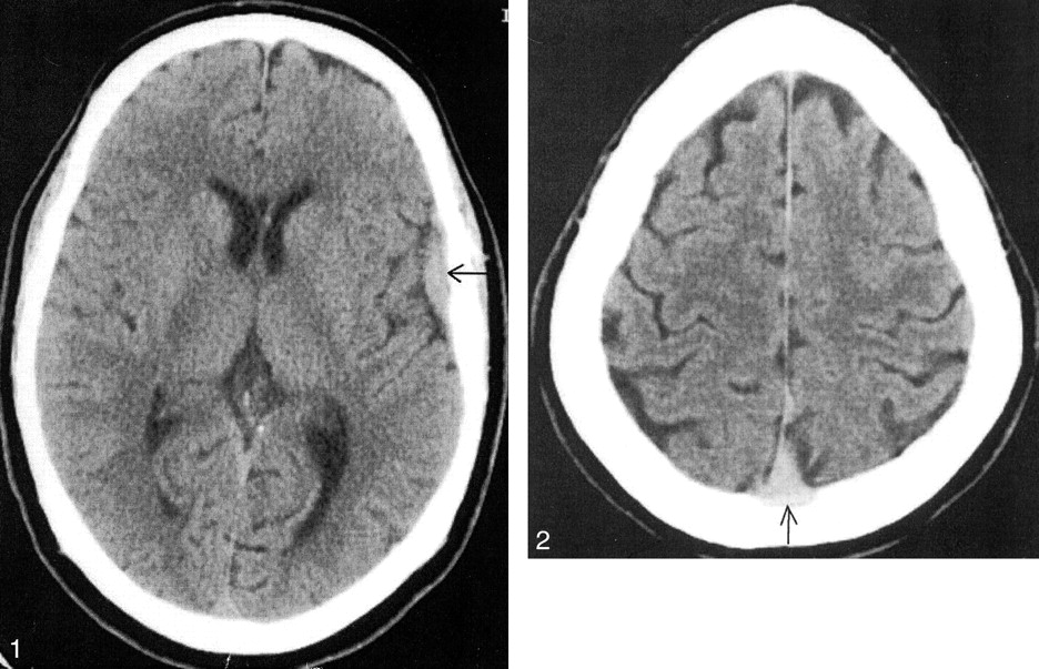
A 65-year-old man with an unremarkable medical history presented to the emergency room for head trauma. A small soft-tissue swelling was noted on his scalp over the left frontal region. The patient also complained of dizziness, headache, and two episodes of nonprojectile vomiting. CT of the brain revealed a convex left frontotemporal area of high attenuation that had the appearance of an extraaxial hematoma (Fig 1). No skull fracture or midline shift was noted.

Axial CT image shows a convex area of high attenuation in the left frontotemporal region (arrow), which has the appearance of an extraaxial hematoma.
Superior images of the brain demonstrated a slightly enlarged triangular area of attenuation posteriorly, which appeared to be greater than the normal attenuation of the superior sagittal sinus (Fig 2). The possibility of superior sagittal sinus thrombosis was therefore entertained, and MR imaging with MR venography of the brain was recommended. Unfortunately, for technical reasons, MR venography could not be performed, and only MR imaging of the brain was performed.

Axial CT image shows a slightly enlarged triangular area of high attenuation seen posteriorly (arrow), suggesting the possibility of superior sagittal sinus thrombosis.
MR imaging of the brain revealed a tubular structure extending from the left sylvian fissure and coursing posteriorly and superiorly over the left parietal convexity to drain into the superior sagittal sinus (Figs 3 and 4). This tubular structure demonstrated a flow void, suggesting that it was a vascular structure. Also, it corresponded to the location of the convex areas of high attenuation seen in the left frontotemporal region on the CT image of the brain. The superior sagittal sinus demonstrated a normal flow void within it, proving that it was not thrombosed. The hyperattenuated superior sagittal sinus that was seen on the CT images was actually a dilated distal portion of this anomalous vessel that appeared to displace the superior sagittal sinus slightly off midline before draining into it (Fig 5). In retrospect, a linear area of high attenuation was seen on the superior images of the CT series, representing the distal portion of this anomalous vessel (Fig 6).
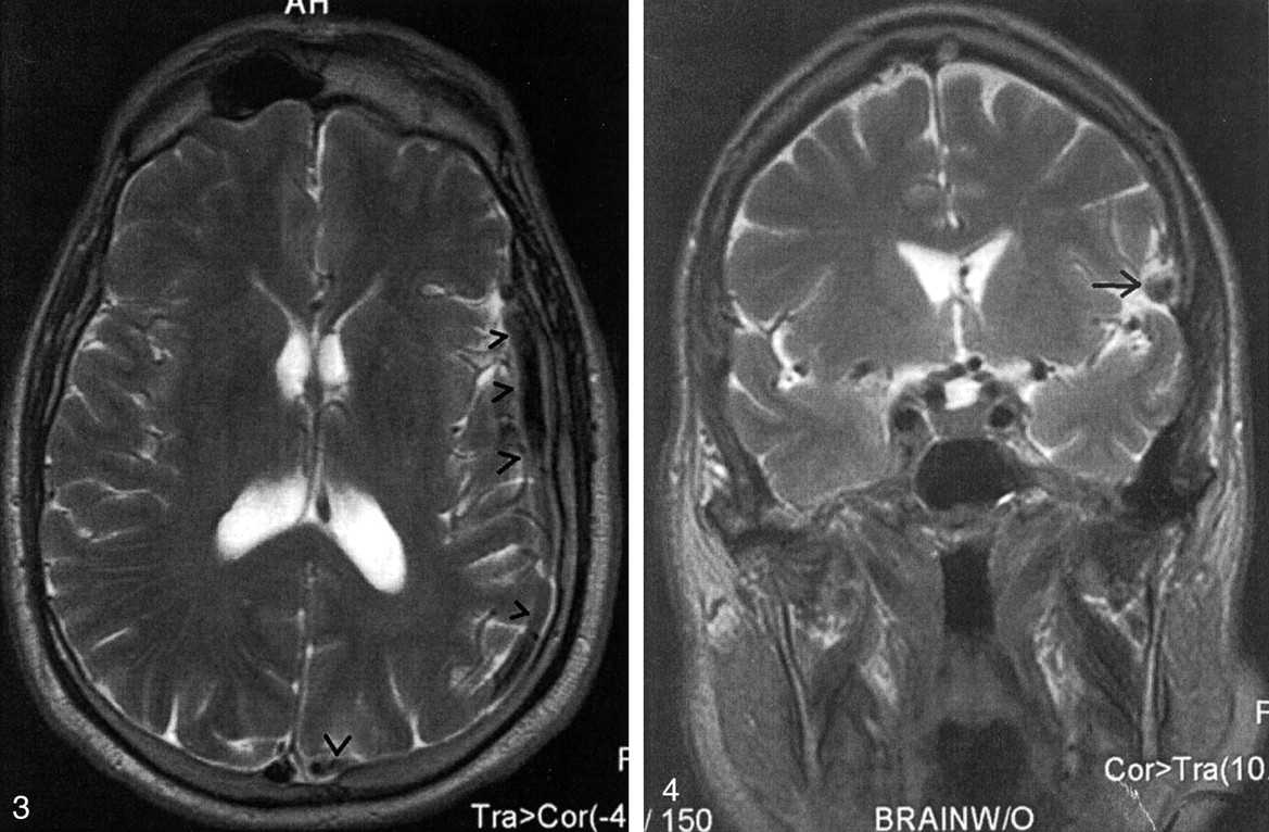
Axial T2-weighted MR image shows a tubular structure demonstrating a flow void extending from the left sylvian fissure and coursing posteriorly over the left parietal convexity to drain into the superior sagittal sinus (arrowheads).

Coronal T2-weighted MR image shows a rounded structure with a flow void in the left sylvian fissure (arrow)

Axial T2-weighted MR image depicts the dilated distal portion of the anomalous vessel (arrow) displacing the superior sagittal sinus slightly off midline before draining into it.

Axial CT depicts a linear area of high attenuation posteriorly (arrow), representing the distal portion of the anomalous vessel just before its entry into the superior sagittal sinus.
Discussion
The SMCV usually runs downward and forward along the sylvian fissure to drain into either the sphenoparietal or the cavernous sinus; however, many variations to the flow have been reported. In their extensive study, Suzuki et al (1) identified seven different drainage patterns of the SMCV: the sphenoparietal, cavernous, emissary, superior petrosal, basal, squamosal, and underdeveloped type of draining veins or sinuses.
In the underdeveloped vein or sinus drainage pattern, the SMCV is absent and the venous drainage of the superficial sylvian area is through a large channel that extends forward, upward, upward and backward, or downward and backward into the superior sagittal sinus or the transverse sinus. We believe our patient had an underdeveloped type of SMCV with an associated large venous channel that extended from the sylvian fissure, coursing backward and upward to eventually drain into the superior sagittal sinus. The increased attenuation of the presumed superior sagittal sinus noted on the superior CT images was actually the distal dilated portion of this vessel that displaced the superior sagittal sinus slightly off midline just before entering it. The diagnosis of an extraaxial hematoma could have led to a neurosurgical evacuation procedure, which may have resulted in significant patient morbidity or death.
Conclusion
The purpose of this case report is to alert the radiologist that an SMCV variant can mimic an extraaxial hematoma. Correct diagnosis is essential in preventing an inappropriate treatment strategy.
References
- Received July 29, 2002.
- Accepted after revision August 11, 2002.
- Copyright © American Society of Neuroradiology
In this issue


Jump to section
Related Articles
Cited By...
- No citing articles found.