Fig 4.
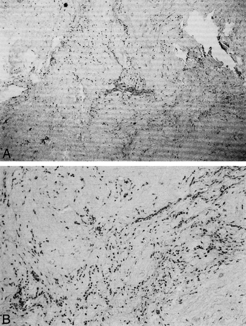
Fig 4.
A, Low-magnification photomicrograph of histologic specimen of the intervertebral disk shows chronic inflammatory infiltrate (hematoxylin & eosin stain; original magnification, × 4).
B, Higher magnification photomicrograph of histologic disk specimen discloses the lymphocytic nature of the infiltrate (hematoxylin & eosin stain; original magnification, × 10).


