
Article Figures & Data
Figures
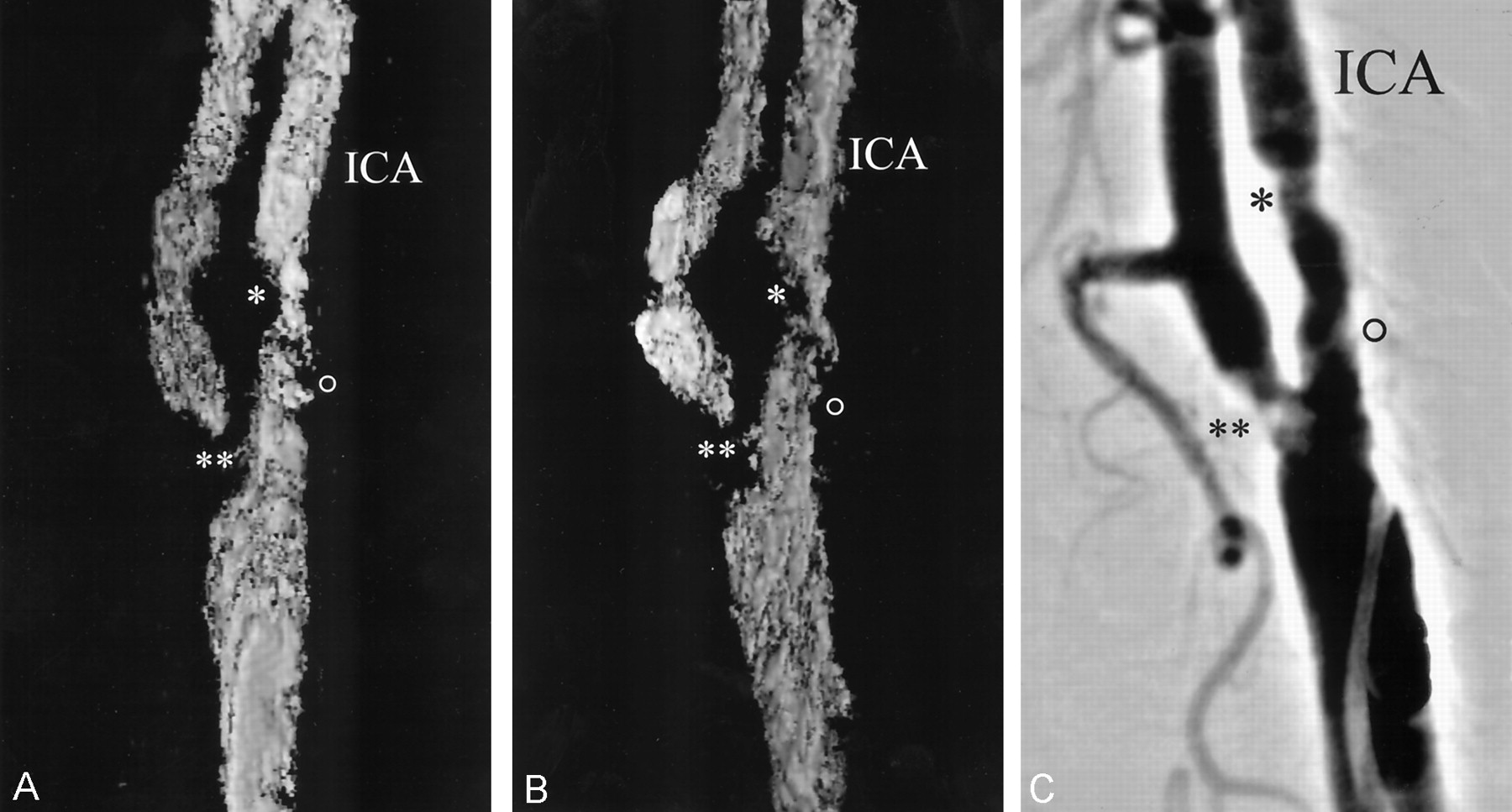
- Fig 1.
Evaluation of reconstruction accuracy of 3D-CDS compared with that of DSA in a patient with moderate ICAS.
Note corresponding findings in two reconstructed color (shown here in black and white) multi-intensity-projection 3D images (A and B) and a DSA image (C) indicate excellent reproducibility. The single asterisk denotes moderate ICAS, the double asterisk denotes high-grade external carotid artery stenosis, and the open circle denotes mild ICAS.
- Fig 2.
Comparison of 3D-CDS (A) and DSA (B) in a patient with high-grade (>82%) ICAS. Stenosis is marked with arrows. An asterisk marks mild ECA stenosis
- Fig 3.
Comparison of 3D-CDS (A) and DSA (B) in a patient with moderate (50–59%) ICAS. Stenosis is marked with arrows. An asterisk marks mild external carotid artery stenosis


Tables
- TABLE 1:
Cut-off points for the quantification of internal carotid artery stenosis by use of CDS according to Nicolaides et al (15) for minimum false-positive and false-negative tests
Stenosis (%)* Duplex Velocity Criteria (m/s) PSVICA EDVICA PSVICA/PSVCCA PSVICA/EDVCCA EDVICA/EDVCCA <47% <150 <80 <2 <10 <2.6 60% 150–250 80–130 2–3.2 7–10 <2.6 70% 150–250 >130 3.2–4 10–15 2.6–5.5 82% >250 >130 >4 15–25 2.6–5.5 90% >250 >130 >4 >25 >5.5 ↵* Diameters determined by angiography according to NASCET criteria.
Note.—CDS indicates color Doppler sonography; PSV, peak systolic velocity; EDV, end diastolic velocity.
- TABLE 2:
CDS and 3D-CDS classification of internal carotid artery stenosis in patients and controls determined among three observers
CDS 3D-CDS (Observer 1) 3D-CDS (Observer 2) 3D-CDS (Observer 3) Controls 16 (33.3%) 10 (20.8%) 8 (16.7%) 12 (25%) ICAS, 30–69% 14 (29.2%) 22 (45.8%) 26 (54.2%) 20 (41.7%) ICAS, 70–99% 18 (37.5%) 16 (33.3%) 14 (29.2%) 16 (33.3%) Note.—CDS indicates color Doppler sonography; 3D-CDS, three-dimensional color Doppler sonography; ICAS, internal carotid artery stenosis.
- TABLE 3:
Sensitivity, specificity, positive predictive values, and negative predictive values for the detection of high-grade internal carotid artery stenosis of 3D-CDS (CDS was criterion standard for all three observers)
Sensitivity Specificity Positive Predictive Value Negative Predictive Value Observer 1 83.3% (15/18) 96.7% (29/30) 93.8% (15/16) 90.6% (29/32) Observer 2 77.8% (14/18) 100% (30/30) 100% (14/14) 88.2% (30/34) Observer 3 83.3% (15/18) 100% (30/30) 100% (15/15) 90.9% (30/33) Note.—CDS indicates color Doppler sonography; 3D-CDS, three-dimensional color Doppler sonography.
In this issue


Jump to section
Related Articles
Cited By...
- 4-Dimensionally Guided 3-Dimensional Color-Doppler Ultrasonography Quantifies Carotid Artery Stenosis With High Reproducibility and Accuracy
- Three-Dimensional Color-Coded Duplex Sonography for Assessment of the Vertebral Artery Origin and Vertebral Artery Stenoses
- Three-Dimensional Assessment of Extracranial Doppler Sonography in Carotid Artery Stenosis Compared With Digital Subtraction Angiography