
Abstract
BACKGROUND AND PURPOSE: Prior reports have described increased signal intensity (SI) of CSF on fluid-attenuated inversion recovery (FLAIR) images of anesthetized patients receiving 100% O2. This appearance can simulate that of diseases. We evaluated the relationship between the concentration of inhaled O2 and the development of increased SI of CSF on FLAIR images.
METHODS: FLAIR was performed in 25 healthy volunteers breathing room air and 100% O2 through a face mask for 5, 10, and 15 minutes. MR imaging, including FLAIR imaging, was performed in 52 patients with no potential meningeal abnormalities under general anesthesia: 21 received an equal mixture of N2O and O2, and 31 received 100% O2. The SI of CSF in volunteers and patients was graded in several locations by using a three-point scale.
RESULTS: SI of CSF significantly increased (P < .05) in various locations, in both volunteers and patients breathing 100% O2, when compared with SI in the same volunteers breathing room air. Hyperintensity of CSF was not significantly different in volunteers receiving 100% O2 through a face mask compared with anesthetized patients receiving 100% O2 through a laryngeal airway or an endotracheal tube. No significant increase in SI occurred in patients receiving 50% O2, when compared with the SI of volunteers breathing room air.
CONCLUSION: Supplemental oxygen at 100% is a main cause of artifactual CSF hyperintensity on FLAIR images, regardless of the anesthetic drug used. This artifact does not develop when 50% O2 is administered.
Reports describe increased signal intensity (SI) in the subarachnoid space on fluid-attenuated inversion recovery (FLAIR) MR images obtained in patients with no clinical or laboratory evidence of CSF or meningeal diseases under general anesthesia. These changes are seen in the basal cisterns and along the cerebral sulci. Initially, these changes were attributed to propofol (1); however, further studies showed that the increased SI of CSF is associated with the administration of 100% oxygen (2, 3).
Understanding the relationship and interaction of different concentrations of O2, inhaled anesthetics, and the SI of CSF on FLAIR images will allow more reliable evaluation of anesthetized patients with suspected meningeal diseases or subarachnoid hemorrhage (SAH). This study was conducted to evaluate the hyperintensity of CSF on FLAIR images of healthy volunteers (control group) and in patients under general anesthesia receiving different concentrations of O2.
Methods
The MR images for this prospective study were obtained between January 2002 and April 2002. The control group was composed of 25 healthy volunteers: 10 women (40%) and 15 men (60%) aged 22–54 years (mean, 28.7 years). FLAIR images were obtained in subjects breathing room air. Then, a non-rebreathing face mask delivering 100% O2 was applied, and FLAIR imaging was repeated 5, 10, and 15 minutes later. Healthy volunteers were examined and their informed consent obtained.
The patient group was composed of 52 patients requiring general anesthesia and undergoing routine MR imaging in a large general hospital. None of the patients had potential meningeal abnormalities. Their ages ranged from 2 months to 72 years (mean, 9.3 years). The group included 23 female (44%) and 29 male (56%) subjects. Twenty-one patients received a mixture of nitrous oxide (N2O) and 50% O2 (subgroup 1), and 31 patients received 100% O2 (subgroup 2). The institutional review board at our hospital approved the imaging of healthy volunteers and waived the need for informed consent from the patients because our study did not interfere with their regular clinical care.
FLAIR images were obtained by using a 1-T magnet and a TR/TE/TI of 11,000/140/2600. The sections ranged from 4 to 6 mm in thickness and were reconstructed at 0.4–0.6-mm increments. A fixed window width of 950 and a window level of 650 were used for documentation on film. One experienced neuroradiologist (A.J.R.) who was blinded to the concentration of O2 and the type of anesthetic used interpreted the images. All cases were randomly reviewed on two separate occasions.
The same face mask was used in the volunteers, and the same ventilator was used in all patients. Particular care was taken to prevent the leakage of room air into the face mask of the healthy volunteers. The patients were ventilated with either a laryngeal mask and airway or an endotracheal tube. The anesthetic drugs were selected at the discretion of the anesthesiologist on a case-by-case basis. Propofol (Propovan; Cristália, Itapira, Brazil), sevoflurane (Sevocris; Cristália), or halothane (Halothano; Cristália) was used. The SI of CSF was graded at several locations: the cerebellopontine angle (CPA); the ambient, quadrigeminal, interpeduncular, prepontine, and suprasellar cisterns; the high-convexity sulci; the Sylvian fissures; and the lateral, third, and fourth ventricles. A three-point grading scale was used: 0 indicated no hyperintensity (complete nulling of CSF SI), I, slightly increased SI or unilateral hyperintensity; and II, significant hyperintensity or bilateral hyperintensity or both.
The Wilcoxon test was used to examine differences in SI in the CSF of the healthy control subjects before and after the administration of O2. The Kruskal-Wallis test, a nonparametric version of the analysis of variance test, was used to compare the control group and both patient subgroups. P values of .05 or less were considered to indicate a significant difference.
Results
In the control group, a statistically significant difference (P = .004) of the SI in several locations was demonstrated when we compared images obtained with room air (Fig 1, Table 1) and those obtained with 100% O2 (Fig 2). The affected locations were the following: the CPA, the ambient, interpeduncular, quadrigeminal, and suprasellar cisterns; the high-convexity sulci; the Sylvian fissures; and the fourth ventricle. There was no significant difference in the SI in the third (P = .102) and lateral (P = .317) ventricles. We also observed no significant difference between the images obtained during 5, 10, and 15 minutes of 100% O2 inhalation.

FLAIR images of a healthy volunteer breathing room air show the expected suppression of the SI of the CSF in the cerebral sulci, fissures, and most of the cisterns.

FLAIR images of the same healthy volunteer as in Figure 1 breathing 100% O2 show hyperintensity in the cerebral sulci, fissures, and cisterns. There is no hyperintensity in the third and lateral ventricles.
SI grades in various CSF spaces in healthy volunteers breathing room air or 100% O2
The SI grades in various CSF spaces in patients breathing 50% and 100% O2 and in healthy volunteers breathing room air are compared in Table 2. Subgroup 1 (N2O/O2) had a statistically different SI only in the CPA cistern (P = .002) and in the prepontine cistern (P = .041), when compared with the SI in the control group breathing room air. Increased SI was seen in these locations, even in healthy subjects breathing room air, as a result of a CSF flow artifact. In fact, the number of individuals with increased SI in the CPA and prepontine cisterns was greater in the control group than in subgroup 1; this finding was due to CSF flow artifact rather than differences in the fraction of inspired oxygen.
SI grades in various CSF spaces in patients breathing 50% and 100% O2 and in healthy volunteers breathing room air
When compared with the controls breathing room air, subgroup 2 (100% O2) had a significantly increased (P = .045) SI in all locations. Similarly, SI was significantly different (P = .022) between subgroup 1 (N2O/O2) and subgroup 2 (100% O2) in all locations. In addition, there was no difference in CSF SI in the control subjects inhaling 100% O2 and in patients in subgroup 2 (100% O2).
Discussion
The mechanism of inversion and recovery causes nulling of the SI of free fluid, making it particularly helpful in the detection of lesions adjacent to the CSF spaces, such as the periventricular and convexity regions. Increases in the protein content or cellularity of the CSF cause a decrease in the T1 relaxation time, with consequent incomplete or absent nulling of SI in the CSF (4). Noguchi et al (5) showed the great value of FLAIR imaging in the evaluation of suspected SAH. Singer et al (6) underscored the importance of FLAIR studies in the diagnosis of neoplastic and inflammatory diseases of the meninges, as well as SAH. Limitations of the FLAIR sequence include magnetic susceptibility artifacts—from the oral cavity, paranasal sinuses, or metallic objects—and CSF pulsation and turbulence artifacts (4, 5).
Filippi et al (1) observed increased SI in the CSF on FLAIR images in children anesthetized with propofol. The authors attributed the increased SI in the convexity region and basal cisterns to the use of propofol, since none of their patients was suspected of having SAH or meningeal disease. The authors offered several possible explanations for the abnormally high SI: incomplete nulling of SI due to propofol (which has a T1 value approaching that of CSF), a transient increase in the protein content within the CSF induced by propofol, and changes in vascular tone caused by propofol that leads to hyperdynamic CSF pulsations. They also suggested that supplemental oxygen could account for the artifact.
Deliganis et al (2) investigated the causes of increased CSF SI on FLAIR images obtained in patients under general anesthesia. They demonstrated the relationship between the increased SI of the CSF and the delivery of oxygen at 100% in eight of nine patients imaged. The single patient in that study who did not have increased SI of the CSF received 50% oxygen. Their study included the imaging of phantoms with varying concentrations of anesthetics and O2. They demonstrated a major reduction in T1 in the phantoms equilibrated with 100% O2. The authors state that tracheal intubation is not necessary to elicit the findings; a facial mask suffices in raising the partial pressure of O2.
By controlling the concentration of O2 delivered to our subjects and by gauging its effect on the SI of the CSF, we hope to help establish parameters for O2 supplementation that will not interfere with FLAIR imaging. In our study, there was a similar increase in the SI of CSF in both healthy volunteers who received 100% O2 through a face mask and in patients who received 100% O2 with an anesthetic drug.
This observation demonstrates that this increase in SI is mostly related to the concentration of inhaled O2, which is in agreement with the results obtained by Deliganis et al (2) and Frigon et al (7). The latter study showed that the type of anesthetic agent is not associated with CSF hyperintensity. Other mechanisms may be involved in the increased SI in the CSF in anesthetized patients. For instance, Fillipi et al (1) described two children with increased SI in the convexity and basal cisterns who received propofol but not supplemental oxygen.
There was no increase in the SI of the CSF in our anesthetized patients who received a mixture of N2O and 50% O2. Their findings were similar to those of healthy volunteers breathing room air. Deliganis et al (2) suggested a similar trend; however their sample was small. Our larger sample allows us to state that patients receiving 50% O2 do not have a significant change in the SI of CSF on FLAIR images. Similarly, Frigon et al (7) showed that an inspired oxygen fraction smaller than 0.60 is not associated with CSF hyperintensity.
In our sample, isolated hyperintensity in the CPA and prepontine cisterns was seen, regardless of the use of supplemental oxygen or anesthetics. This artifact is inherent to the FLAIR sequence and due to the random flow of CSF, which disturbs SI suppression in these locations. This isolated hyperintensity is easily distinguished from the increased SI seen with an elevated partial pressure of O2, with which the hyperintensity is more homogeneous and diffuse (2, 4).
We also found that the increase in SI was less evident within the ventricles than along the cortical sulci and within the cisterns. This finding is in agreement with that of Deliganis et al (2), who hypothesized that O2 diffuses into the cisternal subarachnoid CSF compartment directly from the arterial vascular space through the walls of arteries and arterioles on the pia-arachnoid surface of the brain. They also pointed out that there is a relative lack of major arterial vessels lining the ventricles. Oxygen rapidly diffuses through the blood-CSF barrier, presumably at the pial vessels and through the choroid plexus. A dilutional effect and a less prominent vasculature could at least partly account for the lack of SI increase in the ventricles, as opposed to the sulci and cisterns.
The development of hyperintensity after the administration of O2 is likely due to an increased concentration of oxyhemoglobin in the blood, which causes a paramagnetic effect in the CSF (1). Changes in cerebral blood flow velocity could possibly exacerbate this artifact.
In our current practice, we evaluate the indication for each MR imaging study to be performed under anesthesia to avoid this artifact, or, in a few selected cases, we attempt to use the artifact to our advantage. The delivery of 100% O2 can be used to create a type of noninvasive MR cisternography. We already use this technique in the evaluation of patients with neurocysticercosis (Figs 3 and 4), especially when the subarachnoid space is believed to be involved, to obtain greater contrast of the cyst wall, cortex, and CSF in the cisterns (unpublished data).

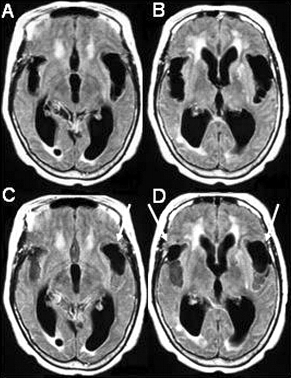
78-year-old female patient evaluated for suspected normal pressure hydrocephalus.
A and B, MRI FLAIR sequence (1.0 T, TR 11,000 ms, TE 140 ms, TI 2,600 ms) shows dilatation of the ventricles and Sylvian fissures.
C and D, FLAIR after 100% O2 for 5 minutes shows increased SI in the sulci and in the posterior aspects of both Sylvian fissures, allowing visualization of hypointense cysts (arrows) due to neurocysticerosis more anteriorly within the Sylvian fissures. Note that there is no increase in the SI of the CSF within the ventricles.

51-year-old male patient with headache.
A and B, FLAIR image shows a large cyst with a scolex in the right Sylvian fissure and a cyst in the suprasellar cistern on the right, which are consistent with neurocysticerosis. The presence of an additional cyst in the perimesencephalic cisterns could not be ruled out due to prominence of these cisterns.
C and D, FLAIR after 100% O2 shows increased SI in the sulci and basal cisterns, confirming the absence of cysts in the perimesencephalic cisterns. In addition, there was improvement in the visualization of the cyst in the right suprasellar cistern and its relationship to the M1 segment of the ipsilateral middle cerebral artery.
Conclusion
Supplemental oxygen at 100% causes an artifactual hyperintensity of the CSF on FLAIR images. This artifact develops during the first 5 minutes of O2 supplementation at 100% and does not significantly change in the following 10 minutes. In our study, this hyperintensity was not dependent on the type of anesthetic drug used and did not develop when 50% O2 was administered. If clinically feasible, patients being evaluated for SAH or meningeal disease should receive no more than 50% oxygen to prevent the artifactual increase in the SI of the CSF expected on FLAIR imaging.
- Received March 5, 2003.
- Accepted after revision July 18, 2003.
- Copyright © American Society of Neuroradiology


