
Article Figures & Data
Figures
- Fig 1.
Superior view of right middle cranial fossa. The superficial middle cerebral vein (arrows) drains into the laterosellar region into the cavernous sinus. The superficial middle cerebral vein is not related with the lesser sphenoid wing during its course and it pierces the dura mater of the lateral wall of the cavernous sinus directly. Transition from arachnoid vein to dural venous sinus is abrupt. ICA signifies internal carotid artery; double arrow, tentorial edge; arrowheads, middle meningeal veins.
- Fig 2.
Dural arteriovenous fistula of the lesser sphenoid wing.
A, Early filling of a selective right external carotid artery injection in the anteroposterior projection. Nourishing branches from the right middle meningeal artery (black arrows) feeds a dilated superficial middle cerebral vein (black arrowhead). The superficial middle cerebral vein drains into a laterocavernous sinus (white arrowhead), which is also directly involved in the dural arteriovenous fistula and is fed by branches of the right middle meningeal artery. Early anterograde filling of the cavernous sinus is observed. There is retrograde filling into the right basal vein (white arrows) and into the right peduncular vein, through the right deep middle cerebral vein.
B, Selective right external carotid injection, later in the venous phase. Retrograde filling of the right superficial cortical veins (black arrow) via the right superficial middle cerebral vein is now observed. The left basal vein (black double arrow) is now also opacified via the peduncular veins, and the straight sinus is clearly delineated. There is infratentorial drainage of the fistula into the anterior and lateral pontomesencephalic veins (black arrowheads). The saccular dilatation of the superficial middle cerebral vein is clearly visible (asterisk).
- Fig 3.
Serial dissection of a fresh specimen.
A, Superior topographic view of the right. The brain has been removed and the bridging veins of the temporal pole, in this case a single superficial middle cerebral vein (black arrowhead), have been sectioned close to the brain surface. The superficial middle cerebral vein is attached to the dura mater overlying the lesser sphenoid wing but keeps the appearance of an arachnoid vein. Note the different appearance between the superficial middle cerebral vein and the middle meningeal vessels (white arrowheads), the latter being embedded in the dura mater. The superficial middle cerebral vein terminates in a laterocavernous sinus (black arrows). Note that in this case the laterocavernous sinus shares both arachnoid and dural characteristics being as translucent as the superficial middle cerebral vein but more greatly embedded in the dura matter. The lateral wall of the cavernous sinus becomes a definite dural venous sinus more posteriorly (white arrows) as it drains into the superior petrosal sinus (asterisk). ICA signifies internal carotid artery.
B, The dura mater of the middle cranial fossa and the ridge of the lesser sphenoid wing have been removed to reveal the sphenoid (small black arrows) and parietal (large black arrow) portions of the anterior branch of the middle meningeal veins and the sinus of the lesser sphenoid wing (black arrowheads). The white arrows demonstrate the location of the sphenoparietal canal (of Trolard), whose roof has been removed. The superficial middle cerebral vein (black double arrow) has been kept in situ and no connections are demonstrated with the sinus of the lesser sphenoid wing. The anterior branch of the middle meningeal artery is seen between the two veins that constitute the anterior branch of the middle meningeal veins.
C, The inner bony plate of the frontal and sphenoid bones has been removed to expose diploic vessels. Part of the roof of the orbit has been removed to expose the superior ophthalmic vein (SOV). The diploic vein of the orbital roof (black arrowheads) is followed anteriorly and exits the skull through the supraorbital foramen (not shown here). The diploic vein of the orbital roof connects with a frontal diploic vein (white arrows) that drains into the superior longitudinal sinus. The diploic vein of the greater sphenoid wing (black arrows) drains into the pterygoid plexus extracranially.
- Fig 4.
Anterior and lateral view of the left convexity of a corrosion cast showing the parietal portion of the anterior branch of the middle meningeal veins. The dual meningeal and diploic nature of the parietal portion of the anterior branch of the middle meningeal veins is demonstrated. The anterior parietal diploic vein (black arrows) and the parietal portion of the anterior branch of the middle meningeal veins may be individualized wherever their course does not overlap. Note how diploic veins enter the venous lacunae of the superior longitudinal sinus at a right angle (white arrow). A parietotemporal diploic vein is demonstrated (white arrowheads); it drained into the middle third of the left transverse sinus.
Fig 5. Corrosion cast specimens illustrating the venous structures in the region of the lesser sphenoid wing.
A, Anteroposterior view of left side of a corrosion cast showing the sinus of the lesser sphenoid wing (arrowheads), the parietal portion of the anterior branch of the middle meningeal veins (large arrow), and the sphenoid portion of the anterior branch of the middle meningeal veins (small arrows). Different branches of the superficial middle cerebral vein (double arrows) are seen behind the sinus of the lesser sphenoid wing. The superficial middle cerebral vein drains into a paracavernous sinus (large asterisk). The sinus of the lesser sphenoid wing is seen to cross over the superior ophthalmic vein (SOV). Only the dorsal aspect of the superior ophthalmic vein was filled in this side. Note the different aspects of the sphenoid and parietal portions of the anterior branch of the middle meningeal veins, in which the former offers a typical aspect of parallel meningeal channels, whereas the latter resembles a diploic vein. A diploic vein of the greater sphenoid wing (small asterisks) is seen to drain into the pterygoid plexus.
B, Superior view of the right side of a corrosion cast in the region of the right lesser sphenoid wing, demonstrating the sinus of the lesser sphenoid wing (white arrowheads), the diploic vein of the orbital roof (asterisk; the anterior portion of this vein is not filled), and the superficial middle cerebral vein (arrows) draining into a lateral wall of a cavernous sinus (not seen). Note how the superficial middle cerebral vein and the sinus of the lesser sphenoid wing are not connected and course on different anatomic planes. The sinus of the lesser sphenoid wing typically crosses over the dorsal portion of the superior ophthalmic vein. The anterior branch of the middle meningeal veins was not filled in this side. SOV signifies superior ophthalmic vein; CS, cavernous sinus; SLS, superior longitudinal sinus.
- Fig 6.
Various venous phases of digital subtraction angiography using selective internal carotid artery injections in three patients with no confirmed cerebrovascular disease.
A, Lateral projection of a late venous phase shows the characteristic venous sinus appearance of the parietal portion of the anterior branch of the middle meningeal veins, seen as two distinct thin channels coursing in parallel (arrowheads).
B, Lateral projection of a late venous phase depicts both the sphenoid (double arrowhead) and parietal (single arrowheads) portions of the anterior branch of the middle meningeal veins. The parietal portion is seen as a wide sinuous channel typical of a diploic channel.
C, Anteroposterior projection demonstrating simultaneously the sinus of the lesser sphenoid wing and the superficial middle cerebral vein. The sinus of the lesser sphenoid wing (black arrowheads) drains medially into the cavernous sinus and is connected laterally to the diploic vein of the orbital roof (black arrow). The superficial middle cerebral artery (white arrows) is seen to drain through emissary veins that drain into the pterygoid plexus (PP).
D, As in 5C, lateral view shows the diploic vein of the orbital roof (black arrows) connecting extracranially with the supraorbital veins (black double arrows). The superficial middle cerebral vein (black arrowheads) is well delineated and drains into the emissary veins of the middle cranial fossa.
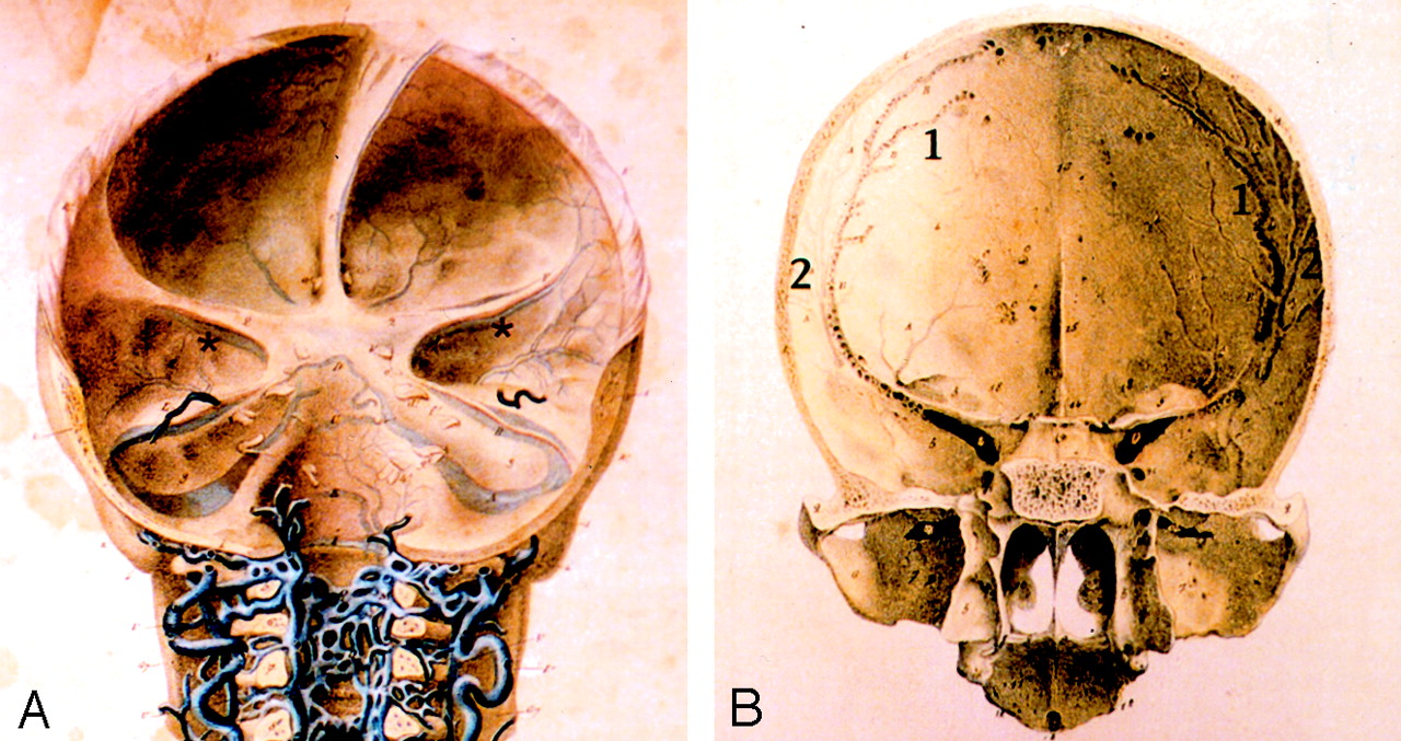
- Fig 7.
Plates from Breschet’s atlas (courtesy of Mme. B. Molitor, Section Histoire de la Médecine, Bibliotèque Inter Universitaire de Médecine, Paris, France).
A, Posterior view of the three cranial fossae. The sphenoid portion of the sphenoparietal sinus, corresponding to the sinus of the lesser sphenoid wing, is shown under the lesser sphenoid wing (brown asterisk). Connections between the sphenoparietal sinus and the anterior branch of the middle meningeal veins are illustrated and were described in the legends of the original text. Note that the superficial middle cerebral artery is not depicted.
B, Posterior view of the anterior and middle cranial fossae, in which the dura mater has been removed. The parietal portion of the sphenoparietal sinus (1) is depicted and artificially distinguished from the anterior branch of the middle meningeal veins (2). The floor of the sulcus of the sphenoparietal sinus is pierced by many small foramina. The legend describes these foramina, which are connections between the sphenoparietal sinus and underlying diploic veins.


