
Article Figures & Data
Figures
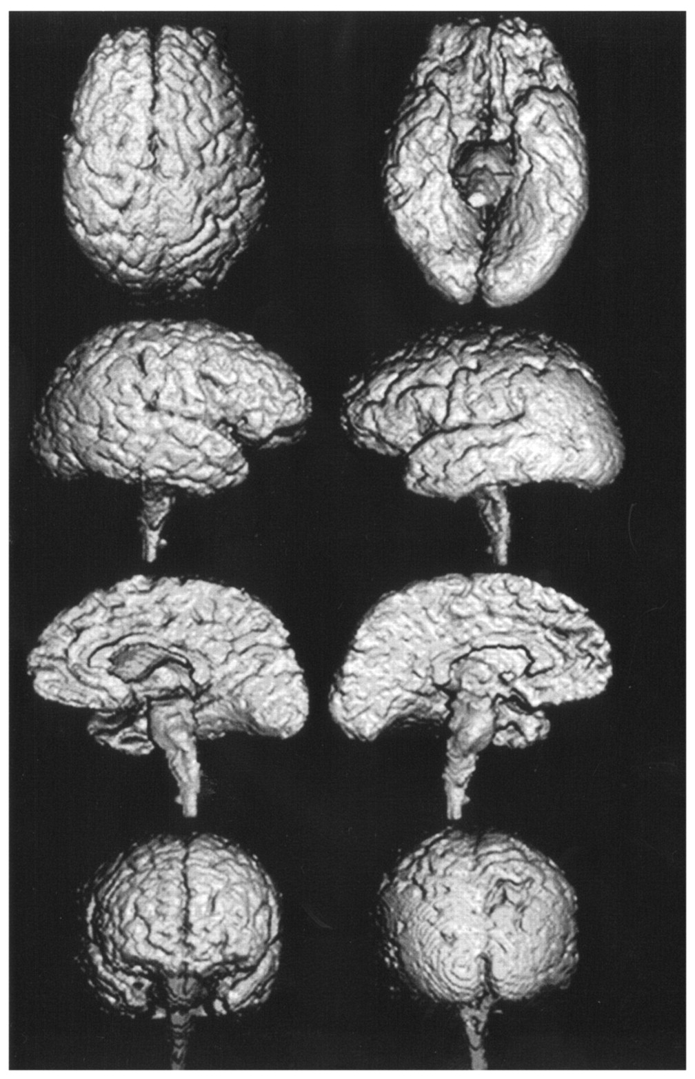
- Fig 1.
3D volume-rendered images reconstructed from thin-section coronal 3D SPGR MR images (TR/TE, 14/3; field of view, 220 mm; matrix, 256 × 256; 124 × 1.5-mm contiguous sections; flip angle, 20°) show mild diffuse atrophy of cerebral cortex and moderate atrophy of brainstem in a 67-year-old woman with PSP (MMSE score of 21).
- Fig 2.
3D volume-rendered images reconstructed from thin-section coronal 3D SPGR MR images (TR/TE, 14/3; field of view, 220 mm; matrix, 256 × 256; 124 × 1.5-mm contiguous section; flip angle, 20°) show severe frontoparietal atrophy in a 63-year-old woman with CBD (MMSE score of 19).
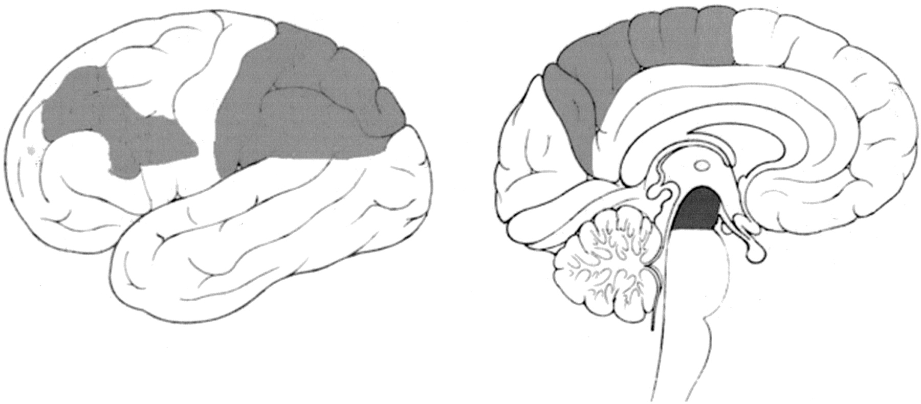
- Fig 3.
Light gray and dark gray areas are regions where atrophy was rated significantly more severe in the CBD and PSP groups, respectively.


Tables
Feature PSP (n = 19) CBD (n = 19) Lim rigidity 14 (74) 18 (95) Axial dystonia or rigidity 17 (89) 7 (37) Postural instability 19 (100) 5 (26) Apraxia 1 (5) 10 (53) Cortical sensory loss 0 (0) 16 (84) Alien limb 0 (0) 8 (42) Myoclonus 0 (0) 12 (63) Postural tremor 1 (5) 9 (47) Hyperreflexia 7 (37) 5 (26) Babinski sign 4 (21) 5 (26) Supranuclear ophthalmoplegia 19 (100) 4 (21) Pseudobulbar palsy 16 (84) 1 (5) Note.—Data in parentheses are percentages.
Region Atrophy Rating PSP CBD Orbitofrontal gyrus 1.8 ± 0.6 1.9 ± 0.7 Anterior inferior frontal gyrus 1.7 ± 0.6 2.1 ± 0.5 Posterior inferior frontal gyrus 1.7 ± 0.6 2.2 ± 0.5* Anterior middle frontal gyrus 1.8 ± 0.8 2.4 ± 0.6* Posterior middle frontal gyrus 1.9 ± 0.8 2.3 ± 0.7 Anterior lateral superior frontal gyrus 2.0 ± 0.8 2.5 ± 0.8 Posterior lateral superior frontal gyrus 1.9 ± 0.7 2.5 ± 0.8 Anterior medial superior frontal gyrus 2.1 ± 0.9 2.7 ± 0.9 Posterior medial superior frontal gyrus 2.1 ± 0.8 2.6 ± 0.8 Lateral precentral gyrus 1.6 ± 0.9 2.1 ± 0.7 Medial precentral gyrus 1.5 ± 0.8 2.1 ± 0.7 Lateral postcentral gyrus 1.7 ± 1.2 2.7 ± 0.7* Medial postcentral gyrus 1.7 ± 1.2 2.7 ± 0.7* Anterior lateral superior parietal lobule 2.1 ± 1.3 3.5 ± 1.1* Posterior lateral superior parietal lobule 2.1 ± 1.1 3.7 ± 1.1* Anterior medial superior parietal lobule 2.2 ± 1.2 3.4 ± 1.2* Posterior medial superior parietal lobule 2.2 ± 1.2 3.5 ± 1.2* Anterior inferior parietal lobule 1.6 ± 1.3 3.2 ± 0.8* Posterior inferior parietal lobule 1.7 ± 1.2 3.7 ± 0.7* Anterior superior temporal gyrus 2.5 ± 0.5 2.6 ± 0.5 Posterior superior temporal gyrus 2.4 ± 0.5 2.5 ± 0.5 Anterior middle temporal gyrus 2.1 ± 0.6 2.2 ± 0.5 Posterior middle temporal gyrus 1.9 ± 0.5 2.1 ± 0.3 Anterior inferior temporal gyrus 2.0 ± 0.5 2.1 ± 0.4 Posterior inferior temporal gyrus 2.0 ± 0.4 2.0 ± 0.4 Anterior parahippocampal gyrus 2.3 ± 0.5 2.2 ± 0.6 Posterior parahippocampal gyrus 2.3 ± 0.5 2.2 ± 0.6 Anterior medial occipital lobe 1.1 ± 0.3 1.1 ± 0.2 Posterior medial occipital lobe 1.1 ± 0.3 1.1 ± 0.2 Anterior lateral occipital lobe 1.1 ± 0.3 1.1 ± 0.2 Posterior lateral occipital lobe 1.1 ± 0.3 1.1 ± 0.2 Midbrain 2.8 ± 1.2* 1.6 ± 1.3 Pons 2.9 ± 1.1 2.2 ± 1.1 * Significantly atrophic compared with the other group (P < .05).
Volume PSP CBD P Value Total intracranial (mL) 1442 ± 124 1393 ± 86 >.05 Whole hemisphere (mL) 1075 ± 130 995 ± 99 <.05 Left hemisphere (mL) 465 ± 52 427 ± 52 <.05 Right hemisphere (mL) 471 ± 50 431 ± 44 <.05 Cerebellar (mL) 117 ± 15 116 ± 14 >.05 Brainstem (mL) 20.6 ± 2.4 22.0 ± 3.1 >.05 Whole hemisphere/total intracranial (%) 74.5 ± 4.4 71.4 ± 4.4 <.05 Left hemisphere/total intracranial (%) 32.2 ± 2.3 30.6 ± 2.6 <.05 Right hemisphere/total intracranial (%) 32.7 ± 2.0 30.9 ± 2.2 <.05 Cerebellar/total intracranial (%) 8.1 ± 0.8 8.3 ± 0.8 >.05 Brainstem/total intracranial (%) 1.4 ± 0.1 1.6 ± 0.2 <.05 Asymmetry index (%) 2.05 ± 1.42 2.84 ± 1.84 <.01 Note.—Data are the mean ± standard deviation.


