
Article Figures & Data
Figures
- Fig 1.
Scatterplot of maximum nCBV (mean normalized rCBV region-of-interest) values versus glial tumor grade. The degree of contrast enhancement on conventional T1-weighted MR images is indicated for each data point. Note that seven (41%) of the 17 high-grade tumors had absent or equivocal enhancement, whereas three (23%) of the 13 low-grade tumors had definite enhancement. At an nCBV cutoff value of 1.5 (horizontal line), no high-grade lesions were incorrectly classified as low grade (100% predictive value for excluding malignancy).
- Fig 2.
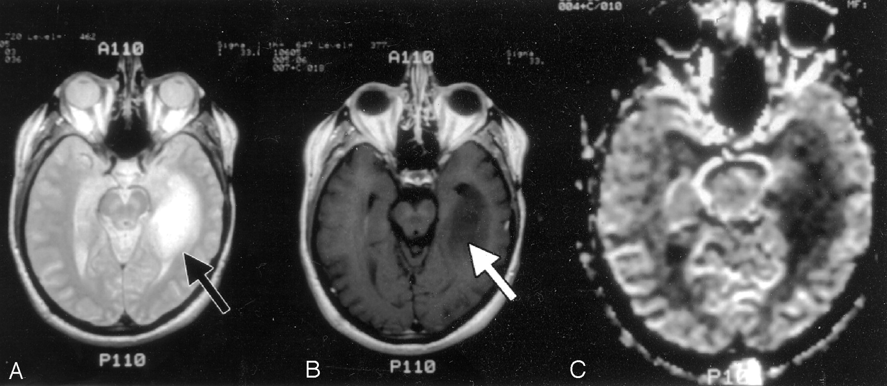
61-year-old man with grade II astrocytoma.
A and B, Axial T2-weighted (A) and contrast-enhanced T1-weighted (B) images demonstrate a mass (arrow) in the left medial temporal lobe that is hyperintense on the T2-weighted image and hypointense, nonenhancing on the T1-weighted image.
C, Correlative rCBV map shows diminished blood volume relative to both gray and white matter in this biopsy-proved low-grade astrocytoma.
- Fig 3.
39-year-old man with grade IV glioma.
A and B, Axial contrast-enhanced T1-weighted (A) and rCBV (B) images through the level of the lateral ventricles demonstrate an enhancing mass. There is elevated blood volume relative to both normal gray and white matter, consistent with biopsy-proved high-grade glioma (arrow). Note the decreased blood volume in the white matter immediately surrounding the lesion, secondary to the space-occupying effects of vasogenic edema.
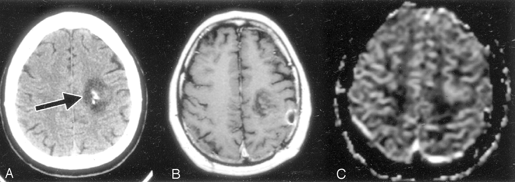
- Fig 4.
65-year-old man with biopsy-proved low-grade oligodendroglioma.
A–C, Axial nonenhanced CT image through the frontal lobes (A), as well as axial contrast-enhanced T1-weighted (B) and rCBV (C) MR images through the same region, demonstrate a calcified left frontal mass (arrow in A). There is trace contrast enhancement and increased blood volume compared with the adjacent gray and white matter. The combination of calcification, relatively mild contrast enhancement, and increased rCBV is not uncommon for oligodendrogliomas of any grade.
- Fig 5.
29-year-old man with mixed malignant oligoastrocytoma.
A–C, Axial T2-weighted (A), contrast-enhanced T1-weighted (B), and rCBV (C) MR images show a lesion in the anterior left mesial temporal lobe that is hyperintense on the T2-weighted image and minimally enhancing on the T1-weighted image. The lesion demonstrates intensely elevated blood volume relative to both gray and white matter (arrow in C).
- Fig 6.
65-year-old man with biopsy-proved anaplastic astrocytoma.
A and B, rCBV map (A) corresponds to the contrast-enhanced T1-weighted MR image (B). Despite only mild enhancement of this high-grade temporal lobe neoplasm on the T1-weighted image, the lesion demonstrates intensely elevated blood volume on the corresponding rCBV map (arrows).
- Fig 7.
ROC curves generated by varying the nCBV cutoff values used to classify the tumor regions of interest (ROIs) shown in Fig 1 as low grade versus high grade. The left curve (diamond-shaped data points) describes the sensitivity and specificity values for low- versus high-grade astrocytomas only. The right curve (square data points) describes the sensitivity and specificity values for low- versus high-grade astrocytomas and oligodendrogliomas grouped together. Of note, the larger area under the left as compared with the right-shifted curve (proportional to accuracy) reflects the superior discriminatory ability of rCBV imaging in distinguishing low- from high-grade astrocytomas than in distinguishing low- from high-grade oligodendrogliomas. At a critical test cutoff value of nCBV = 1.5 for the right-shifted curve (as per Fig 1), sensitivity and specificity for distinguishing low- versus high-grade glial neoplasms are 0.96 and 0.60, respectively. TPF indicates true-positive fraction; FPF, false-positive fraction.
- Fig 8.
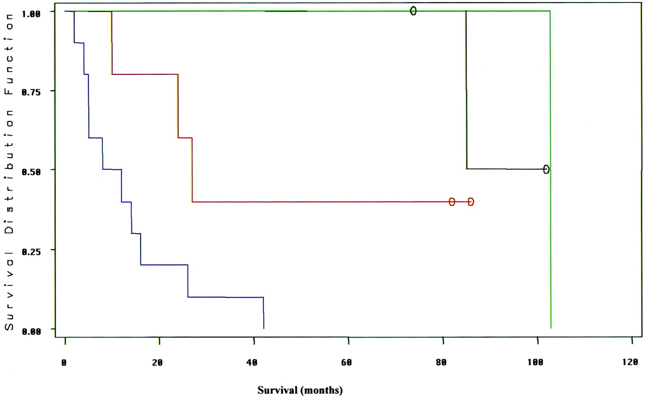
Kaplan-Meir survival curves for the data shown in the Table. Left-shifted curves reflect poor survival, and right-shifted curves reflect good survival. The far left-shifted curve (blue) is for patients with enhancing, high-nCBV (>1.5) tumors; the adjacent left-shifted curve (red) is for patients with nonenhancing, high-nCBV tumors. The far right-shifted curves are for patients with low-nCBV (<1.5) tumors: the rightmost curve (green) is for patients with enhancing tumors and the adjacent curve (black) is for patients with nonenhancing tumors. Although the number of tumors studied was too low to establish a statistically significant difference between enhancement and nCBV as outcome predictors, these data suggest a strong trend toward nCBV being a stronger predictor of survival than is enhancement. Open circles indicate censored data (ie, patients still alive at the time of most recent follow-up.)
- Fig 9.
60-year-old woman with glioblastoma.
A and B, Contrast-enhanced T1-weighted (A) and rCBV (B) MR images show a heterogeneously enhancing anterior right temporal lobe mass. The entire mass, including regions of both minimal and maximal contrast enhancement, demonstrates homogeneously increased rCBV, consistent with biopsy proved high-grade glioma (arrow in B). The elevated blood volume extends even to tumor foci without significant contrast enhancement.


Tables
- TABLE 1:
Survival after baseline MR imaging as a function of patient age, tumor histologic diagnosis, enhancement, and maximal nCBV values in 19 patients with follow-up data
Astrocytoma WHO Grade Age (y) Enhancement nCBV Survival (mo) II/IV* 31 No 0.4 102 II/IV* 23 No 0.8 85 II/IV* 38 No 0.9 74 II/IV 25 Yes 0.9 103 II/IV* 51 No 2.1 82 II/IV* 37 No 2.3 86 III/IV 37 No (trace) 1.6 10 III/IV 71 Yes 1.7 42 IV/IV 52 Yes 1.7 12 IV/IV 69 No 2.2 27 IV/IV 65 Yes 2.3 2 IV/IV 42 No 2.5 24 IV/IV 82 Yes 2.7 5 IV/IV 57 Yes 2.8 14 IV/IV 66 Yes 2.9 4 IV/IV 58 Yes 3.3 26 IV/IV 87 Yes 3.8 5 IV/IV 53 Yes 5.8 16 III/IV 12 Yes 7.3 8 Note.—Survival is sorted by ascending values of nCBV for both the low-grade (II/IV) and high-grade (III/IV and IV/IV) tumor groups.
* Indicates patients still alive at the time of most recent follow-up.
In this issue


Jump to section
Related Articles
Cited By...
- Radiogenomics Provides Insights into Gliomas Demonstrating Single-Arm 1p or 19q Deletion
- Hybrid 18F-Fluoroethyltyrosine PET and MRI with Perfusion to Distinguish Disease Progression from Treatment-Related Change in Malignant Brain Tumors: The Quest to Beat the Toughest Cases
- Effects of Susceptibility Artifacts on Perfusion MRI in Patients with Primary Brain Tumor: A Comparison of Arterial Spin-Labeling versus DSC
- Moving Toward a Consensus DSC-MRI Protocol: Validation of a Low-Flip Angle Single-Dose Option as a Reference Standard for Brain Tumors
- Multisite Concordance of DSC-MRI Analysis for Brain Tumors: Results of a National Cancer Institute Quantitative Imaging Network Collaborative Project
- 3D Pseudocontinuous Arterial Spin-Labeling MR Imaging in the Preoperative Evaluation of Gliomas
- Improving the Grading Accuracy of Astrocytic Neoplasms Noninvasively by Combining Timing Information with Cerebral Blood Flow: A Multi-TI Arterial Spin-Labeling MR Imaging Study
- Mitotic Activity in Glioblastoma Correlates with Estimated Extravascular Extracellular Space Derived from Dynamic Contrast-Enhanced MR Imaging
- Improved Brain Tumor Classification by Sodium MR Imaging: Prediction of IDH Mutation Status and Tumor Progression
- Impact of Software Modeling on the Accuracy of Perfusion MRI in Glioma
- Multimodality Brain Tumor Imaging: MR Imaging, PET, and PET/MR Imaging
- Mean Diffusional Kurtosis in Patients with Glioma: Initial Results with a Fast Imaging Method in a Clinical Setting
- Prognostic Value of Dynamic Susceptibility Contrast-Enhanced and Diffusion-Weighted MR Imaging in Patients with Glioblastomas
- ASFNR Recommendations for Clinical Performance of MR Dynamic Susceptibility Contrast Perfusion Imaging of the Brain
- Three-tesla functional MR language mapping: Comparison with direct cortical stimulation in gliomas
- Role of MRI in Primary Brain Tumor Evaluation
- Advanced Magnetic Resonance Imaging of the Physical Processes in Human Glioblastoma
- Arterial Spin-Labeling Assessment of Normalized Vascular Intratumoral Signal Intensity as a Predictor of Histologic Grade of Astrocytic Neoplasms
- Differentiation between Oligodendroglioma Genotypes Using Dynamic Susceptibility Contrast Perfusion-Weighted Imaging and Proton MR Spectroscopy
- 11C-Methionine Uptake Correlates with Combined 1p and 19q Loss of Heterozygosity in Oligodendroglial Tumors
- Semi-automated and automated glioma grading using dynamic susceptibility-weighted contrast-enhanced perfusion MRI relative cerebral blood volume measurements
- Survival Analysis of Patients with High-Grade Gliomas Based on Data Mining of Imaging Variables
- Imaging Characteristics of Oligodendrogliomas That Predict Grade
- The Added Value of Apparent Diffusion Coefficient to Cerebral Blood Volume in the Preoperative Grading of Diffuse Gliomas
- Does MR Perfusion Imaging Impact Management Decisions for Patients with Brain Tumors? A Prospective Study
- Correlations between Perfusion MR Imaging Cerebral Blood Volume, Microvessel Quantification, and Clinical Outcome Using Stereotactic Analysis in Recurrent High-Grade Glioma
- Quantitative Blood Flow Measurements in Gliomas Using Arterial Spin-Labeling at 3T: Intermodality Agreement and Inter- and Intraobserver Reproducibility Study
- Biology, genetics and imaging of glial cell tumours
- Perfusion CT Imaging of Brain Tumors: An Overview
- Correlation of MR Relative Cerebral Blood Volume Measurements with Cellular Density and Proliferation in High-Grade Gliomas: An Image-Guided Biopsy Study
- In Vivo Correlation of Tumor Blood Volume and Permeability with Histologic and Molecular Angiogenic Markers in Gliomas
- Differentiation among Glioblastoma Multiforme, Solitary Metastatic Tumor, and Lymphoma Using Whole-Tumor Histogram Analysis of the Normalized Cerebral Blood Volume in Enhancing and Perienhancing Lesions
- Combination of high-resolution susceptibility-weighted imaging and the apparent diffusion coefficient: added value to brain tumour imaging and clinical feasibility of non-contrast MRI at 3 T
- Enhancing Fraction in Glioma and Its Relationship to the Tumoral Vascular Microenvironment: A Dynamic Contrast-Enhanced MR Imaging Study
- Optimized Preload Leakage-Correction Methods to Improve the Diagnostic Accuracy of Dynamic Susceptibility-Weighted Contrast-Enhanced Perfusion MR Imaging in Posttreatment Gliomas
- Semiquantitative Assessment of Intratumoral Susceptibility Signals Using Non-Contrast-Enhanced High-Field High-Resolution Susceptibility-Weighted Imaging in Patients with Gliomas: Comparison with MR Perfusion Imaging
- Cerebral Blood Volume Measurements by Perfusion-Weighted MR Imaging in Gliomas: Ready for Prime Time in Predicting Short-Term Outcome and Recurrent Disease?
- Distinguishing Recurrent Intra-Axial Metastatic Tumor from Radiation Necrosis Following Gamma Knife Radiosurgery Using Dynamic Susceptibility-Weighted Contrast-Enhanced Perfusion MR Imaging
- The Blood Oxygen Level-Dependent Functional MR Imaging Signal Can Be Used to Identify Brain Tumors and Distinguish Them from Normal Tissue
- Diagnostic performance of spectroscopic and perfusion MRI for distinction of brain tumors
- Physiologic and Metabolic Magnetic Resonance Imaging in Gliomas
- Dynamic Magnetic Resonance Perfusion Imaging of Brain Tumors