
Article Figures & Data
Figures
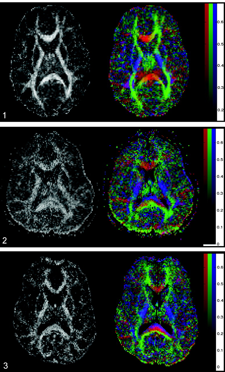
- Fig 1.
Axial FA map (left) and color coded map of mean diffusion direction (right) at the level of the basal ganglia, thalami and internal capsulae in a healthy control subject. Left, Gray-scale FA map displays a high degree of anisotropic diffusion (bright) within the internal capsule and the splenium of the corpus callosum. The cortex and the central gray matter are dark because of their low degree of anisotropic diffusion. Right, Color-coded image displays a predominant left-right-left mean diffusion direction (red) within the center of the splenium of the corpus callosum, an anteroposterior direction (green) within the optic radiations, and a superior-inferior direction (blue) within the posterior internal capsule.
- Fig 2.
Images in a 24-year-old man with severe TBI. Acute GCS, 5. Rankin score at discharge, 3. Left, FA map shows a reduced FA index of the splenium of the corpus callosum (FA = 0.511 ± 0.036, mean control FA = 0.808 ± 0.060) and internal capsule (FA = 0.531 ± 0.036, mean control FA = 0.735 ± 0.066). Right, Color-coded map shows that, within the center of the splenium of the corpus callosum, the normally predominant red voxels are missing and replaced by a mixture of blue and green voxels (compare with Fig 1). This finding suggests that fiber tracts that connect both cerebral hemispheres are injured or disrupted within the center of the splenium.
- Fig 3.
Images in a 37-year-old man with severe TBI. His GCS score at the time of MR imaging was 3, and his Rankin score at discharge was 4. Left, FA map shows a reduced FA index of the corpus callosum (FA = 0.634 ± 0.036). Right, Color-coded map shows a layered blue, red, and green aspect of the splenium of the corpus callosum. This could indicate a partial, selective injury of the most anterior and posterior left-right-left fiber tracts.
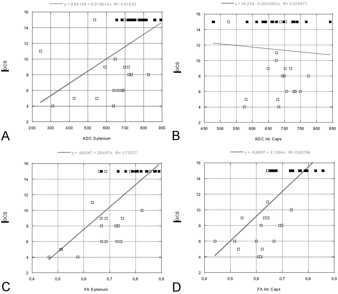
- Fig 4.
Linear regression plots of ADC and FA values of the splenium and internal capsule versus GCS at the time of acute MR imaging (in patients) or at time of comparison MR imaging (control subjects, all with GCS scores of 15). A statistically significant correlation is seen between the FA values of the splenium/internal capsule and GCS, as well as between the ADC values within the splenium and GCS. GCS scores vary between 3 and 15, where 3 represents the worst score, and 15, the best score. Open rectangles indicate patients; solid rectangles, control subjects.
A, ADC splenium versus GCS.
B, ADC internal capsule versus GCS.
C, FA splenium versus GCS.
D, FA internal capsule versus GCS.
- Fig 5.
Linear regression plots of ADC and FA values of the splenium and internal capsule versus Rankin score at the time of discharge (in patients) or at time of comparison MR imaging (control subjects, all with Rankin scores of 0). A statistically significant correlation is seen between the FA values of the splenium/internal capsule and Rankin score, as well as between the ADC values of the splenium and Rankin score. Rankin scores vary between 0 and 5, where 0 represents the best score and 5, the worst score. Open rectangles indicate patients; solid rectangles, control subjects.
A, ADC splenium versus Rankin.
B, ADC internal capsule versus Rankin.
C, FA splenium versus Rankin.
D, FA internal capsule versus Rankin.


Tables
A: ADC values, mm2/sec ROI Left × 10−6 Right × 10−6 P Value Internal capsule 681 ± 81 667 ± 84 .642 Thalamus 715 ± 62 721 ± 59 .785 Putamen 684 ± 80 669 ± 73 .624 B: FA values ROI Left Right P Value Internal capsule 0.731 ± 0.069 0.721 ± 0.077 .761 Thalamus 0.371 ± 0.077 0.383 ± 0.078 .182 Putamen 0.287 ± 0.090 0.286 ± 0.074 .890 Note.—Table lists the mean ADC and FA values ± SD for all bilaterally measured ROIs for all control subjects. Results showed no statistically significant difference between the left- and right-sided ADC and FA measurements. A P value <.005 was considered to indicate a statistically significant difference. Analysis was performed by using a Wilcoxon signed-rank test.
A: ADC values, mm2/sec ROI Control Subjects, × 10−6 Patients, ×10−6 P Value Internal capsule 674 ± 81 683 ± 59 .732 Splenium* 769 ± 61 628 ± 74 .001 Thalamus 718 ± 60 719 ± 62 .975 Putamen 677 ± 75 707 ± 55 .184 B: FA values ROI Control Subjects Patients P Value Internal capsule* 0.725 ± 0.066 0.624 ± 0.072 <.001 Splenium* 0.808 ± 0.060 0.678 ± 0.010 .002 Thalamus 0.377 ± 0.075 0.401 ± 0.040 .147 Putamen 0.286 ± 0.081 0.307 ± 0.040 .257 Note.—Table lists the mean ADC and FA values ± SD for all patients and control subjects within the respective ROIs. Left- and right-sided ADC and FA values were averaged. A statistically significant ADC decrease is seen for the splenium, and a statistically significant FA decrease is seen for the internal capsule. A P value of <.005 was considered to indicate a statistically significant difference. Analysis was performed by using a two-tailed Wilcoxon rank sum test.
* Statistically significant difference.
Measurement GCS Score Rankin Score r Value P Value r Value P Value ADC internal capsule −0.070 .686 0.018 .914 FA internal capsule* 0.657 <.0001 −0.714 <.0001 ADC splenium* 0.515 .0015 −0.599 .0001 FA splenium* 0.725 <.0001 −0.694 <.0001 ADC thalamus −0.165 .342 0.147 .396 FA thalamus −0.163 .348 0.114 .513 ADC putamen −0.314 .065 0.280 .103 FA putamen −0.116 .504 0.187 .281 Note.—Table lists the correlation coefficient r between the measured ADC and FA values and GCS or Rankin score for different anatomic locations. There proved to be a statistically significant correlation between the ADC and FA values measured within the splenium and the GCS and Rankin score, as well as between the FA value of the internal capsule and the GCS or Rankin score. The correlation between the FA values and the GCS or Rankin score was stronger than for the corresponding ADC values. A correlation probability of P < .0025 indicates a statistically significantly correlation between the measured ADC- and FA-values and the GCS/Rankin score.
* Statistically significant correlation.
In this issue


Jump to section
Related Articles
Cited By...
- Multimodal magnetic resonance imaging characterizes clinical outcome in chronic traumatic brain injury
- Neuroprotective Effects of Naltrexone in a Mouse Model of Post-Traumatic Epilepsy
- Visual Cortical Area MT Is Required for Development of the Dorsal Stream and Associated Visuomotor Behaviors
- Visual Cortical Area MT is Required for Development of the Dorsal Stream and Associated Visuomotor Behaviours
- Mild Traumatic Brain Injury Disrupts Functional Dynamic Attractors of Healthy Mental States
- Differential Tractography as a Track-Based Biomarker for Neuronal Injury
- Long-Term White Matter Changes after Severe Traumatic Brain Injury: A 5-Year Prospective Cohort
- A Decade of DTI in Traumatic Brain Injury: 10 Years and 100 Articles Later
- Diffusion Tensor Imaging Detected Optic Nerve Injury Correlates with Decreased Compound Action Potentials after Murine Retinal Ischemia
- Forensic Application of Postmortem Diffusion-Weighted and Diffusion Tensor MR Imaging of the Human Brain in Situ
- Longitudinal changes of structural connectivity in traumatic axonal injury
- Diffusion tensor imaging of acute mild traumatic brain injury in adolescents
- Diffusion Tensor Imaging Reliably Detects Experimental Traumatic Axonal Injury and Indicates Approximate Time of Injury
- Answering the Call: The Influence of Neuroimaging and Electrophysiological Evidence on Rehabilitation
- Proton MR Spectroscopy and MRI-Volumetry in Mild Traumatic Brain Injury
- Whole-Brain N-Acetylaspartate: A Marker of the Severity of Mild Head Trauma
- Diffusion tensor imaging: Scientific advance, clinical tool, or just a pretty picture?