
Abstract
BACKGROUND AND PURPOSE: Although concussion is common among athletes, evidence-based methods for clinical evaluation, treatment, and recovery are lacking. We used a prospective, functional neuroimaging approach to assess sports-related concussion in which imaging was performed before injury so that brain changes resulting from concussion could be better understood.
METHODS: Neurophysiologic correlates of sports-related concussion were investigated in eight college football players by using functional MR imaging. Preseason baseline levels of blood oxygen level–dependent (BOLD) activity were acquired during the performance of a test battery that included mathematical, memory, and sensorimotor coordination tasks. Four players who had a concussion repeated these baseline procedures within 1 week of injury. The remaining control players were retested at the end of the season.
RESULTS: Specific neural signatures of concussion were detected in individual players by comparing postconcussion results to preconcussion baseline values. The validity of these indicators was confirmed by comparing them with the same measures in noninjured control subjects. When compared with control subjects, concussed players had marked within-subject increases in the amplitude and extent of BOLD activity during a finger-sequencing task. Effects were observed primarily in the parietal and lateral frontal and cerebellar regions.
CONCLUSION: Differences in neural functioning were observed in the absence of observed deficits in behavioral performance, suggesting that this approach may increase sensitivity to concussion compared with neuropsychological evaluation alone. Though preliminary, the proposed prospective neuroimaging approach may have great potential for understanding mild traumatic brain injury and identifying mechanisms underlying recovery.
The occurrence of mild traumatic brain injury (MTBI), or concussion, is pervasive among athletes participating in contact sports at all levels of organization (1–4). Although once considered a temporary disruption of function without long-term consequences (5, 6), it is now understood that concussion involves structural changes in the brain (7) that can result in persistent neurobehavioral impairment. This knowledge, along with a recognition of the cumulative effects of multiple concussions (8–10) and the possible deadly consequences of returning to play before recovering from a previous brain injury (11), has amplified the need for an objective, evidence-based approach for assessing and monitoring of MTBI in athletes.
Currently, there is considerable interest in the use of neuroimaging techniques to detect diffuse axonal injuries (12), which are thought to underlie concussion and related cognitive sequelae (13). Concussion occurs when rapid acceleration/deceleration of the head results in the stretching or sheering of nerve fibers. In animals, these forces result in diffuse, distributed damage to white matter, particularly near the gray matter–white matter boundary (14) and in the long white-matter tracts passing through the brain stem (15). However, because of the diffuse nature of these injuries, conventional imaging techniques such as MR imaging and CT have not been reliable for detecting anatomic evidence of injury in humans (16–19).
To date, changes in brain function associated with concussion have not been widely investigated as scientific or diagnostic tools. Neurophysiologic evaluation after concussion can play a critical role not only in furthering our understanding of the anatomy and pathophysiology of MTBI but also in clinically evaluating concussion severity and prognosis and in scientifically evaluating and establishing return-to-play criteria (20). Neurophysiologic studies of the functional correlates of concussion may additionally aid in forming a conceptual link between the known anatomic effects of closed head injury, as reported in the animal literature (14, 15, 21), and the behavioral and cognitive sequelae of MTBI in humans (4).
Because functional MR imaging (fMRI) provides task-specific information about neural function, it is well suited to detect functional abnormalities associated with concussion and can be tailored to address each patient’s specific and most prevalent complaints. In addition, fMRI is sensitive to deficits related to dynamic aspects of neural function that may be evident only under conditions of cognitive load or information processing. fMRI has an added advantage over other brain imaging techniques, such as PET and SPECT, because multiple sessions can be performed on a single subject within a short period of time. This promotes the implementation of prospective, neurophysiologic investigations in which preinjury baseline measures of neural function are obtained for each participant in anticipation of future deleterious events, such as brain injury. The importance of employing prospective methods to study concussion is demonstrated by recent neuropsychological investigations (22–24). Although now recognized as being critical to the evaluation of concussion in athletes, the detection of between-session differences within a single subject is somewhat novel in the area of functional neuroimaging. Nonetheless, it may prove a powerful advance in assessing and monitoring concussion for two primary reasons: First, this within-subject approach allows each player to act as his or her own control, thereby removing (or at least complementing) the need for large normative databases. Second, it may prove more sensitive in identifying pathologic changes within an individual that may be small relative to the typically large between-subject variability observed when physiologic data from larger subpopulations are combined (25–27). Such concussion-related changes in neural activity, while apparent within an individual, would therefore go undetected when compared with so-called normal subjects. Subtle changes in brain activity occurring within individuals can then be combined across individuals to detect consistent patterns of brain injury for a population.
We believe this preliminary study is the first investigation to utilize fMRI in a prospective study of concussion. It was meant primarily as a probe for determining the feasibility of this approach for future studies. A small group of collegiate football players at risk for concussion were studied using a minimal test battery before the start of the season and again if they had a concussion. Control players not receiving a concussion were examined again at the conclusion of the season. Given previous results suggesting a greater demand of neural resources after traumatic brain injury (28, 29), we hypothesized that concussion would result in increased neural activity within task-specific regions involved in higher level cognitive and sensorimotor processes. Furthermore, we hypothesized that this increase would not be apparent in control subjects.
Methods
Subjects
Eight healthy, undergraduate, male intercollegiate football players participated in this study. They were aged 19–23 years, with a mean age of 20 years. The participants played in field positions associated with a high risk of concussion, such as quarterback and running back, among others. All subjects reported being right handed. Informed consent was obtained according to the regulations of the ethical review board of Florida Atlantic University and University MRI.
All subjects (excluding one) underwent the testing procedure before the start of the season to obtain baseline values. Four players subsequently had concussions, as defined by the guidelines of the American Academy of Neurology (30). Three of the players had a concussion (one grade I and two grade II) during regular-season play and were imaged within 1 week of their injury. Because the fourth player (who had a grade II concussion) was not imaged during preseason testing, a baseline measure was obtained at the end of the season after all symptoms had long since subsided. The four players who did not suffer a concussion were retested at the end of the season (after 7 months) during a second baseline session.
Testing Battery
The test battery comprised a series of behavioral and cognitive tasks designed to elicit neural activation and evaluate basic memory, processing, and coordination abilities. The same battery was used during all imaging sessions. All stimuli were delivered to the subjects through a set of goggles attached to the head coil. The test battery consisted of three tasks: a finger sequencing task, a serial calculation task, and a digit symbol task.
Finger Sequencing Task.—
This task provided a test of sensorimotor coordination and memory. Subjects were shown the numbers 2, 3, 4, and 5 arranged in a pseudorandom order. These values corresponded to the second (index) and the fifth (pinky) digits of the hand, respectively. Subjects were required to make sequential finger-to-thumb opposition movements as quickly and accurately as possible following the prescribed sequence. The task was carried out by using the right hand, the left hand, and bimanually in alternating blocks of rest (30 seconds) and movement (30 seconds). An observer rated the subject’s performance, which was subjectively graded on the basis of 1) whether the subject performed the correct sequence, 2) the speed of performance, and 3) the fluidity of the movements.
Serial Calculation Task.—
Subjects were shown a paced addition and subtraction task to assess neural activation related to mental calculation. Following a rest period (30 seconds), a simple equation was displayed. This consisted of a random two-digit number in the range of 70–99 and either “+2” or “−7.” The subject was instructed to perform the calculation and remember the result. Every 2 seconds, the same +2 or −7 instruction was presented. The subject was to repeat the mathematical operation on the previous result and remember the new result. At the end of the presentation sequence (30 seconds), a single number was displayed for 6 seconds. Subjects indicated with simple hand signals whether this number was the same as the their final calculation (probability = 0.75).
Digit Span Task.—
The fMRI version of this working memory task was adapted from the digit span task of the Wechsler Adult Intelligence Scale (WAIS) III. On each trial, subjects were shown a set of five single-digit numbers in a serial fashion. Each digit was viewed for 0.5 second and presented every 3.3 seconds (memory set). After the final number in the memory set was displayed, a probe set of five numbers was presented simultaneously (duration = 1.5 seconds). The subject indicated with hand signals if the memory set and probe set matched exactly (probability that the sets included the same numbers in the same order = 0.6). Five trials were presented in each of four presentation blocks (36 seconds each) interleaved with blocks of rest (30 seconds).
Neuroimaging
Changes in neural activity were evaluated by measuring changes in local blood oxygen level–dependent (BOLD) contrast by using a 1.5-T unit (Signa; GE Medical Systems, Milwaukee, WI). During baseline studies, a spiral sequence with the following parameters was used: TE = 60 msc, flip angle (FA) = 90°, field of view (FOV) = 24 cm, and equivalent matrix size = 128 × 128. Twenty 5-mm-thick axial sections spaced 2.5 mm apart were selected so as to provide coverage of the entire brain. During the postconcussion and postseason studies, echo-planar images were acquired by using a single shot, gradient-echo, echo-planar pulse sequence (TE = 60 ms, FA = 90°, FOV = 24 cm, in-plane resolution = 64 × 64). The section thickness and spacing were the same as those used during spiral imaging.
Before functional imaging, high-resolution anatomical spoiled gradient recalled-echo in the steady-state (SPGR) images (TR/TE/NEX = 325/in phase/2, FA = 90°, FOV = 24 cm, thickness = 5 mm, spacing = 2.5 mm) were obtained at the same section locations as those of the functional images. These images served as the background onto which the functional information was displayed and were also used to coregister the functional scans onto anatomic three-dimensional (3D) SPGR axial images (TR/TE = 34/5, FA = 45°, FOV = 24 or 26 cm, resolution = 256 × 256, thickness = 2 mm) collected at the end of each experimental session. For final analysis, all images were resampled to a common resolution and coordinate space (31).
Analysis of functional data were performed by using AFNI software (32, 33). Preprocessing included motion detection and correction followed by spatial smoothing by convolution with a Gaussian kernel (full width at half maximum = 6 mm) and temporal filtering (low pass, 0.1 Hz). Task-related activity was determined by correlating the time series of each voxel with a model function created by convolving a hemodynamic response function with a vector comprising 1s when a stimulus was present and 0s otherwise.
Within-subject comparisons of BOLD activity were performed by first normalizing the correlation statistic r by using Fisher’s r-to-z transform as follows (Eq 1):  1) where x indicates the imaging session. The difference Zd between z′ scores were computed by the following statistic (Eq 2):
  2) where, in the case of concussed players, z′1 is from the baseline (nonconcussed) session and z′2 is from the concussed session. In the case of the control players, the subscripts 1 and 2 represent the first and second baseline sessions, respectively. The resulting difference statistic was then thresholded at Z > 2.3 and clustered with a minimum volume of 500 mm3.
Results
Behavior
Figure 1 shows performance results of the digit span task and calculation tasks. In general, subjects performed well on both the digit span and addition tasks and poorer for the more difficult subtraction task. Changes in performance across sessions were assessed with two-way repeated-measures analysis of variance tests performed on data from the concussed and control subjects separately by using factors of task (digit span, addition, and subtraction) and session (baseline 1 and either concussed or baseline 2 for the concussed or control groups, respectively). In the concussed group, there was no effect of session and a significant main effect for task (F2,18 = 16.6, P < .001). This was due to the poor performance on the subtraction task compared with the other tasks (baseline = 50% and postconcussion = 56%). For the control group, no significant session or task effects were found, and no interaction was found for either group.

Average performance scores (percentages) for concussed players do not change between baseline (white) and postconcussion (dark gray) sessions. Similarly, no change was observed for the control players when we compared the first baseline session (light gray) with the second baseline session (black). Performance on the digit span and addition tasks were similar and consistently above 80% correct for all sessions. The subtraction task appeared considerably more difficult with percentages of correct values well below those for the other tasks.
Performance measures for the sequencing tasks were less easily quantified. An experimenter inside the MR suite rated the accuracy of the sequence, the speed of the sequence, and the fluidity of movement. All subjects performed movements at an acceptable rate, typically around one to two movements per second. In addition, all subjects performed the sequence in the correct order. Although there was some variability between subjects, performance within a subject, from session to session, was consistent. Thus, like the previous behavioral tests, the sequencing tasks were not particularly sensitive to the presence of concussion.
Functional Imaging
Mean Activation Patterns.—
To characterize the basic pattern of BOLD activity generated for each task, average activation maps were calculated from the baseline conditions of all subjects. In Figure 2, task-related activations are overlaid in color on a 3D representation of one subject’s brain. With the exclusion of addition, the patterns observed were robust and consistent with those reported in the literature for the performance of similar tasks. The relative simplicity of the addition-by-2s task, however, resulted in extremely low levels of task-related activity in individual maps and a general absence of detectable activity in the mean map. As a consequence, data collected during the performance of this task were removed from further analysis.

Significant mean task-related BOLD responses for all subject and all tasks except addition, which failed to show any consistent activity. Maps were derived from the average of the baseline images of each subject. Activity is overlaid in color on a 3D rendering of a single subject’s brain viewed from the top. Activity displayed for the sequencing tasks include contralateral precentral and postcentral gyrus, bilateral middle frontal gyrus (MFG), medial frontal gyrus (medFG), and inferior parietal lobe (IPL). The subtraction and digit span tasks activated similar areas, including prefrontal cortex (PFC), IPL, amd MFG. The digit span task also recruited areas in visual cortex.
The three sequencing tasks (Fig 2) demonstrated similar patterns of activity, with clusters found in medial frontal gyrus (corresponding to the supplementary motor area [SMA]), left middle frontal gyrus, contralateral precentral gyrus (bilateral for bimanual), left inferior frontal gyrus, bilateral superior parietal lobe (extending into the inferior parietal regions), and ipsilateral cerebellum (bilateral for bimanual, not shown). These regions were described previously and are consistent with other reports using similar finger sequencing tasks (34).
The network activated in support of the subtraction task was consistent with reports of the neural basis of numerical calculation (35–37). Activation was found bilaterally in dorsal prefrontal and premotor regions, the anterior portion of the medial frontal gyrus (preSMA), bilateral inferior and superior parietal areas, bilateral occipital gyrus (reflecting frequent visual stimuli), and left declive (not shown).
The digit span task (Fig 2) activated a large bilateral frontoparietal network commonly implicated in working memory tasks (38). This network includes the bilateral dorsal prefrontal cortex, premotor cortex, anterior portion of the SMA, and bilateral inferior parietal lobes. In addition, activity was also found in the bilateral inferior frontal gyrus, thalamus, and both superior and inferior aspects of the cerebellum.
Concussion-Dependent Increases in BOLD Signal Intensity
Although within-subject increases in BOLD signal intensity were observed for most subjects tested on at least a subset of the tasks, increases were considerably greater for concussed players, as shown for two representative subjects in Figure 3. In the control subject, increases in activity were relatively small and, in the case shown, restricted to the SMA and a small region of the left dorsal premotor cortex. In striking contrast, the concussed individual demonstrated large, extensive increases in regions associated with executive functioning, such as working memory and motor planning. These areas included the SMA, bilateral premotor cortex, superior and inferior parietal regions, and bilateral aspects of the cerebellum.

Representative individual Z-score differences between baseline and either a postconcussion session (concussed, left) or postseason baseline sessions (control, right). Colored areas show regions of activity that significantly increased from the baseline value of the bimanual sequencing task. Although both concussed and control subjects demonstrate some increases, those of the concussed player are considerably larger. Activity is significantly increased in the medial frontal gyrus (medFG), middle frontal gyrus (MFG), inferior parietal lobe (IPL), and bilateral cerebellum.
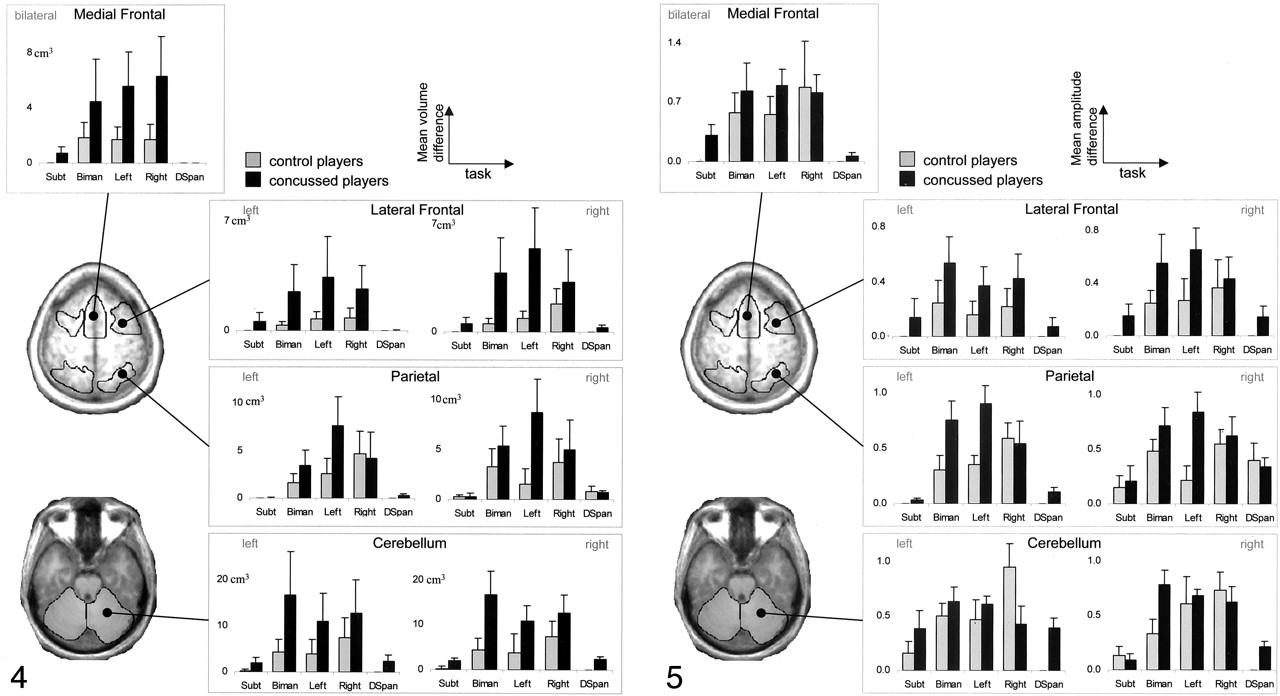
To better quantify within-subject differences observed during all tests in the battery for both groups, we performed a simple region-of-interest (ROI) analysis. Means of both the amplitude and extent (number of voxels) of Z-score differences were calculated in each region and combined across subjects. Nine regions were selected on the basis of a priori knowledge of the neural regions involved in performance of the tasks and post hoc inspection of the difference maps. These regions included a midfrontal ROI (SMA and cingulate motor area) in conjunction with ROIs in the right and left middle frontal gyrus (including prefrontal cortex), bilateral parietal regions (inferior and superior parts), and left and right cerebellum.
Extent of Within-Subject Increases in Activity
Figure 4 shows the mean (across-subject) volume of active tissue in each ROI and tasks for the control and concussed groups. The relatively small number of subjects in this initial work hindered the use of strict statistical quantification of the differences between groups. Therefore, the results were, of necessity, mostly descriptive. Nonetheless, despite the small sample size, clear differences between groups were seen. Overall, the number of voxels showing an increase in activity from baseline was greater for concussed players than for nonconcussed players. For the subtraction and digit span tasks, virtually no increases in activity were observed for control players, with the small differences observed resulting primarily from increases in a single subject. Although increases for the concussed group were larger and more consistent (two subjects had an increase during subtraction and three during digit span), the effects were still small compared with results of the sequencing task.

The mean voxel volume of significant, within-subject BOLD increases in specific ROIs. Error bars show the between-subject standard error. Each plot shows activity changes for each task in an ROI. Stylized images on the left show the regions encompassed by each ROI for two axial sections. The volume of increase was greater for the concussed players (black) than for the control players (gray), particularly for sequencing tasks. Differences between groups were most prominent in frontal and parietal regions. Biman indicates bimanual; DSpan, digit span; Subt, subtraction.
For both groups, postconcussion increases in activation extent associated with the three sequencing tasks were larger and more consistent than those observed for the other tasks. Nonetheless, in the concussed players, the region of increase was larger than that of the control players, a result most pronounced in the left and bimanual sequencing conditions for which large increases (compared with controls) were observed across all ROIs. In the medial frontal ROI, increases were approximately the same for the left, right, and bimanual conditions, with concussed subjects having more than twice the area of increase than controls. In the lateral frontal ROIs, increases are more pronounced bilaterally for the left and bimanual conditions than for the less demanding, right-hand sequencing condition (34). In parietal ROIs, differences are more pronounced for the nondominant left hand, particularly in the right hemisphere. Finally, in the cerebellum, a larger area of increase was observed for concussed players for all sequencing conditions.
Amplitude of Within-Subject Increases in Activity
The mean increase in signal intensity from the baseline session to the postconcussion (concussed group) or postseason (control group) session was calculated using voxels from each ROI that showed a significant difference in Z score. Figure 5 shows the group-averaged values and the between-subject standard error. For both the subtraction and digit span tasks, the amplitude differences mirror the differences in area shown in Figure 4. This is not surprising because (as noted before) the control group had so few significant voxels from which to calculate the mean amplitude.

Amplitude difference (mean voxel intensity) between baseline and postbaseline sessions averaged within ROI and across subjects. Each plot shows activity changes for each task in an ROI. Stylized images on the left show the regions encompassed by the ROI for two axial sections. In general, concussed players (black) showed greater increases in amplitude compared with control subjects (gray); this was particularly evident in the bilateral lateral frontal and parietal regions. Biman indicates bimanual; DSpan, digit span; Subt, subtraction.
The results of the amplitude analysis for the sequencing tasks were in general agreement with the observed changes in activation volume; however, the increases observed were not as pronounced or consistent across ROI as those reported earlier. Differences between control and concussed groups were more prominent in the lateral frontal and parietal ROIs and appeared greater for the left-hand and bimanual conditions than for the right-hand condition. In the left lateral frontal ROI, the concussed group had a greater within-subject increase in mean voxel intensity than the control group for all sequencing tasks. In the right hemisphere, a similar result was seen for the bimanual and left hand conditions. Likewise, the bimanual and left-hand conditions resulted in substantial increase in amplitude for concussed subjects versus control subjects in the bilateral parietal ROI. The fact that frontal and parietal areas showed increases in amplitude and extent may suggest that these regions are more sensitive to the effects of concussion, a conjecture at least partially supported by previous reports (13). Moreover, the lack of difference between the two groups for the right hand indicates that the physiologic effects of concussion may be sensitive to task context; this is revealed more clearly in left-hand and bimanual conditions, variants that are more difficult and require additional neural resources (34).
Discussion
The goal of this study was to evaluate the use of fMRI for investigating concussion in athletes by using a novel prospective approach. These promising preliminary results emphasize the sensitivity of this approach in detecting physiologic correlates of mild concussive injuries. Specific neural signatures of concussion were detected within individual players by comparing postconcussion results with preconcussion baselines. The validity of these indicators was confirmed by comparing the findings with those of noninjured controls undergoing a similar baseline-versus-postseason examination protocol. Concussion-induced differences in neural functioning were observed in the absence of declines in behavioral performance, suggesting that this prospective approach may increase sensitivity to MTBI compared with neuropsychological evaluation alone.
Substantial within-subject increases in the amplitude and extent of the BOLD response were observed in injured subjects compared with noninjured controls. Increases in functional activity in patients with moderate-to-severe brain injury have been postulated to reflect the recruitment of additional neural resources in response to moderate processing loads (28, 39). Axonal damage after concussion is thought to reduce processing efficiency, thereby imposing additional demands on the neural networks involved in task performance. In turn, this apparent intrinsic increase in cognitive load enhances hemodynamic activity similar to that found in healthy subjects performing memory (38) and sensorimotor coordination tasks (34, 40–45) of increasing difficulty. In cases of mild concussions, such as those reported here and elsewhere (28, 39) in which little if any decrement in performance is observed, compensation in the neural network recruited during cognitive engagement seems to be required to maintain preinjury performance levels.
Concussion-related within-subject increases in BOLD activity during sequencing tasks were most dominant, in terms of extent and amplitude, in the bilateral inferior-superior parietal region and dorsal-lateral and frontal cortex. These areas comprise a network often identified in functional imaging studies of working memory in healthy populations (46, 47). Activity in this network during sequencing reflects the memory demands that stem from the need to perform online storage of a prescribed numerical pattern to produce the appropriate behavioral performance. Concussion-induced increases in activity in this network agree with previous functional imaging results of brain injury in which activations of injured and noninjured subjects were compared by using tasks involving working memory (28, 29, 39, 48). Physiologic evidence of working memory impairment is also in line with common complaints of short-term memory problems in those with traumatic brain injury.
In addition to its putative role in working memory, the parietal cortex is involved in the sensorimotor transformation of visual space (49, 50) and in the storage and manipulation of numerical information (36, 51). The other ROIs investigated, the supplementary motor cortex (52–54) and cerebellum (55–57), are also intimately involved in the preparation, performance, and timing of complex motor tasks. Activation of this network during finger sequencing likely reflects the process of translating visual numerical instruction into activation of the appropriate spatial sequence and the production of the correctly ordered motor response (34). Increased activity in these regions may reflect deficits in coordinated motor action, a finding compatible with previous work showing behavioral deficits in motor control and coordination as well as associated neurophysiologic changes (58). Taken together, these initial results provide preliminary support for the use of within-subject approaches in the study of the neurophysiologic consequences of MTBI and suggest that such approaches may be sensitive to processing deficits in both memory and motor tasks.
Among the tasks used in the testing battery, the motor sequencing tasks were the most sensitive to concussion. The addition task produced little or no significant increases in BOLD signal intensity during both baseline and follow-up sessions. This is likely attributable to the simplicity and automaticity of the task, which, because it places relatively little demand on neural resources, generates subsequently small hemodynamic changes. Subtraction, on the contrary, produced robust activity in regions consistent with those reported in the literature (35, 59). However, within-subject increases after concussion were rare with this task and present in only a single subject. Results from behavioral studies have suggested that the serial-7s task is too difficult and therefore not sensitive to concussion (60). Rather, it may be more valid as a test of calculation ability than of cognitive deficits (61). In fMRI, the subtraction task, by virtue of its difficulty, may result in maximum signal intensity by saturation of the neural response or by oxygen saturation of hemoglobin (62), thereby precluding activity changes sensitive to concussion.
A somewhat surprising finding was that within-subject increases in activity were also observed in control players, albeit to a lesser degree than in concussed players. Although the cause of these changes is unclear, BOLD signal intensity is an indirect measure of neural activity that depends on the hemodynamic response to cerebral oxygen metabolism, and as such, it is influenced by changes in a number of physiologic factors (62, 63). For instance, evidence from a rat study suggests that chronic exercise may increase metabolic capacity (64). In addition, characteristics of the MR machines or nonspecific effects related to the intervening academic semester (eg, changes in diet, sleeping patterns, arousal levels, and mental stimulation or lack thereof from classroom activity) might also affect the hemodynamic response.
We must also recognize the possibility that the increases observed here may reflect actual changes in neural function resulting from the accumulation of minor, subconcussive impacts. Despite no reports of concussion, control players likely had multiple, minor head impacts during the season. Whereas the negative effects of exposure to repeated concussive blows are well accepted (8, 65), the effect of multiple, subconcussive impacts is still a subject of debate and empirical inquiry. To date, however, the dominant view is that multiple, minor blows over time, as in boxing or soccer, can result in severe cognitive impairment (but see reference 66). This issue can be better addressed in future studies with close monitoring of players’ histories in conjunction with concurrent imaging of both concussed and control players.
Conclusions
fMRI shows promise as a valuable diagnostic and research tool in the assessment of concussion injuries in athletes. The data presented here represent the initial stages in developing a comprehensive research protocol for detecting, assessing, and tracking sports-related MTBI. This approach may also be pertinent in evaluating disorders arising from brain injuries such as post-traumatic stress disorder. The results are encouraging, providing a solid basis for the use of within-subject, prospective measures of physiologic changes associated with MTBI. In addition, these results demonstrate that physiologic markers of concussion can be detected within days after injury, indicating the usefulness of this approach in tracking progress and identifying mechanisms of recovery.
Footnotes
Supported by NIMH, Neurosciences Research Branch, grants MH42900 and MH01386.
References
- Received April 8, 2003.
- Accepted after revision October 2, 2003.
- Copyright © American Society of Neuroradiology
In this issue


Jump to section
Related Articles
Cited By...
- Structural abnormalities in thalamo-prefrontal tracks revealed by high angular resolution diffusion imaging predict working memory scores in concussed children
- Role of advanced neuroimaging, fluid biomarkers and genetic testing in the assessment of sport-related concussion: a systematic review
- Concussion is confusing us all
- What evidence exists for new strategies or technologies in the diagnosis of sports concussion and assessment of recovery?
- Impaired cerebral haemodynamic function associated with chronic traumatic brain injury in professional boxers
- Abnormal whole-brain functional networks in homogeneous acute mild traumatic brain injury
- Contributions of neuroimaging, balance testing, electrophysiology and blood markers to the assessment of sport-related concussion
- The effect of telephone counselling on reducing post-traumatic symptoms after mild traumatic brain injury: A randomised trial
- A validation of the post concussion symptom scale in the assessment of complex concussion using cognitive testing and functional MRI
- Summary and agreement statement of the 2nd International Conference on Concussion in Sport, Prague 2004
- Summary and agreement statement of the 2nd International Conference on Concussion in Sport, Prague 2004