Advertisement

Main menu

  • Home
  • Content
    • Current Issue
    • Accepted Manuscripts
    • Article Preview
    • Past Issue Archive
    • Video Articles
    • AJNR Case Collection
    • Case of the Week Archive
    • Case of the Month Archive
    • Classic Case Archive
  • Special Collections
    • AJNR Awards
    • ASNR Foundation Special Collection
    • Most Impactful AJNR Articles
    • Photon-Counting CT
    • Spinal CSF Leak Articles (Jan 2020-June 2024)
  • Multimedia
    • AJNR Podcasts
    • AJNR SCANtastic
    • Trainee Corner
    • MRI Safety Corner
    • Imaging Protocols
  • For Authors
    • Submit a Manuscript
    • Submit a Video Article
    • Submit an eLetter to the Editor/Response
    • Manuscript Submission Guidelines
    • Statistical Tips
    • Fast Publishing of Accepted Manuscripts
    • Graphical Abstract Preparation
    • Imaging Protocol Submission
    • Author Policies
  • About Us
    • About AJNR
    • Editorial Board
    • Editorial Board Alumni
  • More
    • Become a Reviewer/Academy of Reviewers
    • Subscribers
    • Permissions
    • Alerts
    • Feedback
    • Advertisers
    • ASNR Home

User menu

  • Alerts
  • Log in

Search

  • Advanced search
American Journal of Neuroradiology

American Journal of Neuroradiology

ASHNR American Society of Functional Neuroradiology ASHNR American Society of Pediatric Neuroradiology ASSR
  • Alerts
  • Log in

Advanced Search

  • Home
  • Content
    • Current Issue
    • Accepted Manuscripts
    • Article Preview
    • Past Issue Archive
    • Video Articles
    • AJNR Case Collection
    • Case of the Week Archive
    • Case of the Month Archive
    • Classic Case Archive
  • Special Collections
    • AJNR Awards
    • ASNR Foundation Special Collection
    • Most Impactful AJNR Articles
    • Photon-Counting CT
    • Spinal CSF Leak Articles (Jan 2020-June 2024)
  • Multimedia
    • AJNR Podcasts
    • AJNR SCANtastic
    • Trainee Corner
    • MRI Safety Corner
    • Imaging Protocols
  • For Authors
    • Submit a Manuscript
    • Submit a Video Article
    • Submit an eLetter to the Editor/Response
    • Manuscript Submission Guidelines
    • Statistical Tips
    • Fast Publishing of Accepted Manuscripts
    • Graphical Abstract Preparation
    • Imaging Protocol Submission
    • Author Policies
  • About Us
    • About AJNR
    • Editorial Board
    • Editorial Board Alumni
  • More
    • Become a Reviewer/Academy of Reviewers
    • Subscribers
    • Permissions
    • Alerts
    • Feedback
    • Advertisers
    • ASNR Home
  • Follow AJNR on Twitter
  • Visit AJNR on Facebook
  • Follow AJNR on Instagram
  • Join AJNR on LinkedIn
  • RSS Feeds

AJNR is seeking candidates for the AJNR Podcast Editor. Read the position description.

OtherBRAIN

Primitive Vertebrobasilar System Associated with a Ruptured Aneurysm

Kenji Yagi, Koichi Satoh, Junichiro Satomi and Shinji Nagahiro
American Journal of Neuroradiology May 2004, 25 (5) 781-783;
Kenji Yagi
  • Find this author on Google Scholar
  • Find this author on PubMed
  • Search for this author on this site
Koichi Satoh
  • Find this author on Google Scholar
  • Find this author on PubMed
  • Search for this author on this site
Junichiro Satomi
  • Find this author on Google Scholar
  • Find this author on PubMed
  • Search for this author on this site
Shinji Nagahiro
  • Find this author on Google Scholar
  • Find this author on PubMed
  • Search for this author on this site
  • Article
  • Figures & Data
  • Info & Metrics
  • Responses
  • References
  • PDF
Loading

Abstract

Summary: Persistent primitive lateral vertebrobasilar (PPLVB) anastomosis is a rare remnant of early fetal development whose occurrence, to our knowledge, has been reported only once. We now report a new variant in which the basilar artery was fed primarily by a PPLVB anastomosis because on one side the vertebrobasilar junction was incompletely developed and on the other it was aplastic. Aneurysmal rupture in this case may be attributable to excessive hemodynamic stress.

Fenestration and duplication are the unusual variations and configurations of previously reported intracranial vertebrobasilar systems (1–8). Remnants of a primitive vertebrobasilar anastomosis can result in rare duplications of the vertebrobasilar junction (2, 3, 5, 7, 8). The long tract of this rare anomalously persistent vessel was named persistent primitive lateral vertebrobasilar (PPLVB) anastomosis by De Caro et al (3). We now report a new variant in which the basilar artery (BA) was fed primarily by a PPLVB anastomosis.

Case Report

This 62-year-old man was admitted with sudden and transient loss of consciousness. He had undergone percutaneous transluminal coronary angioplasty for myocardiosclerosis 10 years earlier. On admission, he was alert but suffered severe headache and recurrent vomiting. Neurologic examination disclosed no deficits.

A CT scan revealed subarachnoid hemorrhage (SAH), and the patient underwent emergent four-vessel angiography. Bilateral carotid angiograms disclosed no anomalies. The left vertebral artery (VA) terminated at the posterior inferior cerebellar artery (PICA) (Fig 1) and the basilar artery (BA) was fed via an anomalous artery, recognized as a PPLVB anastomosis arising from the right VA. It passed laterally in parallel with the basilar trunk and connected vertically with the middle of the trunk at the level of the origin of the left anterior inferior cerebellar artery (AICA) (Fig 2). The BA was fed from the caudal and the cranial direction from the basilar-PPLVB anastomosis juncture; the flow was antegrade in the upper trunk and retrograde in the lower trunk. No normal junctions of the right VA and BA were demonstrated. There was a saccular aneurysm at the left basilar AICA contralateral to the basilar-PPLVB anastomosis jointure.

Fig 1.
  • Download figure
  • Open in new tab
  • Download powerpoint
Fig 1.

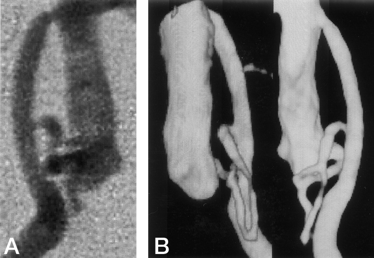
Left vertebral angiogram, lateral view. The left VA terminates at the PICA.

Fig 2.
  • Download figure
  • Open in new tab
  • Download powerpoint
Fig 2.

Right vertebral angiogram.

A, Early arterial phase.

B, Late arterial phase.

No normal vertebrobasilar junction is visualized. The BA is fed only via the PPLVB anastomosis. Flow is into the upper and lower BA via the BA-PPLVB anastomosis jointure. Note the saccular aneurysm at the left basilar anterior inferior cerebellar artery.

The patient underwent successful endovascular embolization of the aneurysm via the PPLVB anastomosis. He was discharged without any neurologic deficit.

Follow-up angiograms, obtained approximately 4 months later, again confirmed complete obliteration of the aneurysm. While the BA was primarily fed through the PPLVB anastomosis, 3D angiograms confirmed flow from the right VA to the BA (Fig 3). Some flow was delivered via anomalous, thin, meandering vessels that were duplicated at the level of the lower BA (Fig 4).

Fig 3.
  • Download figure
  • Open in new tab
  • Download powerpoint
Fig 3.

Schematic representation of the vertebrobasilar system. The arrows indicate the anomalous flow. The PPLVB anastomosis supplies most of the basilar artery flow with some flow delivered by anomalous, thin, hypoplastic arteries (asterisk).

Fig 4.
  • Download figure
  • Open in new tab
  • Download powerpoint
Fig 4.

Right vertebral angiogram depicting the vertebrobasilar system.

A, Digital subtraction angiogram in the anteroposterior view.

B, Three-dimensional angiograms in the left posterolateral oblique and posteroanterior view. Note the two indirect connections between the BA and the left VA, one through the PPLVB anastomosis, and the other through thin, serpentine arteries.

Discussion

Fenestration and duplication are documented vertebrobasilar system anomalies (2, 3, 5, 8) with some types of fenestration or duplication occurring at the vertebrobasilar junction (Fig 5). They derive from different fetal origins between the BA and the VA or the vertebobasilar junction.

Fig 5.
  • Download figure
  • Open in new tab
  • Download powerpoint
Fig 5.

Schema of fenestration or duplication in intracranial vertebrobasilar systems.

A, Basilar fenestration.

B, Duplication of the VA.

C, Premedullary duplication of the VA.

D, PPLVB anastomosis.

As shown in Figure 5A, the BA develops from the fusion of the primitive longitudinal neural artery. Incomplete fusion results in BA fenestration (1, 2, 4, 8) and a pathologic structural defect. Muscular gaps have been documented in the tunica media of the medial walls at the two ends of the fenestration (1, 4). These gaps and increased hemodynamic stress have been suggested as factors in the high incidence of aneurysms at the proximal end (1, 2, 4, 8).

According to Padget (9), the VA are formed during the 32nd–40th day of gestation: seven primitive cervical segmental arteries connect with each other and to the longitudinal neural arteries via bilateral channels that parallel the longitudinal neural arteries on each side to form the primitive VA. At this stage of development, there is a transient primitive vertebrobasilar anastomosis between the longitudinal neural artery and the first segmental arteries. It has been suggested that remnants of these anastomotic vessels result in intracranial VA or vertebrobasilar artery duplication (2, 7, 8).

On the basis of their angiographic studies, Lasjaunias et al (5, 10) presented schemas of 14 types of variation of the vertebrobasilar system. One type is the result of duplication of only the VA (Fig 5B), and another consists of duplication of the VA and the lower BA where a short segment of the persistent primitive vertebrobasilar anastomosis remains (Fig 5C). The latter is called premedullary duplication of the VA. De Caro et al (3) were the first to report another duplication formed with the remnant of the primitive long tract anastomosis between the right VA and the middle BA and named it PPLVB anastomosis (Fig 5D). To our knowledge, ours is only the second report of a PPLBV anastomosis.

In our case, both vertebrobasilar junctions were aplastic or hypoplastic, and the BA was primarily fed via the PPLVB anastomosis. The junction ipsilateral with the anastomosis manifested a thin, serpentine, anomalous, and duplicated artery, a variation that has not been reported elsewhere. We posit that in our patient vertebrobasilar connections failed to develop and primitive anastomotic arteries involuted during the 32nd–40th days of gestation.

An aneurysm developed at the site involved with this particular variation of the vertebrobasilar system. Although VA duplication results in a relatively high incidence of anomalies, including aneurysms (6, 7), we posit that our patient’s aneurysm development was related to the presence of a PPLVB anastomosis. The increased hemodynamic stress from the PPLVB anastomosis may have induced aneurysm formation and subsequent rupture, because the anastomosis supplied most of the blood flow to the BA and connected to it vertically.

The authors report the new vertebrobasilar variant accompanied with a ruptured aneurysm.

References

  1. ↵
    Black SPW, Ansbacher LE. Saccular aneurysm associated with segmental duplication of the basilar artery. J Neurosurg 1984;61:1005–1008
    PubMed
  2. ↵
    De Caro R, Parenti A, Munari PF. Fenestration of the vertebrobasilar junction. Acta Neurochir (Wien) 1991;108:85–87
    PubMed
  3. ↵
    De Caro R, Parenti A, Munari PF. Persistent primitive lateral vertebrobasilar anastomosis. Acta Neurochir (Wien) 1996;138:592–594
    PubMed
  4. ↵
    De Caro R, Serafini MT, Galli S, et al. Anatomy of segmental duplication in the human basilar artery: possible site of aneurysm formation. Clin Neuropathol 1995;14:303–309
    PubMed
  5. ↵
    Lasjaunias P, Braun JP, Hasso AN, et al. True and false fenestration of the vertebral artery. J Neuroradiol 1980;7:157–166
    PubMed
  6. ↵
    Miyazaki S, Kamata K, Yamaura A. Multiple aneurysms of the vertebrobasilar system associated with fenestration of the vertebral artery. Surg Neurol 1981;15:192–195
    CrossRefPubMed
  7. ↵
    Mizukami M, Tomita T, Mine T, Mihara H. Bypass anomaly of the vertebral artery associated with cerebral aneurysm and arteriovenous malformation. J Neurosurg 1972;37:204–209
    PubMed
  8. ↵
    Sanders WP, Sorek PA, Mehta BA. Fenestration of intracranial arteries with special attention to associated aneurysms and other anomalies. AJNR Am J Neuroradiol 1993;14:675–680
    Abstract/FREE Full Text
  9. ↵
    Padget DH. The development of cranial arteries in the human embryo. Contrib Embryonol Carneg Inst 1948;221:205–262
  10. ↵
    Lasjaunias P, Guibert-Tranier F, Braun JP. The pharyngocerebellar artery or ascending pharyngeal artery origin of the posterior inferior cerebellar artery. J Neuroradiol 1981;8:317–325
    PubMed
  • Received June 23, 2003.
  • Accepted after revision August 23, 2003.
  • Copyright © American Society of Neuroradiology
View Abstract
PreviousNext
Back to top

In this issue

American Journal of Neuroradiology: 25 (5)
American Journal of Neuroradiology
Vol. 25, Issue 5
1 May 2004
  • Table of Contents
  • Index by author
Advertisement
Print
Download PDF
Email Article

Thank you for your interest in spreading the word on American Journal of Neuroradiology.

NOTE: We only request your email address so that the person you are recommending the page to knows that you wanted them to see it, and that it is not junk mail. We do not capture any email address.

Enter multiple addresses on separate lines or separate them with commas.
Primitive Vertebrobasilar System Associated with a Ruptured Aneurysm
(Your Name) has sent you a message from American Journal of Neuroradiology
(Your Name) thought you would like to see the American Journal of Neuroradiology web site.
CAPTCHA
This question is for testing whether or not you are a human visitor and to prevent automated spam submissions.
Cite this article
Kenji Yagi, Koichi Satoh, Junichiro Satomi, Shinji Nagahiro
Primitive Vertebrobasilar System Associated with a Ruptured Aneurysm
American Journal of Neuroradiology May 2004, 25 (5) 781-783;

Citation Manager Formats

  • BibTeX
  • Bookends
  • EasyBib
  • EndNote (tagged)
  • EndNote 8 (xml)
  • Medlars
  • Mendeley
  • Papers
  • RefWorks Tagged
  • Ref Manager
  • RIS
  • Zotero
0 Responses
Respond to this article
Share
Bookmark this article
Primitive Vertebrobasilar System Associated with a Ruptured Aneurysm
Kenji Yagi, Koichi Satoh, Junichiro Satomi, Shinji Nagahiro
American Journal of Neuroradiology May 2004, 25 (5) 781-783;
del.icio.us logo Twitter logo Facebook logo Mendeley logo
  • Tweet Widget
  • Facebook Like
  • Google Plus One

Jump to section

  • Article
    • Abstract
    • Case Report
    • Discussion
    • References
  • Figures & Data
  • Info & Metrics
  • Responses
  • References
  • PDF

Related Articles

  • No related articles found.
  • PubMed
  • Google Scholar

Cited By...

  • No citing articles found.
  • Crossref
  • Google Scholar

This article has not yet been cited by articles in journals that are participating in Crossref Cited-by Linking.

More in this TOC Section

  • Usefulness of Quantitative Susceptibility Mapping for the Diagnosis of Parkinson Disease
  • White Matter Alterations in the Brains of Patients with Active, Remitted, and Cured Cushing Syndrome: A DTI Study
  • Qualitative and Quantitative Analysis of MR Imaging Findings in Patients with Middle Cerebral Artery Stroke Implanted with Mesenchymal Stem Cells
Show more BRAIN

Similar Articles

Advertisement

Indexed Content

  • Current Issue
  • Accepted Manuscripts
  • Article Preview
  • Past Issues
  • Editorials
  • Editor's Choice
  • Fellows' Journal Club
  • Letters to the Editor
  • Video Articles

Cases

  • Case Collection
  • Archive - Case of the Week
  • Archive - Case of the Month
  • Archive - Classic Case

Special Collections

  • AJNR Awards
  • ASNR Foundation Special Collection
  • Most Impactful AJNR Articles
  • Photon-Counting CT
  • Spinal CSF Leak Articles (Jan 2020-June 2024)

More from AJNR

  • Trainee Corner
  • Imaging Protocols
  • MRI Safety Corner

Multimedia

  • AJNR Podcasts
  • AJNR Scantastics

Resources

  • Turnaround Time
  • Submit a Manuscript
  • Submit a Video Article
  • Submit an eLetter to the Editor/Response
  • Manuscript Submission Guidelines
  • Statistical Tips
  • Fast Publishing of Accepted Manuscripts
  • Graphical Abstract Preparation
  • Imaging Protocol Submission
  • Evidence-Based Medicine Level Guide
  • Publishing Checklists
  • Author Policies
  • Become a Reviewer/Academy of Reviewers
  • News and Updates

About Us

  • About AJNR
  • Editorial Board
  • Editorial Board Alumni
  • Alerts
  • Permissions
  • Not an AJNR Subscriber? Join Now
  • Advertise with Us
  • Librarian Resources
  • Feedback
  • Terms and Conditions
  • AJNR Editorial Board Alumni

American Society of Neuroradiology

  • Not an ASNR Member? Join Now

© 2025 by the American Society of Neuroradiology All rights, including for text and data mining, AI training, and similar technologies, are reserved.
Print ISSN: 0195-6108 Online ISSN: 1936-959X

Powered by HighWire